CARDIAC SEPTUM AR ATLAS
Interactive 3D Version Available
Explore this anatomy in full 360° rotation with our interactive 3D viewer
View in 3D →What is the Cardiac Septum?
The heart is divided into chambers by muscular walls known as cardiac septa. These include the interatrial septum, which separates the left and right atria, and the interventricular septum, which divides the left and right ventricles. These septa are crucial for maintaining the unidirectional flow of blood, preventing the mixing of oxygen-rich and oxygen-poor blood between the chambers.
INTERATRIAL SEPTUM
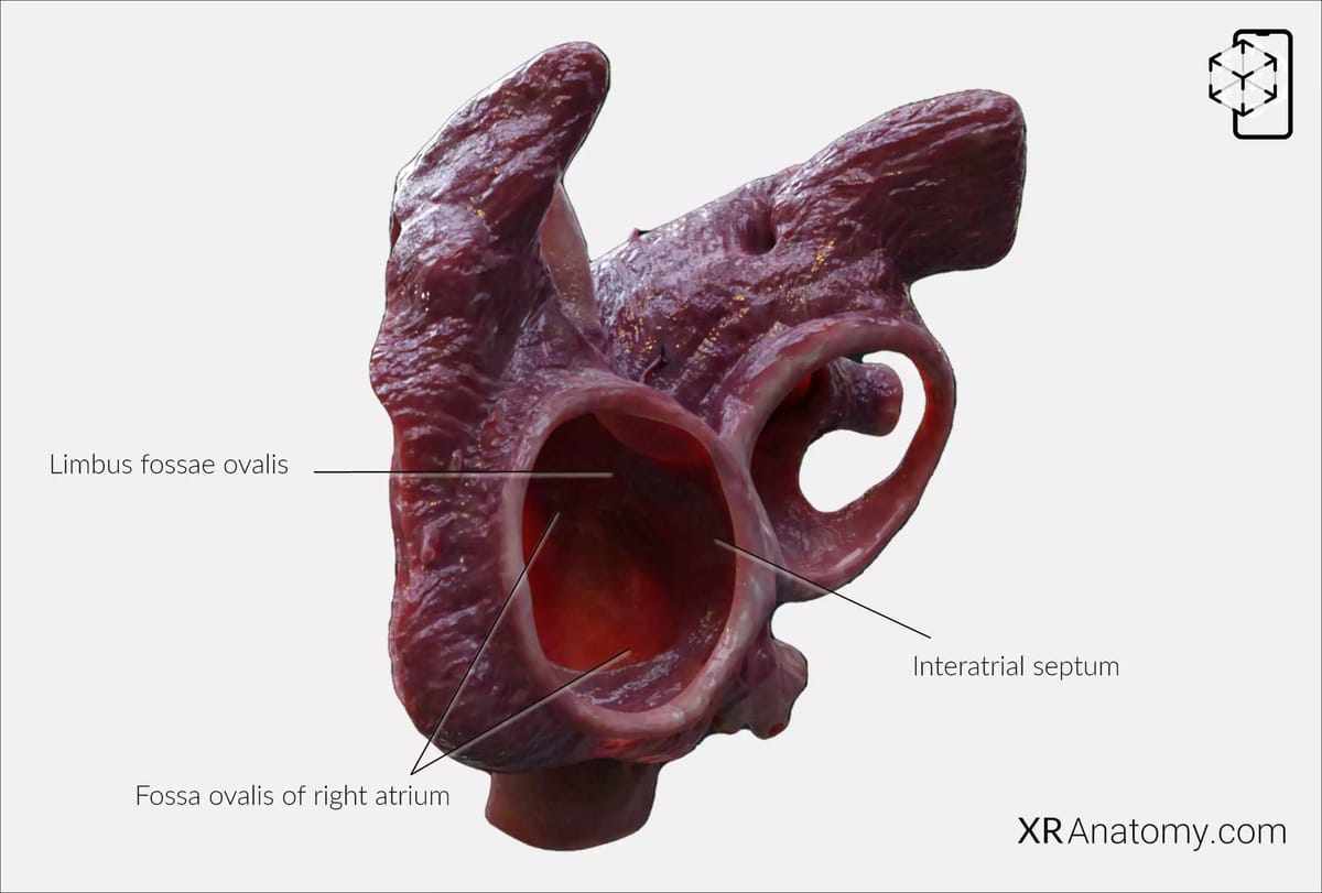
The interatrial septum is a thin, delicate wall that separates the right and left atria of the heart. This septum plays a vital role in ensuring that oxygenated and deoxygenated blood in the atria do not mix, maintaining efficient circulation.
Within this septum lies the fossa ovalis of the right atrium, a depression marking the site of the former foramen ovale—a crucial opening in fetal circulation that allows blood to bypass the non-functional fetal lungs. After birth, the foramen ovale typically closes, leaving behind the fossa ovalis as a remnant.
Surrounding the fossa ovalis is the limbus fossae ovalis, a prominent ridge that borders the oval fossa. This margin is more pronounced above and along the edges.
ATRIOVENTRICULAR SEPTUM
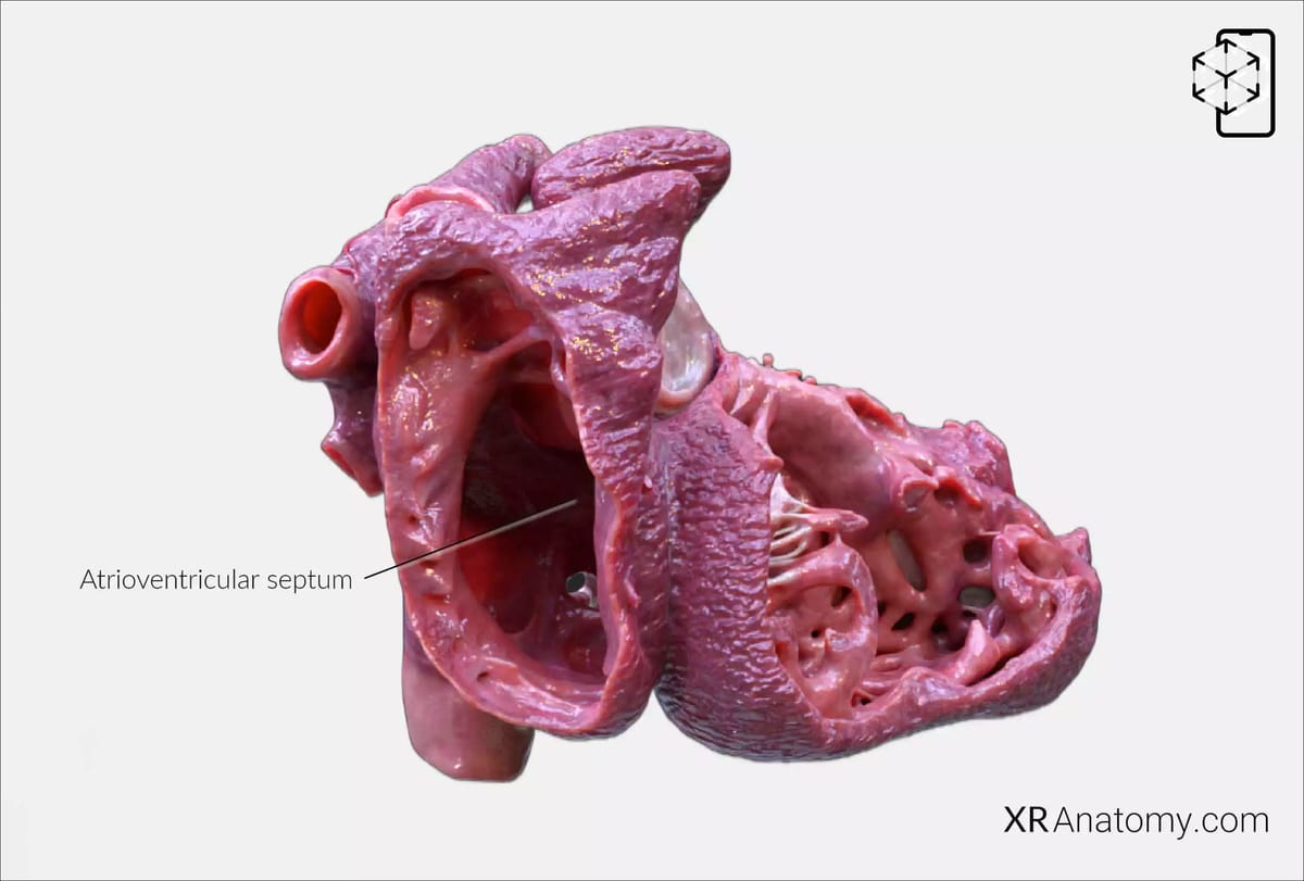
Due to a slight offset in the attachment of the atrioventricular valves—the tricuspid and mitral valves—a portion of the ventricular septum lies between the atria and ventricles. This area is known as the atrioventricular septum. Located posterior to the hinge of the tricuspid valve, it plays a critical role in maintaining the structural separation and proper electrical conduction between the upper and lower chambers of the heart.
INTERVENTRICULAR SEPTUM
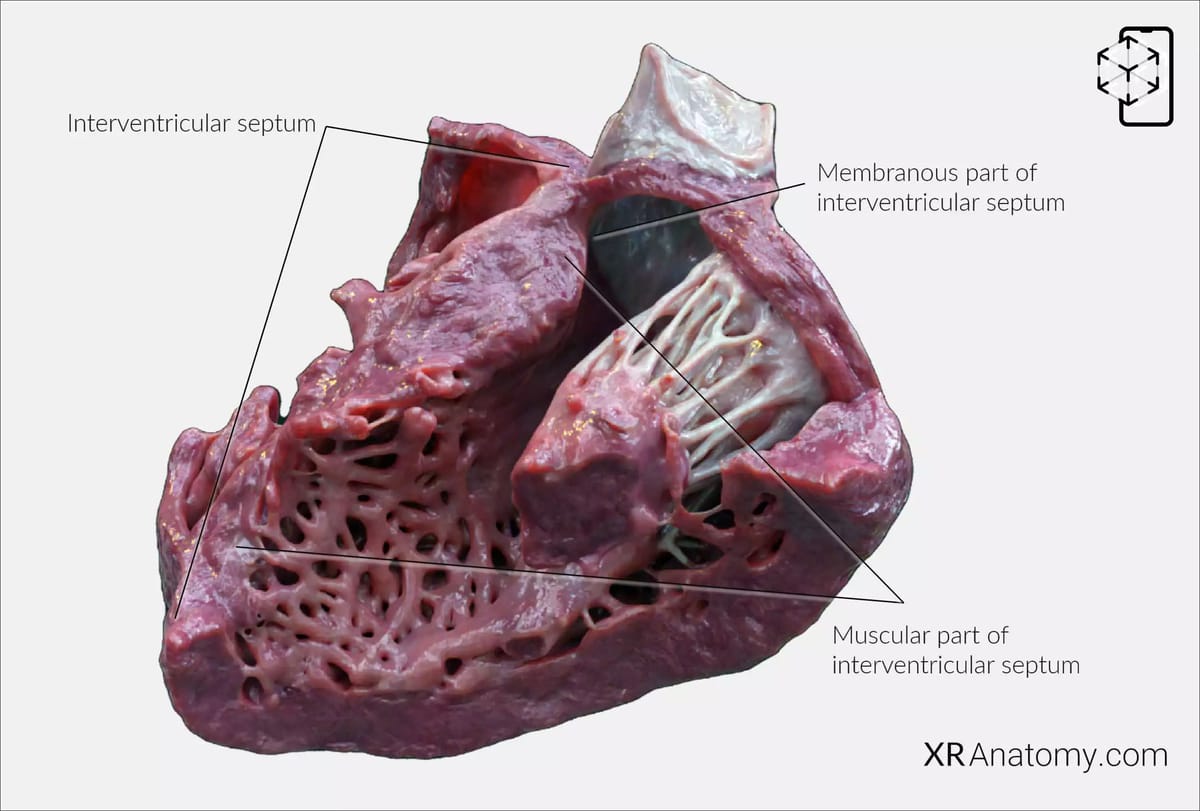
The interventricular septum is a robust, muscular wall that divides the left and right ventricles of the heart. It is essential for maintaining the separation of oxygen-rich blood in the left ventricle from oxygen-poor blood in the right ventricle, ensuring efficient and unidirectional blood flow through the circulatory system. This septum consists of two distinct parts: a thick muscular portion and a thin membranous portion.
The muscular part of the interventricular septum makes up the majority of the septum's structure, providing the necessary strength to withstand the high pressures generated during ventricular contractions.
The membranous part of the interventricular septum, composed primarily of fibrous tissue, represents the thinner section near the atrioventricular valves. This area is clinically significant as it is a common site for congenital defects known as ventricular septal defects, which can lead to abnormal blood flow between the ventricles if not corrected.
BIBLIOGRAPHY
1. Gray H, Lewis W. Angiology. In: Anatomy of the Human Body. 1918. p. 526–542.
2. Gosling JA, Harris PF, Humpherson JR, Whitmore I, Willan PLT. Human anatomy: color atlas and textbook. 6th ed. 2017. 45–58 p.
3. Anderson RH, Spicer DE, Hlavacek AM, Cook AC, Backer CL. (2013). Anatomy of the cardiac chambers. In Wilcox’s Surgical Anatomy of the Heart (4th ed., pp. 13–50). Cambridge University Press.
4. Fritsch H, Kuehnel W. Color Atlas of Human Anatomy. Vol. Volume 2, Color Atlas and Textbook of Human Anatomy. 2005. 10–42 p.
5. Moore K, Dalley A, Agur A. Clinically Oriented Anatomy. Vol. 7ed, Clinically Oriented Anatomy. 2014. 132–151 p.
6. Ho SYen. Anatomy for Cardiac Electrophysiologists: A Practical Handbook. Cardiotext Pub; 2012. 5–27 p.
7. Standring S, editor. Gray's Anatomy: The Anatomical Basis of Clinical Practice. 41st ed. London: Elsevier; 2016.
8. Moore KL, Agur AMR, Dalley AF. Essential Clinical Anatomy. 5th ed. Philadelphia: Wolters Kluwer; 2015.
