HEART SURFACES AND BORDERS AR ATLAS
TABLE OF CONTENTS
What are the Borders of the Heart?
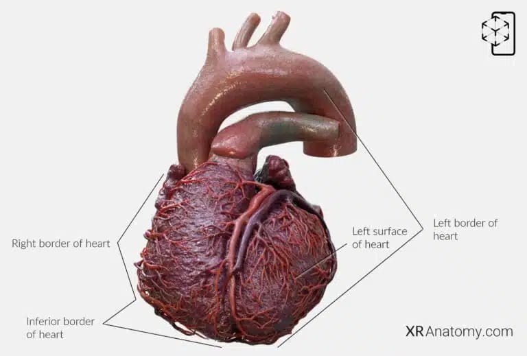
The heart, a vital organ responsible for pumping blood throughout the body, has several distinct borders and surfaces that are significant in understanding its anatomy and relationship with surrounding structures.
The right border of the heart is formed superiorly by the superior vena cava and mainly by the right atrium inferiorly. This border faces the right lung and is important in radiographic imaging to assess right atrial enlargement.
The inferior border of the heart, nearly horizontal in orientation, is primarily formed by the right ventricle. It rests on the diaphragm and separates the anterior (sternocostal) surface from the diaphragmatic (inferior) surface.
The left border of the heart outlines the heart's left side, including the curves formed by the aortic arch, pulmonary trunk, and left auricle, as well as the border of the left ventricle extending down to the apex of the heart. This border faces the left lung and is important in imaging to evaluate the size and shape of the left ventricle.
The left surface of the heart, also known as the pulmonary surface, is directed towards the left lung. It is mainly formed by the left ventricle and part of the left atrium.
HEART BASE
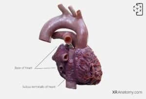
The base of the heart is the heart's posterior aspect, oriented posteriorly and to the right. It is mainly formed by the left atrium, which receives the pulmonary veins returning oxygenated blood from the lungs.
Running vertically along the right atrium is the sulcus terminalis of the heart, a groove that extends from the front of the superior vena cava to the front of the inferior vena cava. Internally, this corresponds to the crista terminalis, marking the an important junction of the right atrial wall.
The anterior surface of the heart, also known as the sternocostal surface, faces anteriorly towards the sternum and ribs. It is composed primarily of the right atrium and predominantly of the right ventricle, with a small contribution from the left ventricle.
Facing the right lung, the right surface of the heart is formed mainly by the right atrium. It extends between the superior and inferior vena cava and is important in considering right-sided heart conditions.
The anterior interventricular sulcus is a groove on the anterior surface of the heart marking the position where the right and left ventricles meet. It contains the anterior interventricular artery (left anterior descending artery) and the great cardiac vein.
The apex of the heart is the tip of the left ventricle, oriented anteriorly, downward, and to the left. It lies in the fifth intercostal space at the midclavicular line and is crucial in auscultation for heart sounds, particularly the mitral valve.
HEART: INFERIOR SURFACE

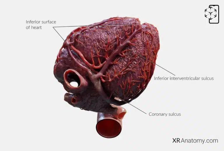
The inferior surface of the heart, also known as the diaphragmatic surface, faces downward and rests upon the diaphragm. It is composed mainly of the left ventricle and partly of the right ventricle. This surface is important clinically as it relates to the inferior wall of the heart, which can be affected in certain types of myocardial infarctions.
Running along this surface is the inferior interventricular sulcus, a groove marking the junction between the right and left ventricles on the diaphragmatic surface. It contains the posterior interventricular artery (usually a branch of the right coronary artery) and the middle cardiac vein.
Encircling the heart and demarcating the atria from the ventricles is the coronary sulcus, also known as the atrioventricular groove. This groove houses important vessels.
Related Structures
BIBLIOGRAPHY
1. Gray H, Lewis W. Angiology. In: Anatomy of the Human Body. 1918. p. 526–542.
2. Gosling JA, Harris PF, Humpherson JR, Whitmore I, Willan PLT. Human anatomy: color atlas and textbook. 6th ed. 2017. 45–58 p.
3. Anderson RH, Spicer DE, Hlavacek AM, Cook AC, Backer CL. (2013). Anatomy of the cardiac chambers. In Wilcox’s Surgical Anatomy of the Heart (4th ed., pp. 13–50). Cambridge University Press.
4. Fritsch H, Kuehnel W. Color Atlas of Human Anatomy. Vol. Volume 2, Color Atlas and Textbook of Human Anatomy. 2005. 10–42 p.
5. Moore K, Dalley A, Agur A. Clinically Oriented Anatomy. Vol. 7ed, Clinically Oriented Anatomy. 2014. 132–151 p.
6. Ho SYen. Anatomy for Cardiac Electrophysiologists: A Practical Handbook. Cardiotext Pub; 2012. 5–27 p.
7. Standring S, editor. Gray's Anatomy: The Anatomical Basis of Clinical Practice. 41st ed. London: Elsevier; 2016.
8. Moore KL, Agur AMR, Dalley AF. Essential Clinical Anatomy. 5th ed. Philadelphia: Wolters Kluwer; 2015.
