ROOT OF AORTA AR ATLAS
Interactive 3D Version Available
Explore this anatomy in full 360° rotation with our interactive 3D viewer
View in 3D →TABLE OF CONTENTS
ROOT OF AORTA
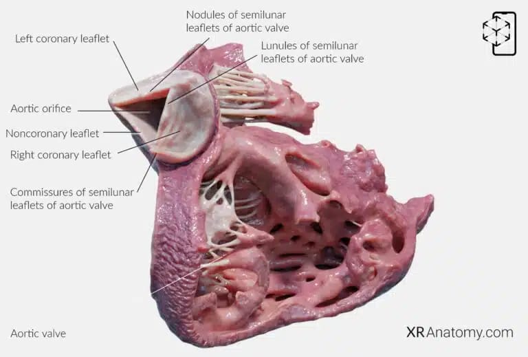
The root of the aorta is a critical structure forming the initial segment of the aorta as it emerges from the left ventricle. It encompasses the portion of the left ventricular outflow tract that supports the leaflets of the aortic valve. This area includes the aortic valve leaflets, the sinuses of Valsalva, and other components that facilitate the efficient flow of blood from the heart into the systemic circulation.
AORTIC VALVE
The aortic valve is a semilunar valve located between the left ventricle and the ascending aorta. It regulates blood flow from the heart into the aorta, ensuring that oxygen-rich blood is delivered to the body while preventing backflow into the left ventricle during diastole. The valve consists of three crescent-shaped leaflets: the right coronary leaflet, the left coronary leaflet, and the noncoronary leaflet. Each leaflet is associated with specific anatomical features and plays a role in the valve's function.
The aortic orifice is the opening through which blood exits the left ventricle to enter the aorta. The three leaflets of the aortic valve guard this orifice, opening and closing with each heartbeat to maintain proper blood flow.
The right coronary leaflet is associated with the right coronary artery,
The left coronary leaflet is associated with the left coronary artery.
The noncoronary leaflet does not give rise to a coronary artery but is essential in completing the valve structure and function.
AORTIC SINUSES
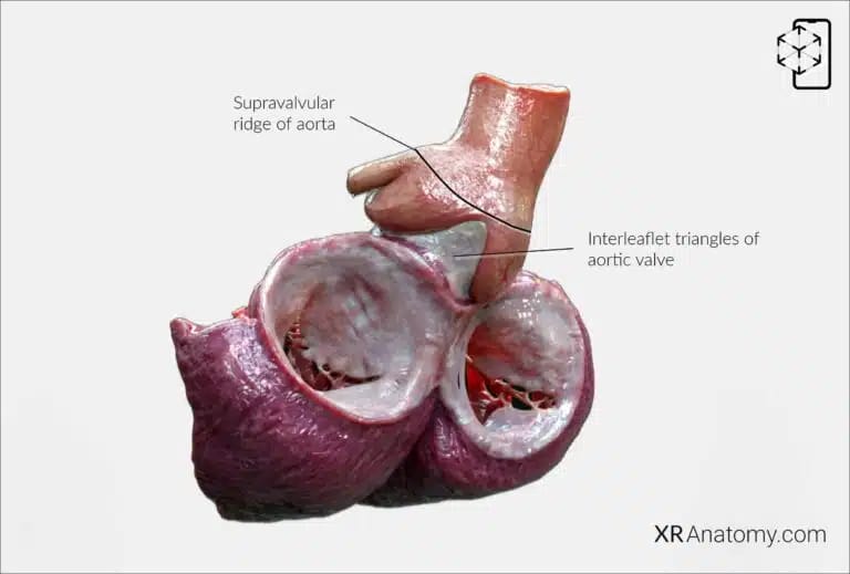
The aortic sinuses, also known as the sinuses of Valsalva, are three small dilations of the aortic wall located just above the aortic valve leaflets. Composed of elastic tissue, these sinuses are positioned behind each of the valve leaflets. They help in the closure of the valve and facilitate efficient blood flow into the coronary arteries. The sinuses are named based on their association with the coronary arteries: the right coronary sinus gives rise to the right coronary artery, the left coronary sinus gives rise to the left coronary artery, and the noncoronary sinus does not give rise to a coronary artery.
INTERLEAFLET TRIANGLES
The interleaflet triangles are fibrous structures located between the attachments of the aortic valve leaflets. These triangular areas correspond to the spaces where adjacent leaflets meet and are integral to the structural integrity of the aortic root. They allow for flexibility and proper movement of the valve leaflets during the cardiac cycle, contributing to the efficient opening and closing of the aortic valve.
The supravalvular ridge is a fibrous ring that marks the transition from the aortic root to the ascending aorta. It provides structural support and maintains the integrity of the aorta as it begins its course upward.
BIBLIOGRAPHY
1. Gray H, Lewis W. Angiology. In: Anatomy of the Human Body. 1918. p. 526–542.
2. Gosling JA, Harris PF, Humpherson JR, Whitmore I, Willan PLT. Human anatomy: color atlas and textbook. 6th ed. 2017. 45–58 p.
3. Anderson RH, Spicer DE, Hlavacek AM, Cook AC, Backer CL. (2013). Anatomy of the cardiac chambers. In Wilcox’s Surgical Anatomy of the Heart (4th ed., pp. 13–50). Cambridge University Press.
4. Fritsch H, Kuehnel W. Color Atlas of Human Anatomy. Vol. Volume 2, Color Atlas and Textbook of Human Anatomy. 2005. 10–42 p.
5. Moore K, Dalley A, Agur A. Clinically Oriented Anatomy. Vol. 7ed, Clinically Oriented Anatomy. 2014. 132–151 p.
6. Ho SYen. Anatomy for Cardiac Electrophysiologists: A Practical Handbook. Cardiotext Pub; 2012. 5–27 p.
7. Standring S, editor. Gray's Anatomy: The Anatomical Basis of Clinical Practice. 41st ed. London: Elsevier; 2016.
8. Moore KL, Agur AMR, Dalley AF. Essential Clinical Anatomy. 5th ed. Philadelphia: Wolters Kluwer; 2015.
