ROOT OF PULMONARY TRUNK AR ATLAS
Interactive 3D Version Available
Explore this anatomy in full 360° rotation with our interactive 3D viewer
View in 3D →ROOT OF PULMONARY TRUNK
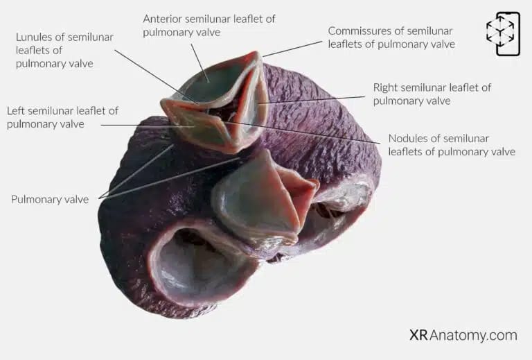
The root of the pulmonary trunk is the initial segment of the pulmonary artery as it arises from the right ventricle of the heart. This portion supports the pulmonary valve and is crucial in directing deoxygenated blood from the right ventricle to the lungs for oxygenation. The root includes structures such as the pulmonary valve leaflets and the proximal part of the pulmonary trunk, ensuring smooth blood flow and proper valve function.
PULMONARY VALVE
The pulmonary valve is a semilunar valve located between the right ventricle and the pulmonary artery. It plays a vital role in regulating blood flow from the heart to the lungs. Composed of three semilunar leaflets—the right, left, and anterior semilunar leaflets—it opens during ventricular systole to allow deoxygenated blood to flow into the pulmonary artery and closes during diastole to prevent backflow into the right ventricle.
The opening of the pulmonary trunk is the passage through which blood is ejected from the right ventricle into the pulmonary artery during ventricular contraction. This opening is guarded by the pulmonary valve, ensuring unidirectional flow.
The right semilunar leaflet is one of the three cusps of the pulmonary valve, named based on its anatomical position relative to the other leaflets.
Similarly, the left semilunar leaflet is named for its position on the left side of the valve,
The anterior semilunar leaflet is located at the front. Together, these leaflets function harmoniously to facilitate proper valve operation.
SINUSES OF PULMONARY TRUNK
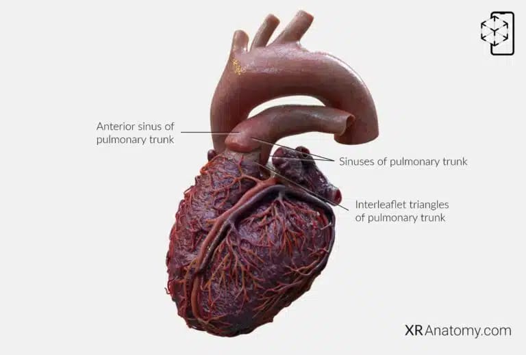
The sinuses of the pulmonary trunk are three small dilations located at the base of the pulmonary trunk, just behind each of the semilunar leaflets of the pulmonary valve. These sinuses are important in the valve's function.
The right sinus is named based on its anatomical position relative to the aorta. Similarly, the left sinus is located opposite the left side of the aorta. The anterior sinus is situated at the front, facing the sternum. These sinuses contribute to the efficient operation of the pulmonary valve by facilitating its opening and closing during the cardiac cycle.
INTERLEAFLET TRIANGLES
The interleaflet triangles of the pulmonary trunk are triangular areas of the arterial wall located between the attachments of the pulmonary valve leaflets. These specific regions contribute to the flexibility and movement of the valve leaflets, allowing them to open and close effectively during the cardiac cycle.
The supravalvular ridge of the pulmonary trunk is a circular region situated at the level of the commissures (points where the leaflets meet) of the semilunar leaflets of the pulmonary valve.
BIBLIOGRAPHY
1. Gray H, Lewis W. Angiology. In: Anatomy of the Human Body. 1918. p. 526–542.
2. Gosling JA, Harris PF, Humpherson JR, Whitmore I, Willan PLT. Human anatomy: color atlas and textbook. 6th ed. 2017. 45–58 p.
3. Anderson RH, Spicer DE, Hlavacek AM, Cook AC, Backer CL. (2013). Anatomy of the cardiac chambers. In Wilcox’s Surgical Anatomy of the Heart (4th ed., pp. 13–50). Cambridge University Press.
4. Fritsch H, Kuehnel W. Color Atlas of Human Anatomy. Vol. Volume 2, Color Atlas and Textbook of Human Anatomy. 2005. 10–42 p.
5. Moore K, Dalley A, Agur A. Clinically Oriented Anatomy. Vol. 7ed, Clinically Oriented Anatomy. 2014. 132–151 p.
6. Ho SYen. Anatomy for Cardiac Electrophysiologists: A Practical Handbook. Cardiotext Pub; 2012. 5–27 p.
7. Standring S, editor. Gray's Anatomy: The Anatomical Basis of Clinical Practice. 41st ed. London: Elsevier; 2016.
8. Moore KL, Agur AMR, Dalley AF. Essential Clinical Anatomy. 5th ed. Philadelphia: Wolters Kluwer; 2015.
