CARDIAC VEINS AR ATLAS
Interactive 3D Version Available
Explore this anatomy in full 360° rotation with our interactive 3D viewer
View in 3D →CARDIAC VEINS
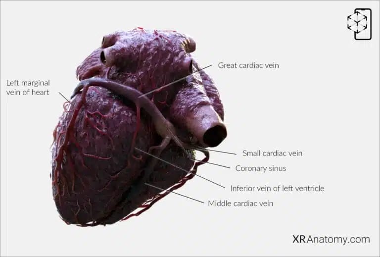
Cardiac veins: These are a network of veins that drain blood from the heart's muscle (myocardium) back into the right atrium. (1,3)
CORONARY SINUS
Coronary sinus: The primary vein of the heart, where the larger veins draining the myocardium empty into. It is situated between the left atrial wall and the ventricular myocardium. It drains into the right atrium. (1,2)
Great cardiac vein: A major vein that runs within the left atrioventricular groove alongside the left circumflex artery, draining blood from the left ventricular apex, anterior interventricular septum, anterior portions of both ventricles, and part of the left atrium. (4)
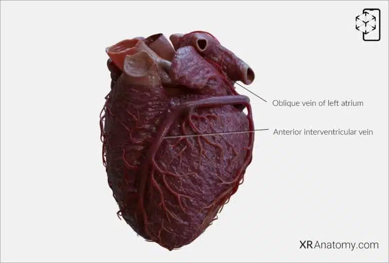
Anterior interventricular vein: Travels in the anterior interventricular groove with the left anterior descending coronary artery (LAD), draining blood from the left ventricular apex, anterior interventricular septum, anterior portions of both ventricles. (4)
Left marginal vein of heart: Drains the lateral and posterior/inferior wall of the left ventricle. (2,4)
Inferior vein of left ventricle: A tributary that drains the inferior wall of the left ventricle. It usually joins the great cardiac vein or the coronary sinus. (1,2,4)
Oblique vein of left atrium: A small vessel that collects blood from the inferior wall of the left atrium. It typically merges with either the great cardiac vein or the coronary sinus. (4)
Middle cardiac vein: Ascends with the inferior interventricular artery and drains into the coronary sinus. (1,2,4)
Small cardiac vein: Runs alongside the right coronary artery in the right atrioventricular groove and typically drains into the coronary sinus. This vein primarily drains the inferior wall of the right ventricle. (1,2,3)
Right marginal vein of heart: Runs alongside the right marginal artery, following the lateral wall of the right ventricle. (4)
ANTERIOR INTERVENTRICULAR VEIN
ANTERIOR CARDIAC VEINS
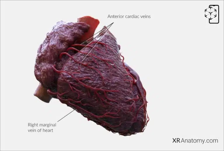
Anterior cardiac veins: A variable group of veins that drain a significant portion of the right ventricle, and they directly empty into the right atrium above the right atrioventricular groove. (4)
BIBLIOGRAPHY
1. Gray H, Lewis W. Angiology. In: Anatomy of the Human Body. 1918. p. 526–542.
2. Gosling JA, Harris PF, Humpherson JR, Whitmore I, Willan PLT. Human anatomy: color atlas and textbook. 6th ed. 2017. 45–58 p.
3. Anderson RH, Spicer DE, Hlavacek AM, Cook AC, Backer CL. (2013). Anatomy of the cardiac chambers. In Wilcox’s Surgical Anatomy of the Heart (4th ed., pp. 13–50). Cambridge University Press.
4. Fritsch H, Kuehnel W. Color Atlas of Human Anatomy. Vol. Volume 2, Color Atlas and Textbook of Human Anatomy. 2005. 10–42 p.
5. Moore K, Dalley A, Agur A. Clinically Oriented Anatomy. Vol. 7ed, Clinically Oriented Anatomy. 2014. 132–151 p.
6. Ho SYen. Anatomy for Cardiac Electrophysiologists: A Practical Handbook. Cardiotext Pub; 2012. 5–27 p.
7. Standring S, editor. Gray's Anatomy: The Anatomical Basis of Clinical Practice. 41st ed. London: Elsevier; 2016.
8. Moore KL, Agur AMR, Dalley AF. Essential Clinical Anatomy. 5th ed. Philadelphia: Wolters Kluwer; 2015.
