LEFT ATRIOVENTRICULAR VALVE AR ATLAS
Interactive 3D Version Available
Explore this anatomy in full 360° rotation with our interactive 3D viewer
View in 3D →TABLE OF CONTENTS
LEFT ATRIOVENTRICULAR VALVE

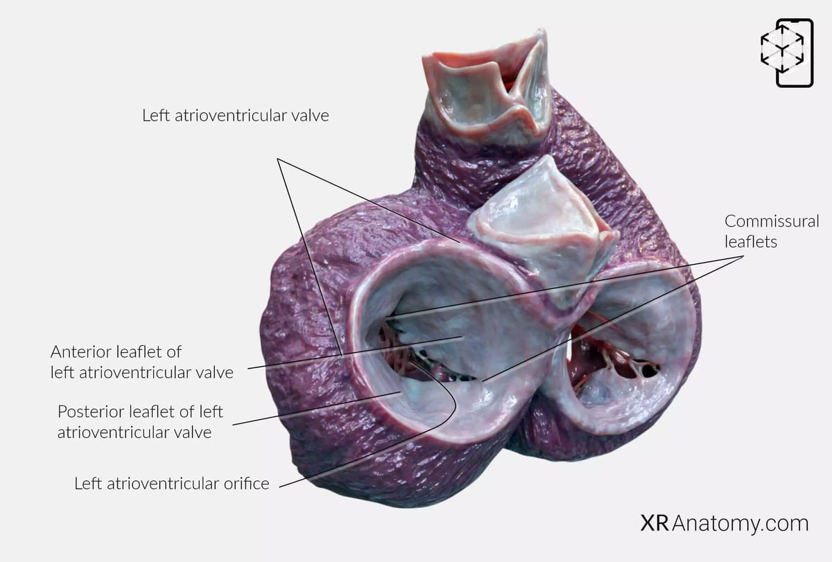
The left atrioventricular valve, commonly known as the mitral valve, plays a crucial role in the heart's function by regulating blood flow between the left atrium and the left ventricle. It ensures that oxygen-rich blood moves efficiently from the left atrium to the left ventricle without backflow during ventricular contraction. The valve surrounds the left atrioventricular orifice, an opening that facilitates this unidirectional flow. Structurally, the mitral valve is characterized by its two leaflets: the larger anterior leaflet and the smaller posterior leaflet.
- The anterior leaflet is positioned close to the aortic valve and is often subdivided into three sections—A1, A2, and A3—in surgical contexts to allow for precise identification and treatment during procedures.
- The posterior leaflet, while smaller, is similarly divided into three scallops—P1, P2, and P3—to aid in surgical descriptions and interventions.
- At the points where the anterior and posterior leaflets converge are the commissural leaflets. These smaller leaflets are located at the angles of the valve and are essential in sealing the orifice during ventricular contraction.
A crucial aspect of the mitral valve's function is the presence of a tension apparatus consisting of papillary muscles and chordae tendineae. This apparatus prevents the valve leaflets from inverting or prolapsing into the atrium during ventricular systole (contraction). The papillary muscles contract simultaneously with the ventricular myocardium, tightening the chordae tendineae and securing the leaflets in place. This coordinated action ensures efficient closure of the valve.
BIBLIOGRAPHY
1. Gray H, Lewis W. Angiology. In: Anatomy of the Human Body. 1918. p. 526–542.
2. Gosling JA, Harris PF, Humpherson JR, Whitmore I, Willan PLT. Human anatomy: color atlas and textbook. 6th ed. 2017. 45–58 p.
3. Anderson RH, Spicer DE, Hlavacek AM, Cook AC, Backer CL. (2013). Anatomy of the cardiac chambers. In Wilcox’s Surgical Anatomy of the Heart (4th ed., pp. 13–50). Cambridge University Press.
4. Fritsch H, Kuehnel W. Color Atlas of Human Anatomy. Vol. Volume 2, Color Atlas and Textbook of Human Anatomy. 2005. 10–42 p.
5. Moore K, Dalley A, Agur A. Clinically Oriented Anatomy. Vol. 7ed, Clinically Oriented Anatomy. 2014. 132–151 p.
6. Ho SYen. Anatomy for Cardiac Electrophysiologists: A Practical Handbook. Cardiotext Pub; 2012. 5–27 p.
7. Standring S, editor. Gray's Anatomy: The Anatomical Basis of Clinical Practice. 41st ed. London: Elsevier; 2016.
8. Moore KL, Agur AMR, Dalley AF. Essential Clinical Anatomy. 5th ed. Philadelphia: Wolters Kluwer; 2015.


