RIGHT VENTRICLE AR ATLAS
Interactive 3D Version Available
Explore this anatomy in full 360° rotation with our interactive 3D viewer
View in 3D →RIGHT VENTRICLE
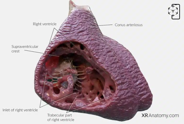
The right ventricle is one of the heart's four chambers, located anteriorly and forming the majority of the heart's anterior surface. It receives deoxygenated blood from the right atrium and pumps it into the pulmonary circulation via the pulmonary artery. Structurally, it has a triangular shape and consists of three main components: the inlet, the trabecular part, and the outlet. These components work together to ensure efficient blood flow to the lungs for oxygenation.
INLET OF RIGHT VENTRICLE
The inlet of the right ventricle includes the tricuspid valve apparatus, which comprises the tricuspid valve leaflets, chordae tendineae, and papillary muscles. This area ensures unidirectional blood flow from the right atrium into the right ventricle while preventing backflow during ventricular contraction. The supraventricular crest is a muscular ridge situated between the pulmonary valve and the tricuspid valve. It separates the inflow and outflow tracts of the right ventricle, playing a crucial role in directing blood flow efficiently through the heart.
The conus arteriosus, also known as the infundibulum, is the outflow component of the right ventricle. It is a conical pouch located in the upper part of the right ventricle that provides support for the pulmonary valve and ensures smooth blood flow into the pulmonary artery.
TRABECULAR PART OF RIGHT VENTRICLE
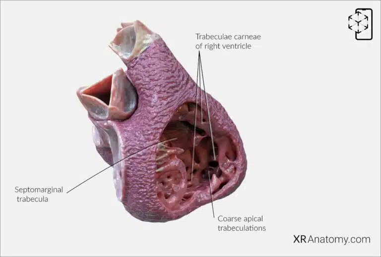
The trabecular part of the right ventricle is characterized by a network of muscular ridges known as trabeculae carneae. This area is thinner and extends down to the apex of the heart. The coarse trabeculations increase the surface area of the ventricular wall, enhancing its contractile ability and contributing to efficient blood flow.
The septomarginal trabecula, also known as the moderator band. It is a significant muscular band that extends from the interventricular septum to the anterior papillary muscle. This structure contains part of the right bundle branch of the heart's conducting system, facilitating coordinated contraction of the ventricle.
- The parietal limb of the septomarginal trabecula, or the posteroinferior limb, proceeds posteriorly. It supports the muscular septum, and the medial papillary muscle typically originates from this section, contributing to the stability and function of the tricuspid valve.
- The anterior limb of the septomarginal trabecula runs up to the attachment of the pulmonary valve leaflets. It is integral to the structure of the right ventricle, ensuring proper alignment and function of the outflow tract.
At the apex of the right ventricle, the coarse apical trabeculations are robust muscle bundles. These structures contrast with the finer trabeculations of the left ventricle and play a vital role in the mechanical efficiency of the right ventricle during contraction.
PAPILLARY MUSCLES OF RIGHT VENTRICLE

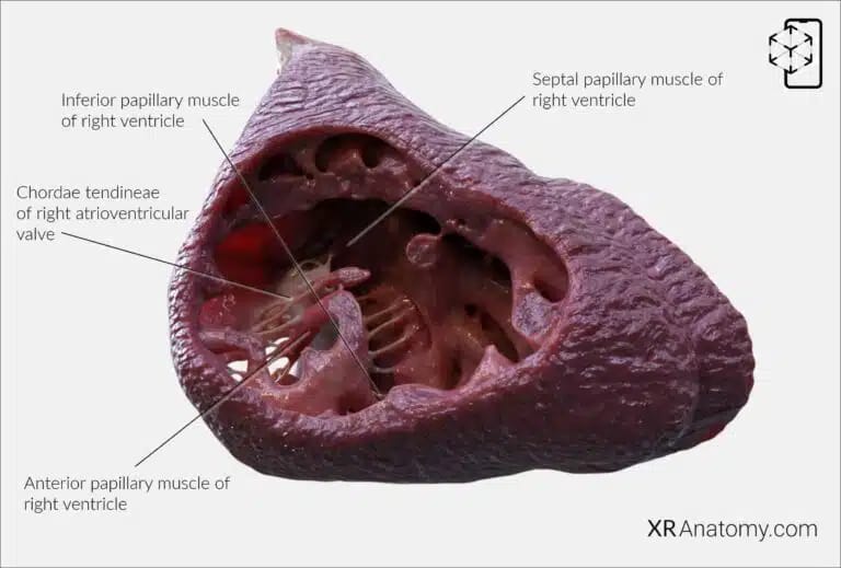
The papillary muscles of the right ventricle are conical muscular projections that extend into the ventricular lumen, connecting to the tricuspid valve leaflets via chordae tendineae. They are crucial in preventing valve prolapse during ventricular contraction by anchoring the valve leaflets.
There are three primary papillary muscles in the right ventricle:
- - The anterior papillary muscle is the largest and most significant. It originates from the anterior wall of the right ventricle and is supported by the septomarginal trabecula. This muscle connects to the anterior and posterior cusps of the tricuspid valve, ensuring their proper function.
- - The inferior (posterior) papillary muscle arises from the inferior wall of the right ventricle. It attaches to the posterior and septal cusps of the tricuspid valve, aiding in their stabilization during contraction.
- - The septal papillary muscle is the smallest and may vary in presence and size. It originates from the interventricular septum and connects to the anterior and septal cusps of the tricuspid valve.
The chordae tendineae of the right atrioventricular valve are fibrous cords that link the papillary muscles to the valve leaflets. They prevent the inversion of the leaflets into the atrium during systole, maintaining efficient valve closure and preventing regurgitation. Additionally, the ventricle contains chordae tendineae spuriae, or false chordae. These fibrous-muscular bundles are distinct from true chordae as they do not connect to the atrioventricular valves. They are more commonly observed in the left ventricle but can be present in the right ventricle.
BIBLIOGRAPHY
1. Gray H, Lewis W. Angiology. In: Anatomy of the Human Body. 1918. p. 526–542.
2. Gosling JA, Harris PF, Humpherson JR, Whitmore I, Willan PLT. Human anatomy: color atlas and textbook. 6th ed. 2017. 45–58 p.
3. Anderson RH, Spicer DE, Hlavacek AM, Cook AC, Backer CL. (2013). Anatomy of the cardiac chambers. In Wilcox’s Surgical Anatomy of the Heart (4th ed., pp. 13–50). Cambridge University Press.
4. Fritsch H, Kuehnel W. Color Atlas of Human Anatomy. Vol. Volume 2, Color Atlas and Textbook of Human Anatomy. 2005. 10–42 p.
5. Moore K, Dalley A, Agur A. Clinically Oriented Anatomy. Vol. 7ed, Clinically Oriented Anatomy. 2014. 132–151 p.
6. Ho SYen. Anatomy for Cardiac Electrophysiologists: A Practical Handbook. Cardiotext Pub; 2012. 5–27 p.
7. Standring S, editor. Gray's Anatomy: The Anatomical Basis of Clinical Practice. 41st ed. London: Elsevier; 2016.
8. Moore KL, Agur AMR, Dalley AF. Essential Clinical Anatomy. 5th ed. Philadelphia: Wolters Kluwer; 2015.
