ZYGOMATIC BONE
TABLE OF CONTENTS
Interactive 3D Version Available
Explore this anatomy in full 360° rotation with our interactive 3D viewer
View in 3D →What is the Zygomatic Bone?
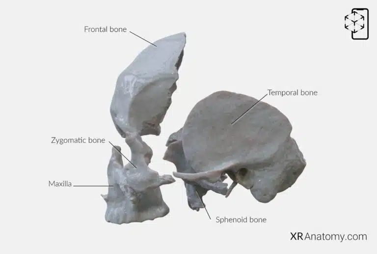
The , commonly known as the cheekbone, plays a crucial role in the facial skeleton by forming the prominence of the cheeks and contributing to the lateral wall and floor of the orbit. four bones, creating a strong and stable structure that supports facial expressions and protects vital sensory organs. These articulations are with: The frontal bone, The sphenoid bone, The maxilla, The temporal bone.
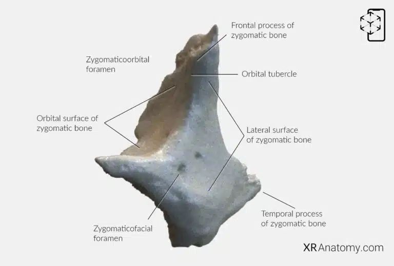
The zygomatic bone is a paired, diamond-shaped bone that forms the prominence of the cheeks and part of the lateral wall and floor of the orbit. Its is convex and smooth, constituting the outer aspect of the cheek. This surface is pierced by the , a small opening that transmits the zygomaticofacial nerve and vessels to the facial structures. Extending superiorly from the bone is the , a thin, elongated projection that articulates with the frontal bone. This process contributes to the formation of the lateral wall of the orbit and provides attachment points for facial muscles. The , located on the frontal process, is a bony prominence that serves as an attachment site for the lateral palpebral ligament and parts of the orbicularis oculi muscle, playing a role in eyelid movement. Projecting posteriorly, the extends to meet the zygomatic process of the temporal bone, together forming the zygomatic arch. This arch is significant in the attachment of muscles involved in mastication.
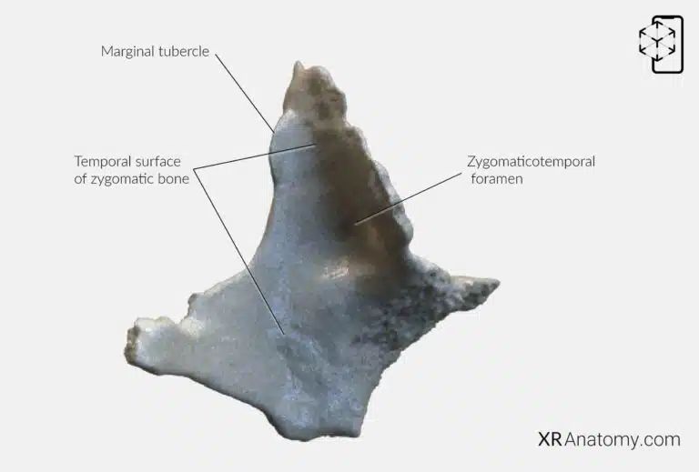
The temporal surface of the zygomatic bone is the inner aspect that faces the temporal bone, contributing to the formation of the temporal and infratemporal fossae. This surface is concave and houses the , which transmits the zygomaticotemporal nerve, supplying sensation to the skin of the temple region. On the posterior margin of the frontal process lies the , a raised area that provides additional surface area for muscle attachment and contributes to the bone's articulation with adjacent structures.
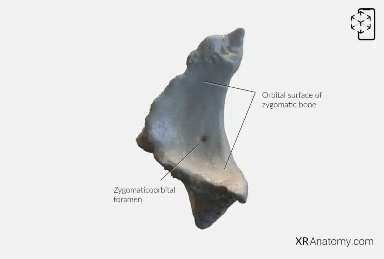
The forms a significant part of the lateral wall and floor of the orbit, contributing to the protection of the eye. This smooth surface features an opening known as that allows passage of the zygomatic nerve branches into the orbit.
BIBLIOGRAPHY
1. Henry G, Warren HL. Osteology. In: Anatomy of the Human Body. 20th ed. Philadelphia: Lea & Febiger; 1918. p. 129–97.
2. Sampson HW, Montgomery JL, Henryson GL. Atlas of the human skull. College Station: Texas A & M University Press; 2007.
4. Saylam C, Özer MA, Ozek C, Gurler T. Anatomical Variations of the Frontal and Supraorbital Transcranial Passages. Journal of Craniofacial Surgery. 2003;14(1):10–2.
5. Hosemann W, Gross R, Goede U, Kuehnel T. Clinical anatomy of the nasal process of the frontal bone (spina nasalis interna). Otolaryngology – Head and Neck Surgery. 2001;125(1):60–5.
6. Steele DG, Bramblett CA. The anatomy and biology of human skeleton. Texas A&M University Press; 1988.
7. Tersigni-Tarrant MTA, Shirley NR. Human osteology. Vol. 4, Forensic Anthropology: An Introduction. 2012. 33–68 p.
8. Monjas-Cánovas I, García-Garrigós E, Arenas-Jiménez JJ, Abarca-Olivas J, Sánchez-Del Campo F, Gras-Albert JR. Radiological Anatomy of the Ethmoidal Arteries: CT Cadaver Study. Acta Otorrinolaringologica (English Edition). 2011;62(5):367–74.
9. Pereira G, Lopes P, Santos A, Krebs. Morphometric aspects of the jugular foramen in dry skulls of adult individuals in Southern Brazil. Vol. 27, J. Morphol. Sci. 2010.
12. Kunc V, Fabik J, Kubickova B, Kachlik D. Vermian fossa or median occipital fossa revisited: Prevalence and clinical anatomy. Annals of Anatomy. 2020 May 1;229:151458.
14. Standring S. The skull. In: Gray’s anatomy: the anatomical basis of clinical practice. 2021st ed. Elsevier Health Sciences; 2021. p. 558–73.
15. Rhoton AL. Chapter 1 Overview of Temporal Bone. Neurosurgery. 2007;
17. Tóth M, Moser G, Patonay L, Oláh I. Development of the anterior chordal canal. Annals of Anatomy. 2006;
18. Carpenter G, Knipe H. Tympanic part of temporal bone. Radiopaedia.org. 2014 Mar 23;
19. Eckerdal O. The petrotympanic fissure: A link connecting the tympanic cavity and the temporomandibular joint. Cranio – Journal of Craniomandibular Practice. 1991;9(1):15–22.
21. Singh R, Kishore Gupta N, Kumar R. Morphometry and Morphology of Foramen Petrosum in Indian Population. Basic Sciences of Medicine. 2020;2020(1):8–9.
23. Piagkou M, Xanthos T, Anagnostopoulou S, Demesticha T, Kotsiomitis E, Piagkos G, et al. Anatomical variation and morphology in the position of the palatine foramina in adult human skulls from Greece. Journal of Cranio-Maxillofacial Surgery. 2012 Oct 1;40(7):e206–10.
