Anatomy
Cervical Vertebrae
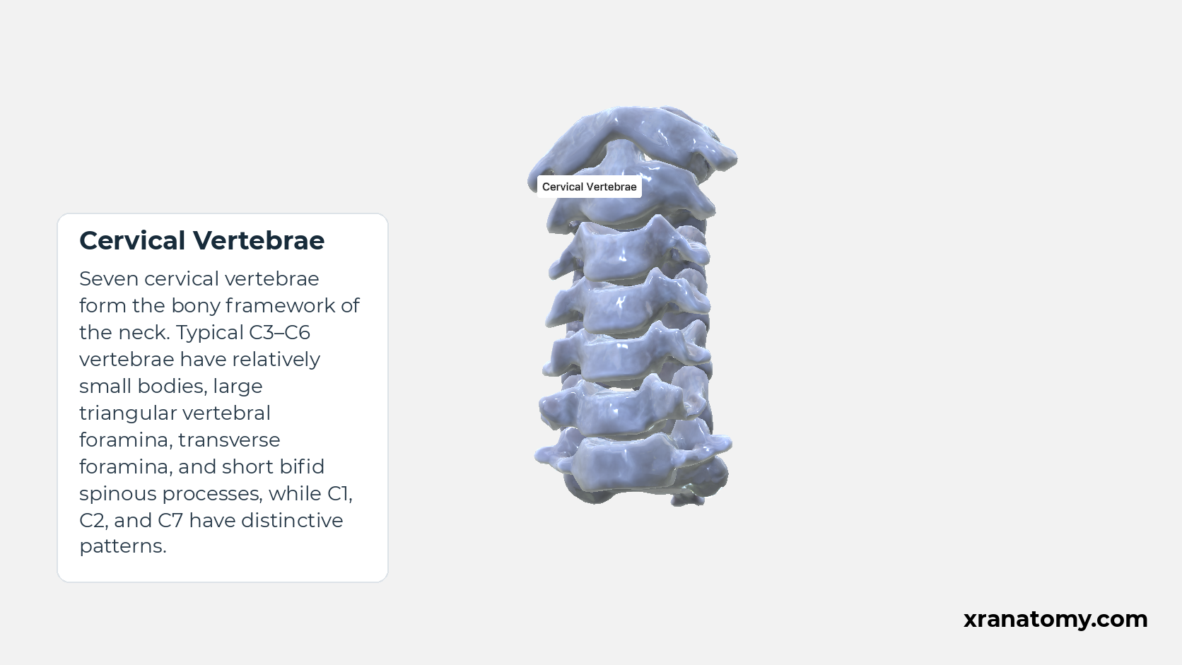
Seven cervical vertebrae form the bony framework of the neck. Typical C3–C6 vertebrae have relatively small bodies, large triangular vertebral foramina, transverse foramina, and short bifid spinous processes, while C1, C2, and C7 have distinctive patterns.
Cervical Overview
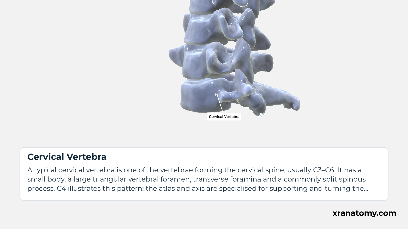
Cervical Vertebra
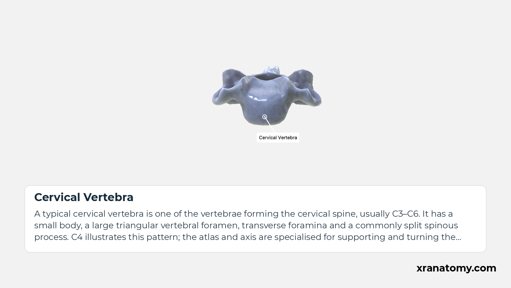
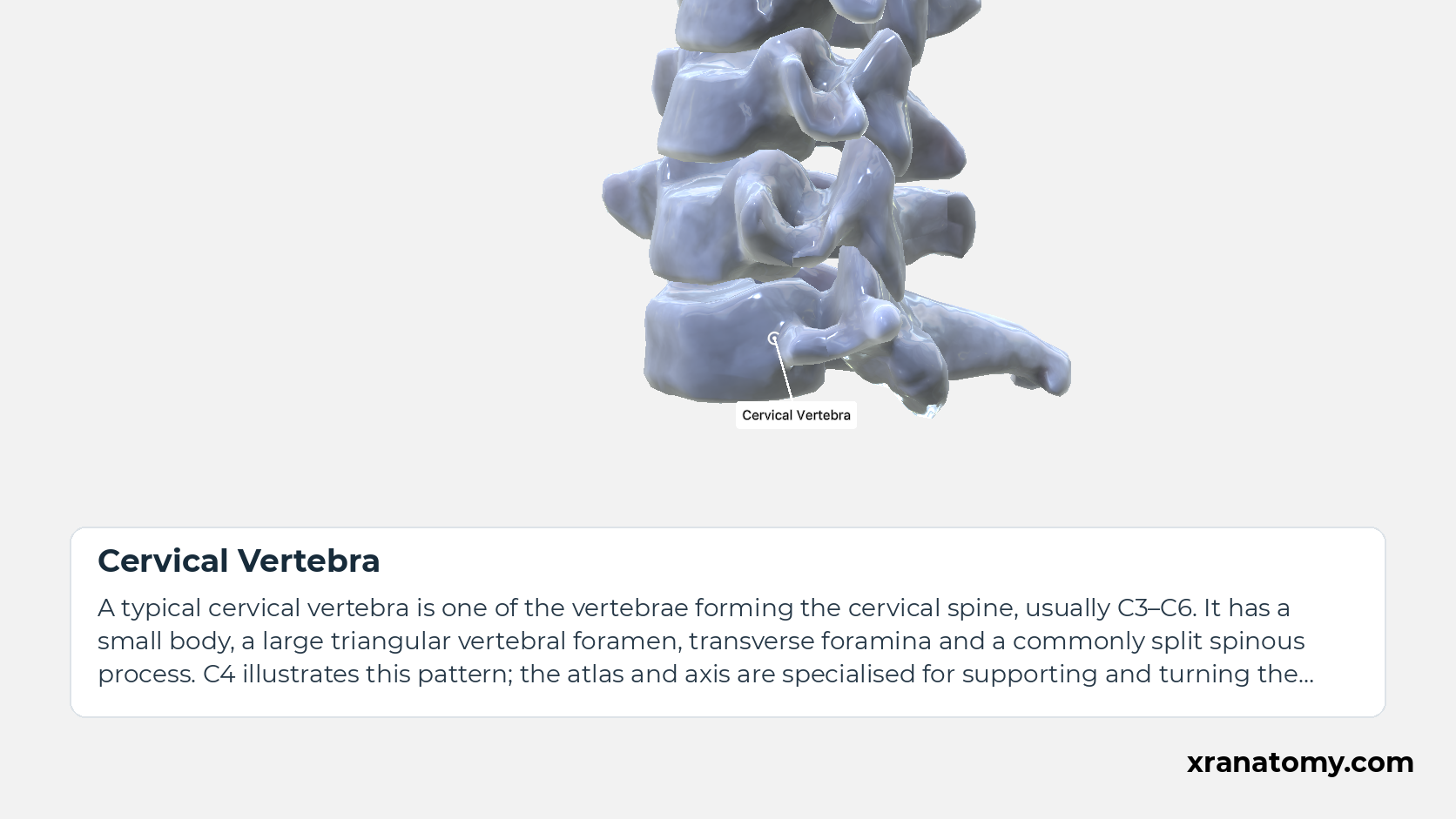
A typical cervical vertebra is one of the vertebrae forming the cervical spine, usually C3–C6. It has a small body, a large triangular vertebral foramen, transverse foramina and a commonly split spinous process. C4 illustrates this pattern; the atlas and axis are specialised for supporting and turning the head.Show less
Study Cervical Vertebrae; Typical C3; C6 Overview in this fixed image. Use the thumbnails to move between the 2D views; each view highlights one structure. The image does not rotate.
Illustration by XR Anatomy.
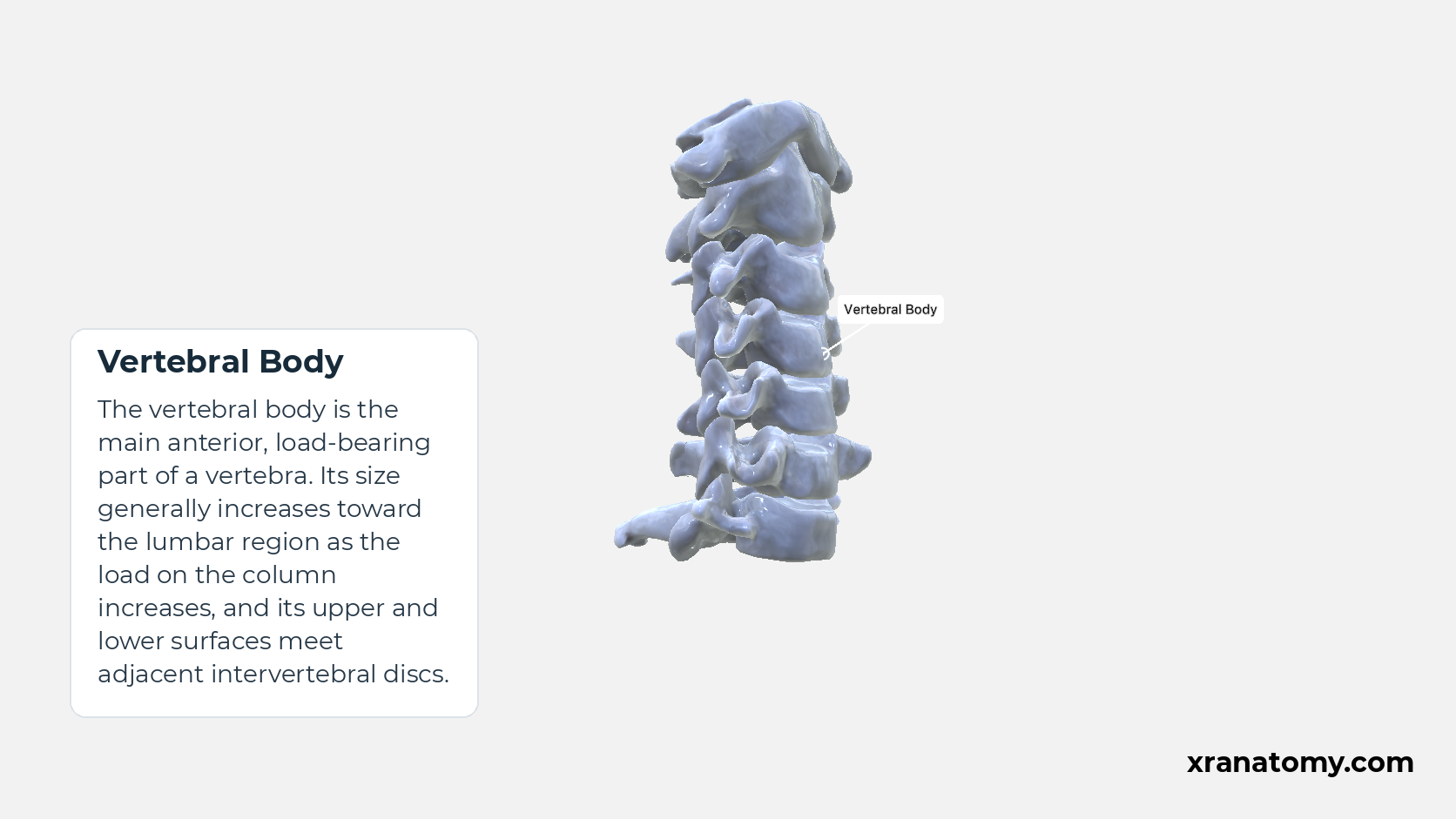
Vertebral Body
The vertebral body is the main anterior, load-bearing part of a vertebra. Its size generally increases toward the lumbar region as the load on the column increases, and its upper and lower surfaces meet adjacent intervertebral discs.
Illustration by XR Anatomy.
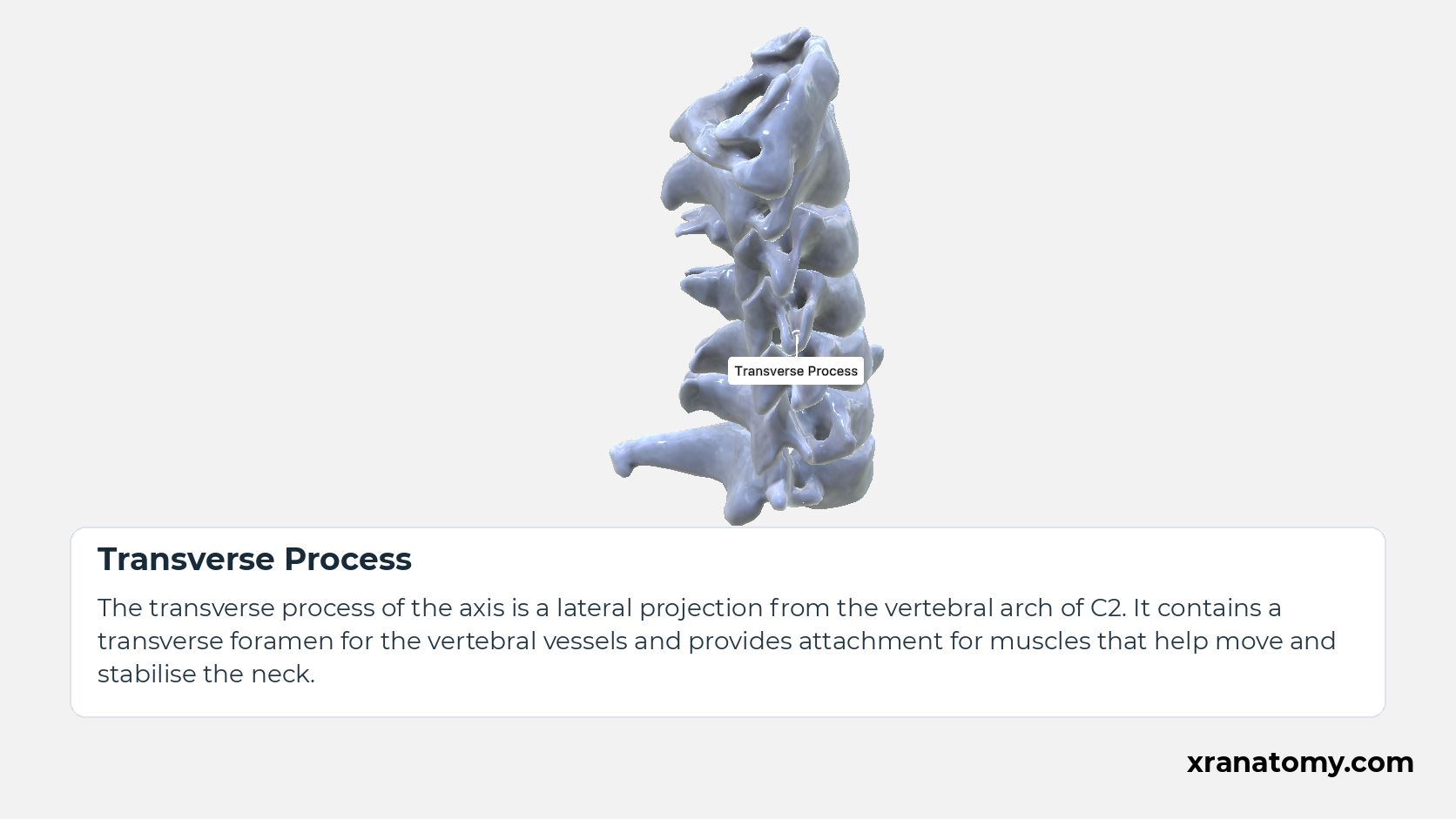
Transverse Process
The transverse process of the axis is a lateral projection from the vertebral arch of C2. It contains a transverse foramen for the vertebral vessels and provides attachment for muscles that help move and stabilise the neck.
Illustration by XR Anatomy.
Cervical Vertebrae
Seven cervical vertebrae form the bony framework of the neck. Typical C3–C6 vertebrae have relatively small bodies, large triangular vertebral foramina, transverse foramina, and short bifid spinous processes, while C1, C2, and C7 have distinctive patterns.Show less
Illustration by XR Anatomy.
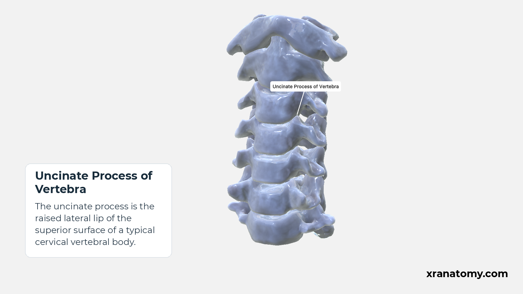
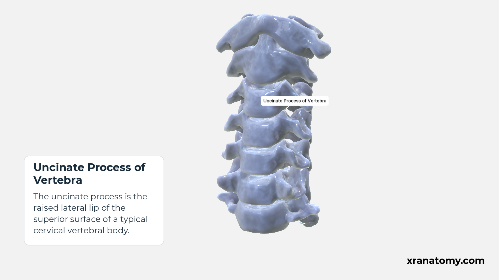
Uncinate Process of Vertebra
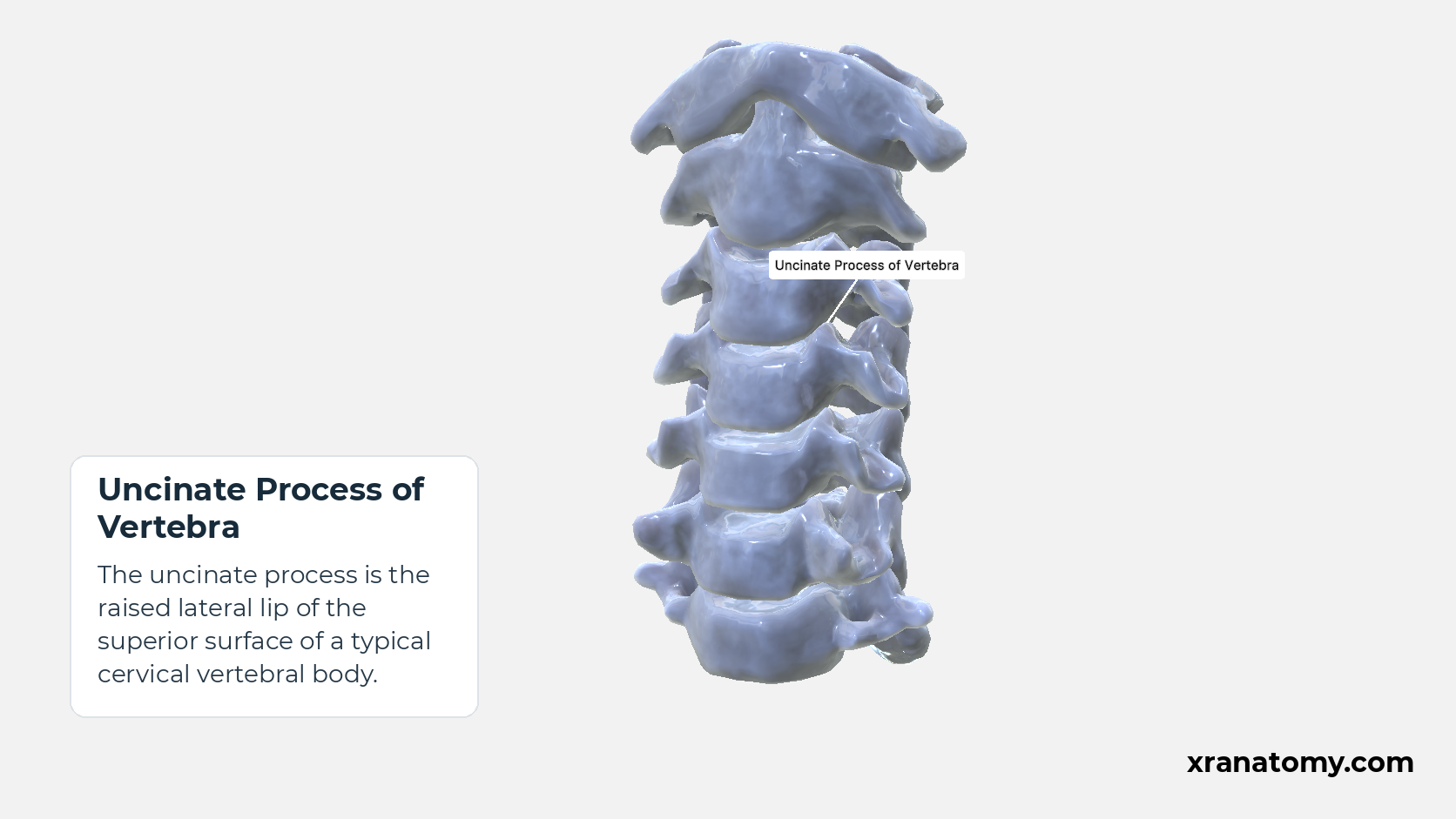
The uncinate process is the raised lateral lip of the superior surface of a typical cervical vertebral body.
Illustration by XR Anatomy.
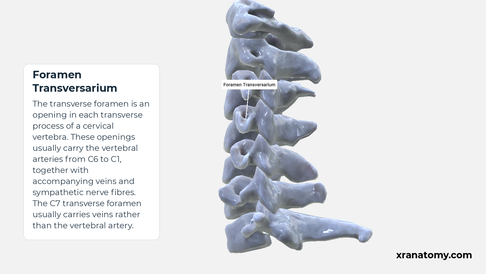
Foramen Transversarium
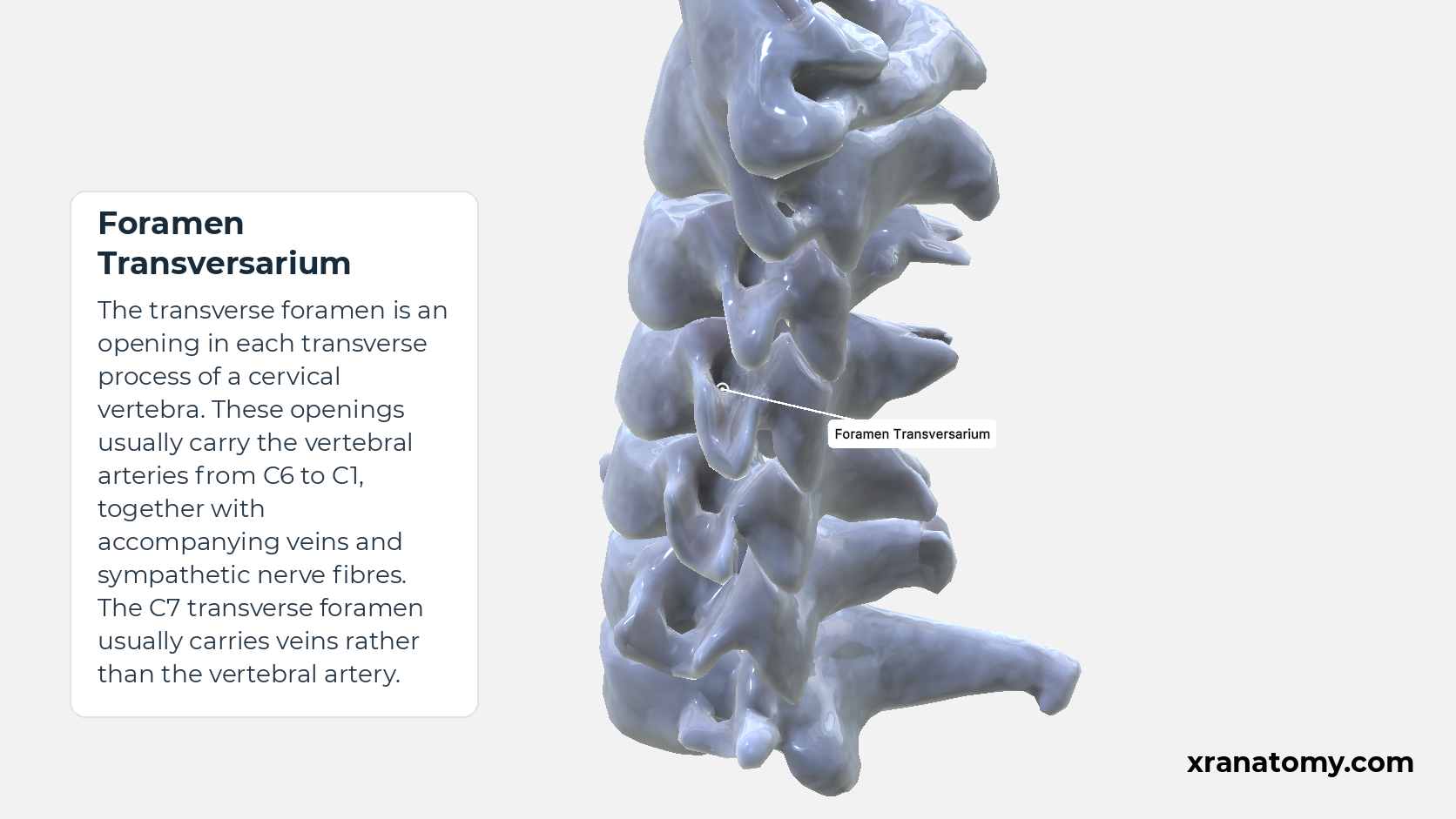
The transverse foramen is an opening in each transverse process of a cervical vertebra. These openings usually carry the vertebral arteries from C6 to C1, together with accompanying veins and sympathetic nerve fibres. The C7 transverse foramen usually carries veins rather than the vertebral artery.Show less
Illustration by XR Anatomy.
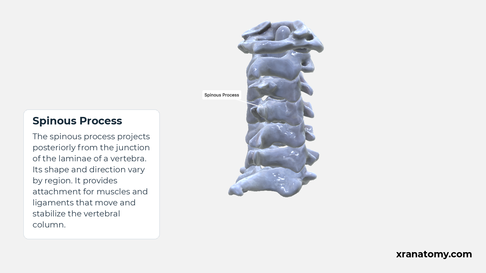
Spinous Process
The spinous process projects posteriorly from the junction of the laminae of a vertebra. Its shape and direction vary by region. It provides attachment for muscles and ligaments that move and stabilize the vertebral column.
Illustration by XR Anatomy.
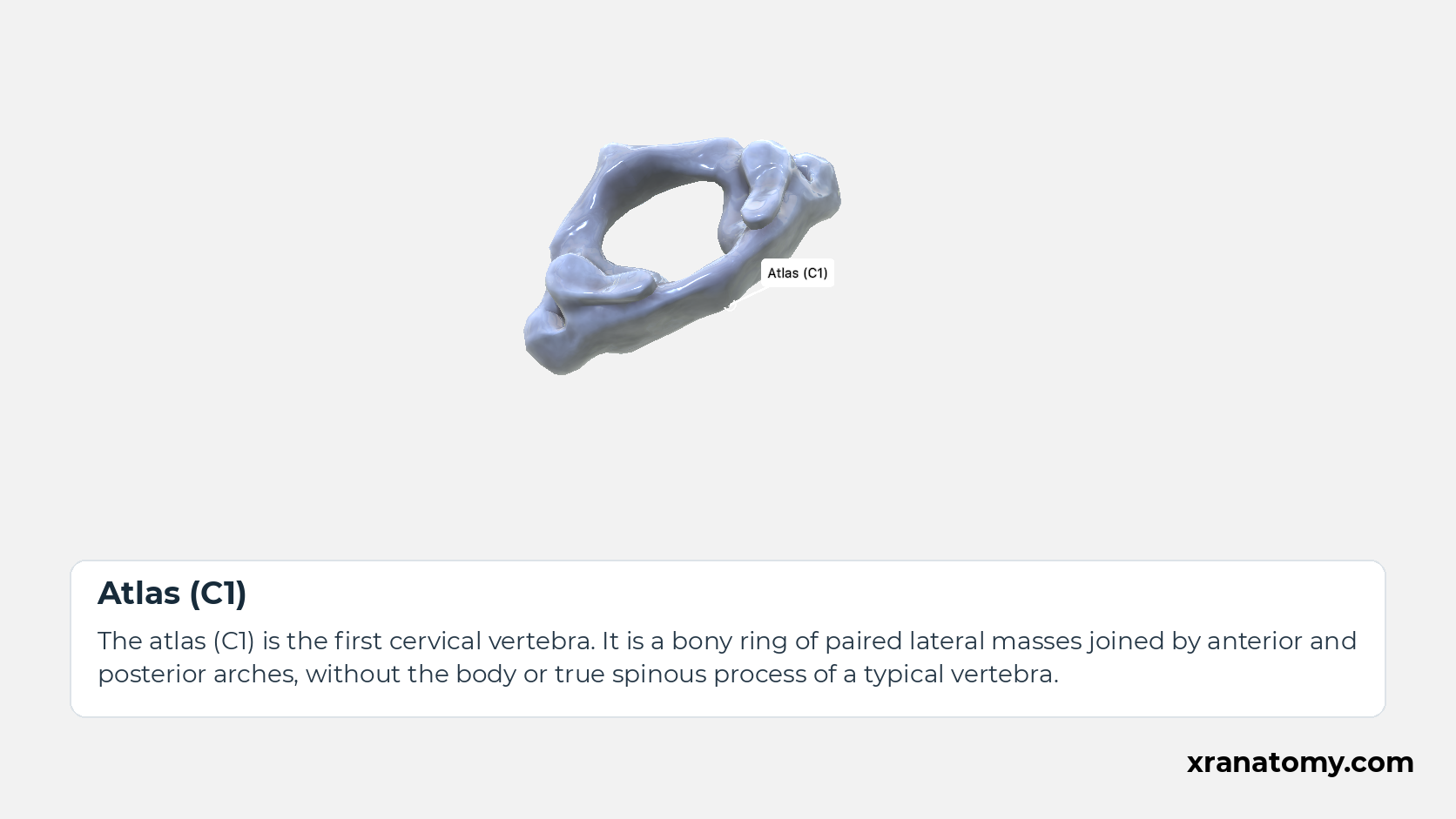
Atlas (C1)
The atlas (C1) is the first cervical vertebra. It is a bony ring of paired lateral masses joined by anterior and posterior arches, without the body or true spinous process of a typical vertebra.
Illustration by XR Anatomy.
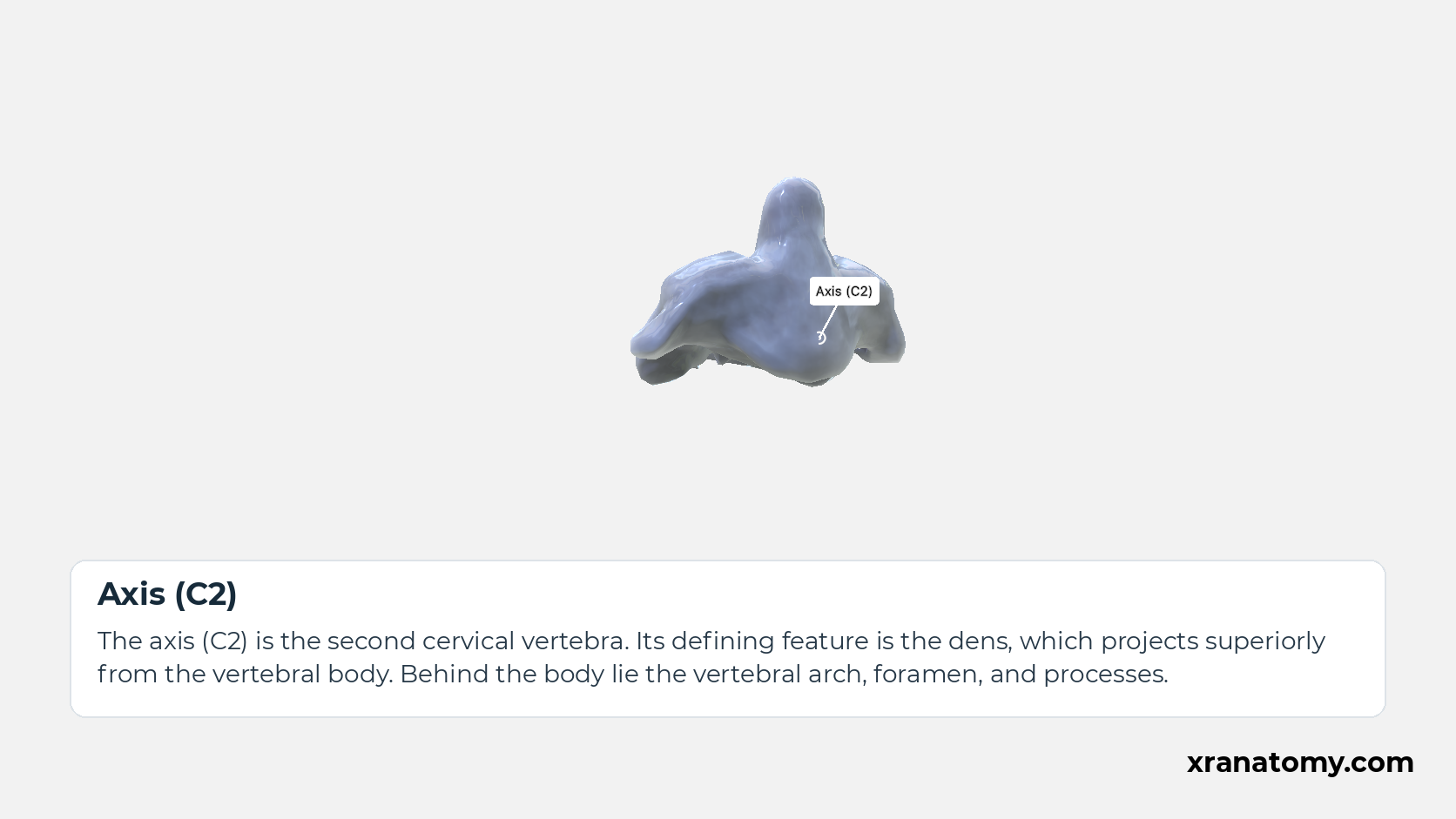
Axis (C2)
The axis (C2) is the second cervical vertebra. Its defining feature is the dens, which projects superiorly from the vertebral body. Behind the body lie the vertebral arch, foramen, and processes.
Illustration by XR Anatomy.
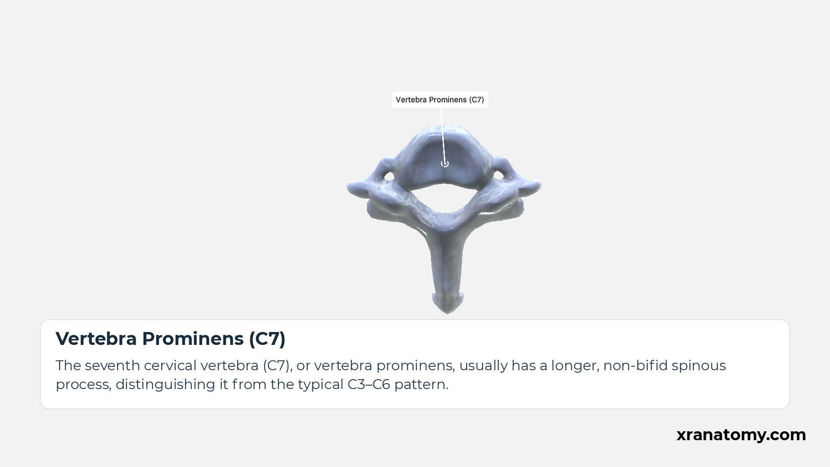
Vertebra Prominens (C7)
The seventh cervical vertebra (C7), or vertebra prominens, usually has a longer, non-bifid spinous process, distinguishing it from the typical C3–C6 pattern.
Illustration by XR Anatomy.
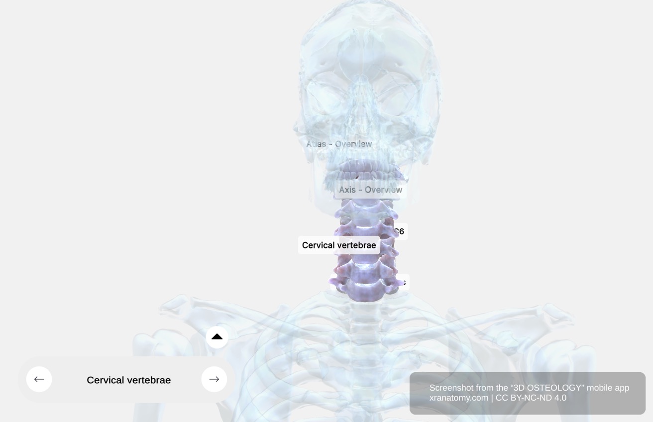 ↺ drag to rotate · scroll to zoom
Cervical Vertebrae; Typical C3; C6 Overview
Cervical Vertebrae — Typical C3–C6 Overview
Drag the model or use the viewer controls to rotate and zoom the column, then read the sections below for each feature.
Sourced from 3D Osteology.
The cervical region contains seven vertebrae. C1 is the atlas, C2 is the axis, C7 is the vertebra prominens, and C3–C6 share the typical cervical pattern described below.
Practice · Active recall · 1 question Recall: Cervical Overview
The cervical region contains vertebrae.
seven
The cervical region contains seven vertebrae. C1 is the atlas, C2 is the axis, C7 is the vertebra prominens, and C3–C6 share the typical cervical pattern described below.
Review this sectionHow well did you know it? · press 1–4
Nice work — revisit in a few days.
General Features
Cervical Vertebra
A typical cervical vertebra is one of the vertebrae forming the cervical spine, usually C3–C6. It has a small body, a large triangular vertebral foramen, transverse foramina and a commonly split spinous process. C4 illustrates this pattern; the atlas and axis are specialised for supporting and turning the head.Show less
Study Cervical Vertebra; C4 in this fixed image. Use the thumbnails to move between the 2D views; each view highlights one structure. The image does not rotate.
Illustration by XR Anatomy.
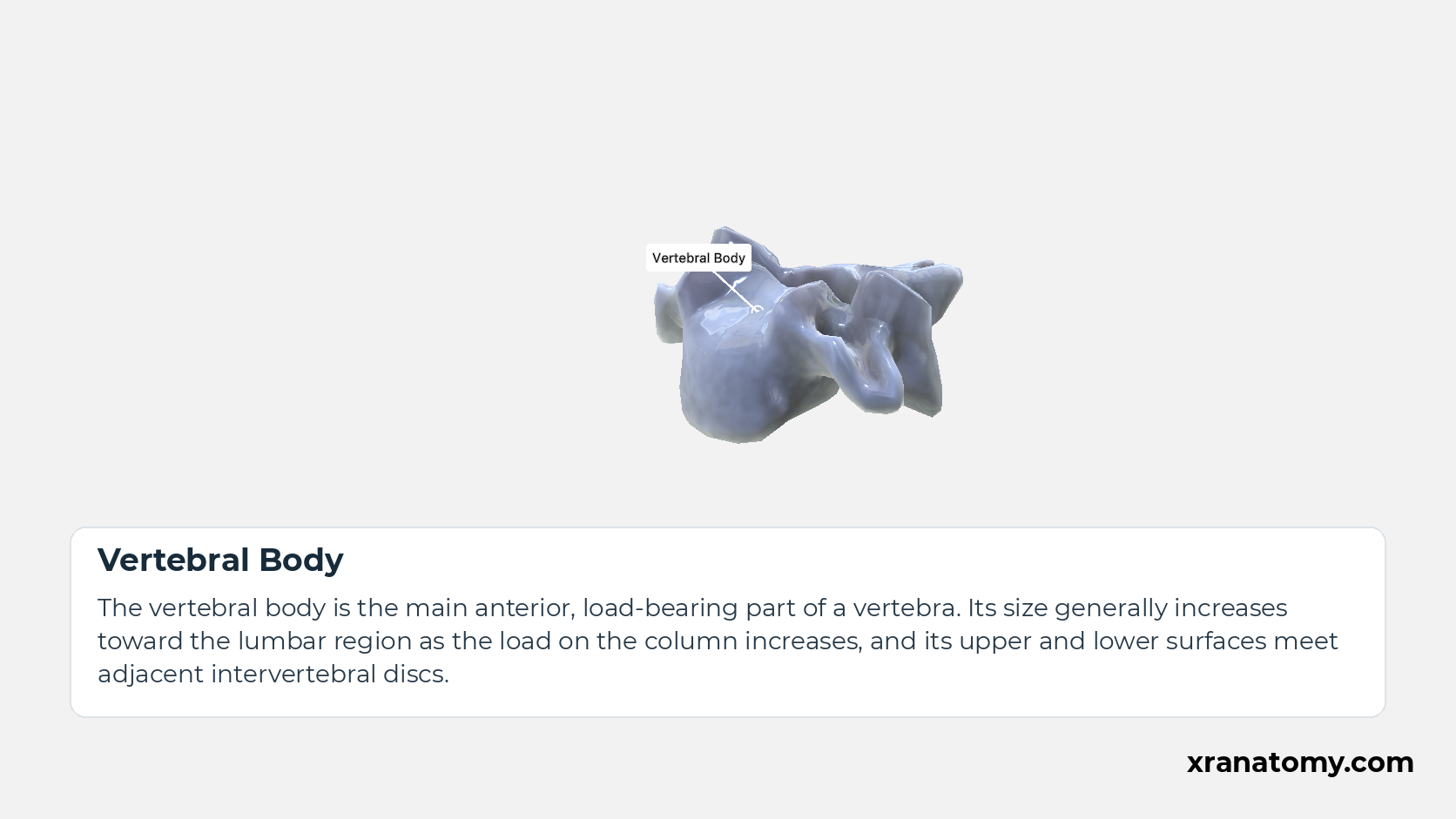
Vertebral Body
The vertebral body is the main anterior, load-bearing part of a vertebra. Its size generally increases toward the lumbar region as the load on the column increases, and its upper and lower surfaces meet adjacent intervertebral discs.
Illustration by XR Anatomy.
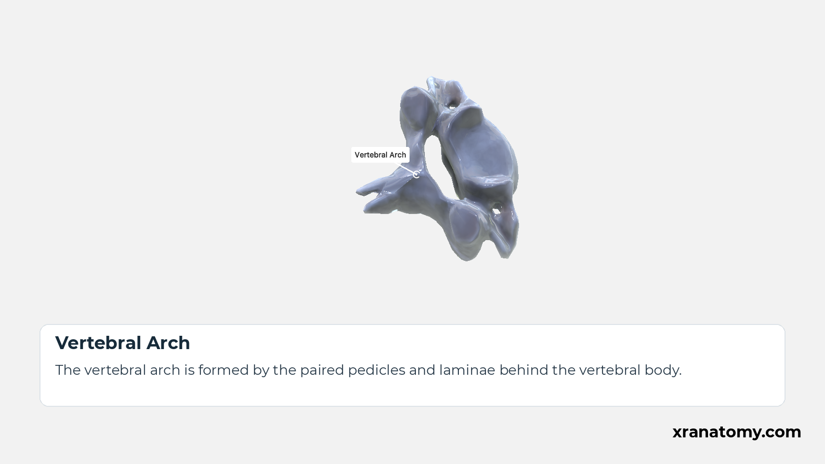
Vertebral Arch
The vertebral arch is formed by the paired pedicles and laminae behind the vertebral body.
Illustration by XR Anatomy.
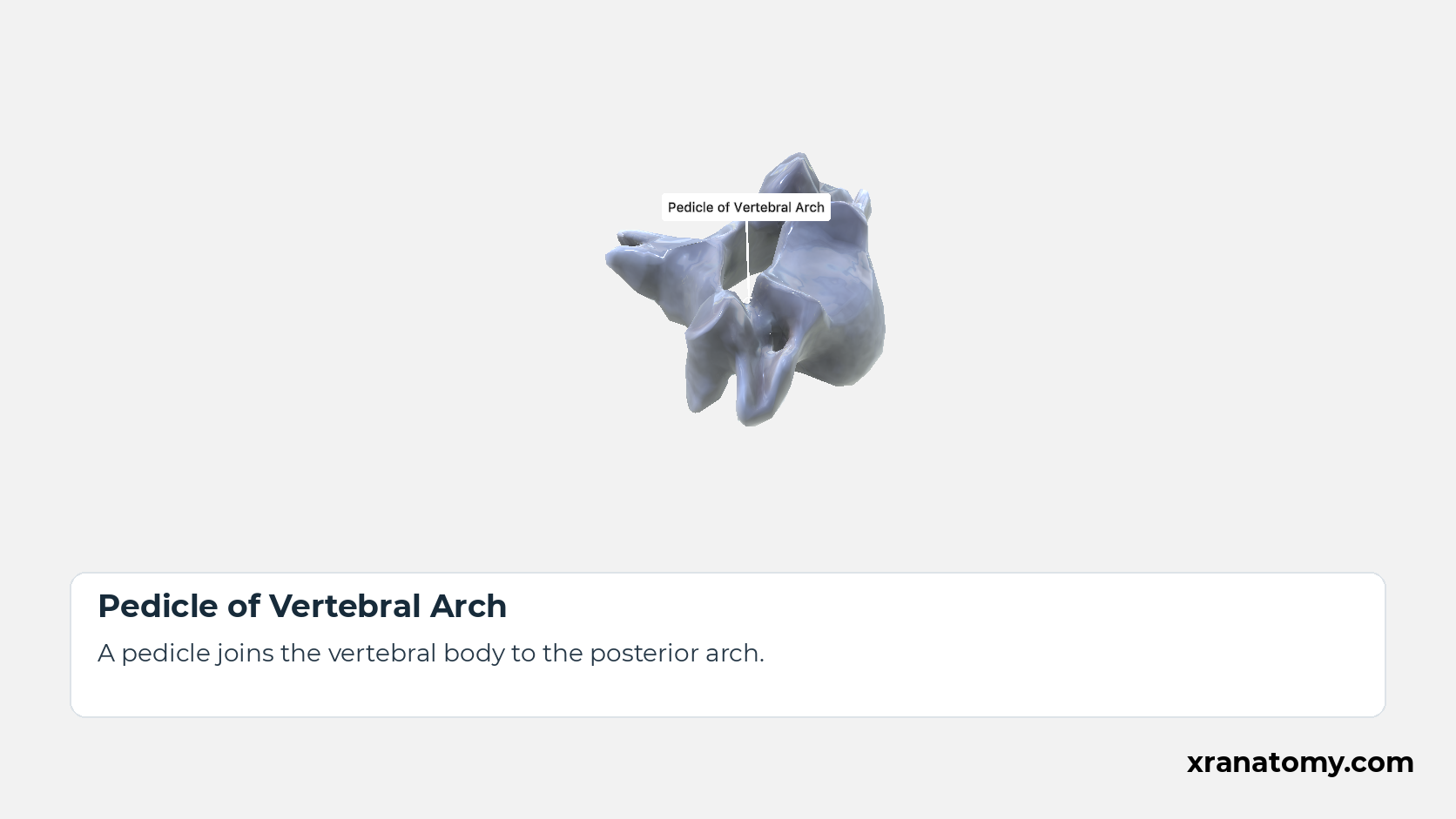
Pedicle of Vertebral Arch
A pedicle joins the vertebral body to the posterior arch.
Illustration by XR Anatomy.
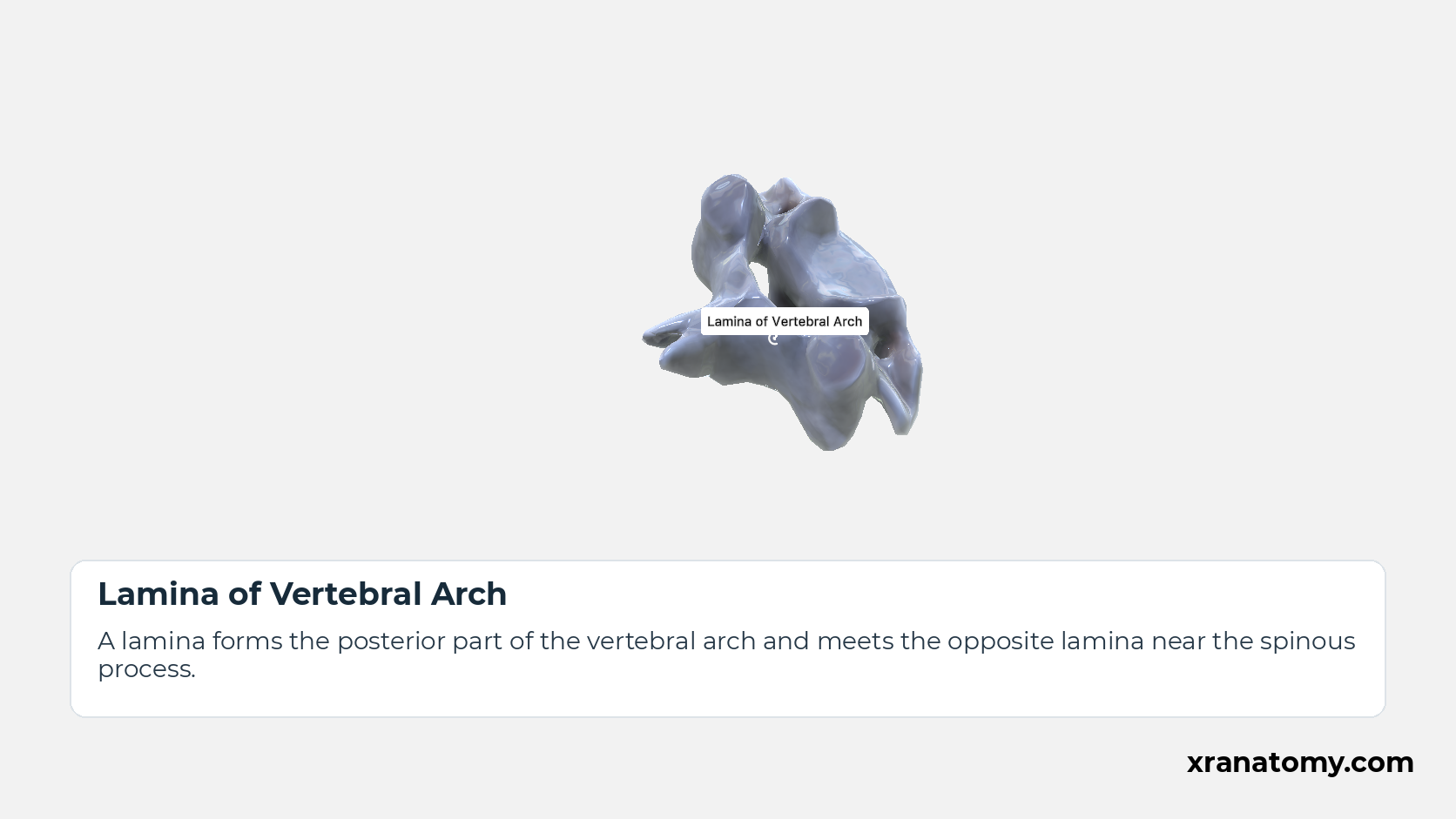
Lamina of Vertebral Arch
A lamina forms the posterior part of the vertebral arch and meets the opposite lamina near the spinous process.
Illustration by XR Anatomy.
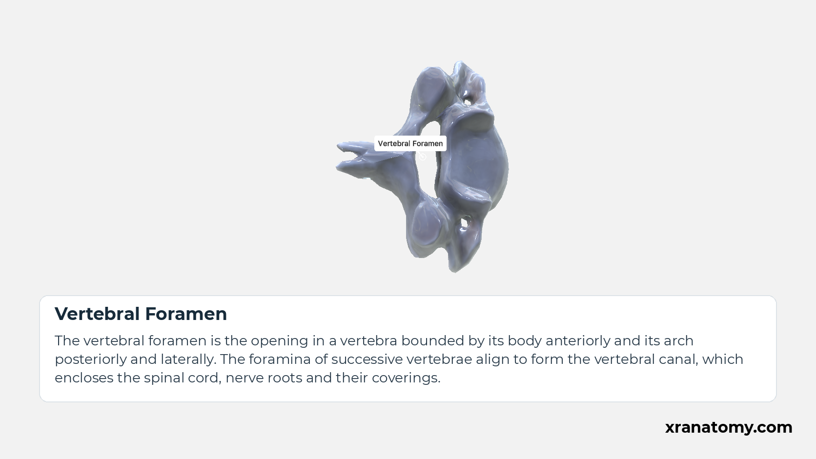
Vertebral Foramen
The vertebral foramen is the opening in a vertebra bounded by its body anteriorly and its arch posteriorly and laterally. The foramina of successive vertebrae align to form the vertebral canal, which encloses the spinal cord, nerve roots and their coverings.Show less
Illustration by XR Anatomy.
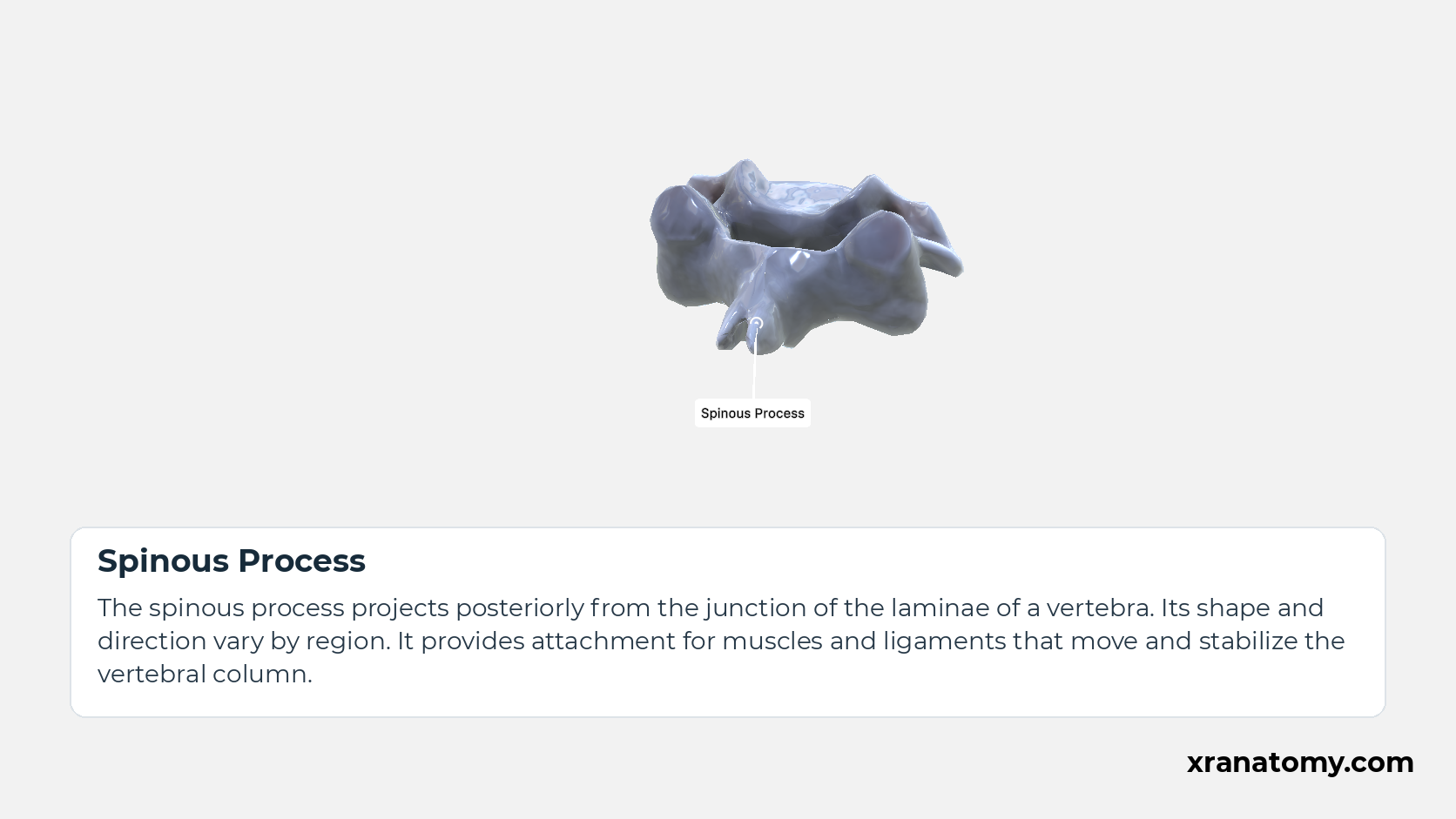
Spinous Process
The spinous process projects posteriorly from the junction of the laminae of a vertebra. Its shape and direction vary by region. It provides attachment for muscles and ligaments that move and stabilize the vertebral column.
Illustration by XR Anatomy.
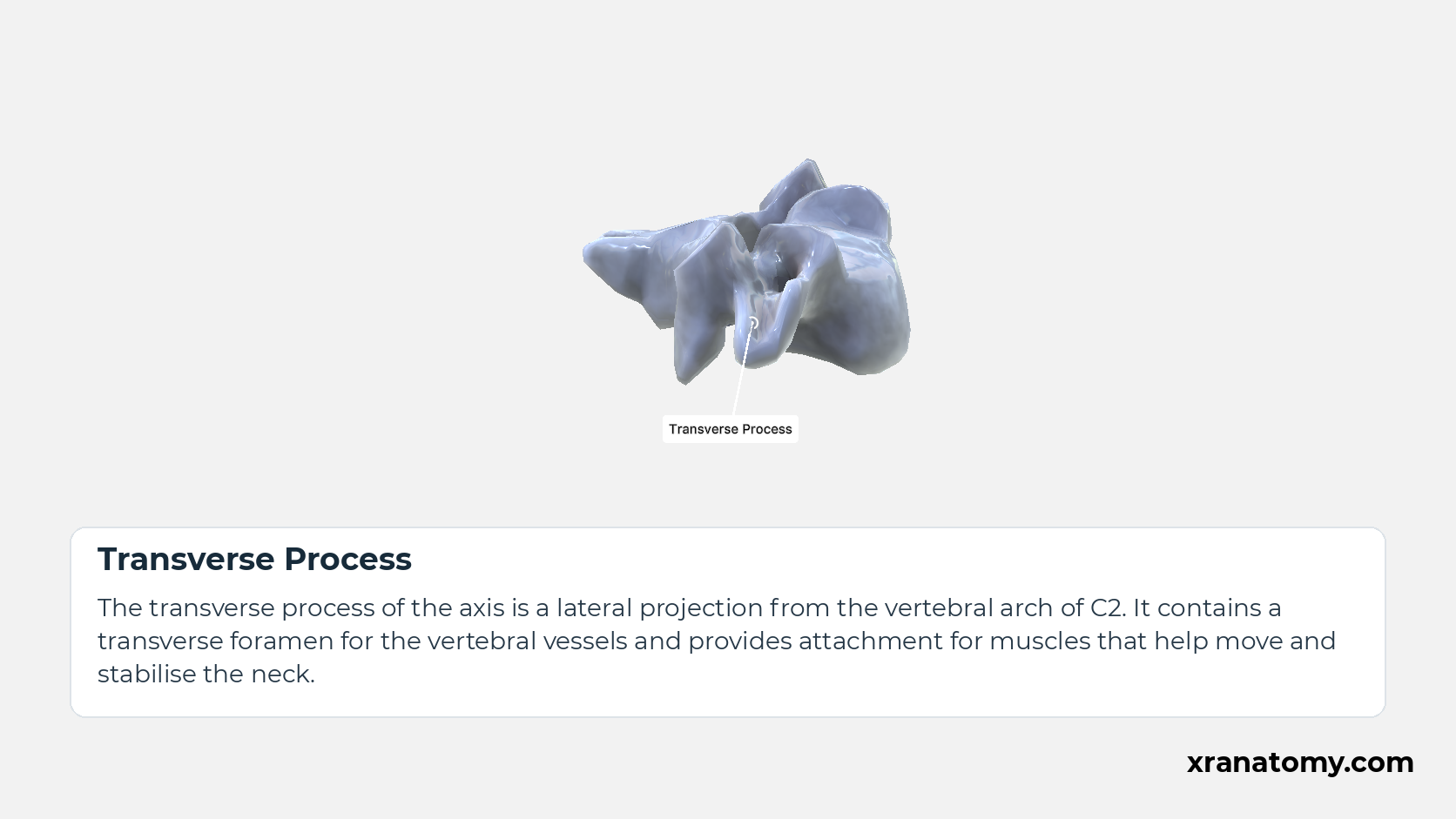
Transverse Process
The transverse process of the axis is a lateral projection from the vertebral arch of C2. It contains a transverse foramen for the vertebral vessels and provides attachment for muscles that help move and stabilise the neck.
Illustration by XR Anatomy.
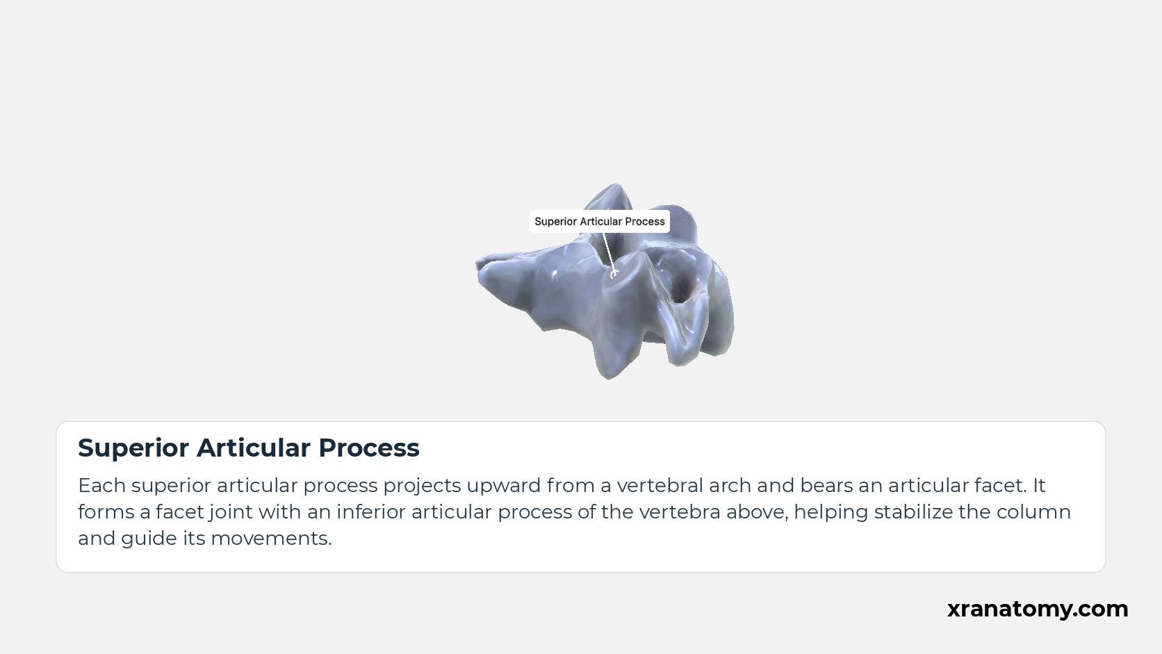
Superior Articular Process
Each superior articular process projects upward from a vertebral arch and bears an articular facet. It forms a facet joint with an inferior articular process of the vertebra above, helping stabilize the column and guide its movements.
Illustration by XR Anatomy.
Inferior Articular Process
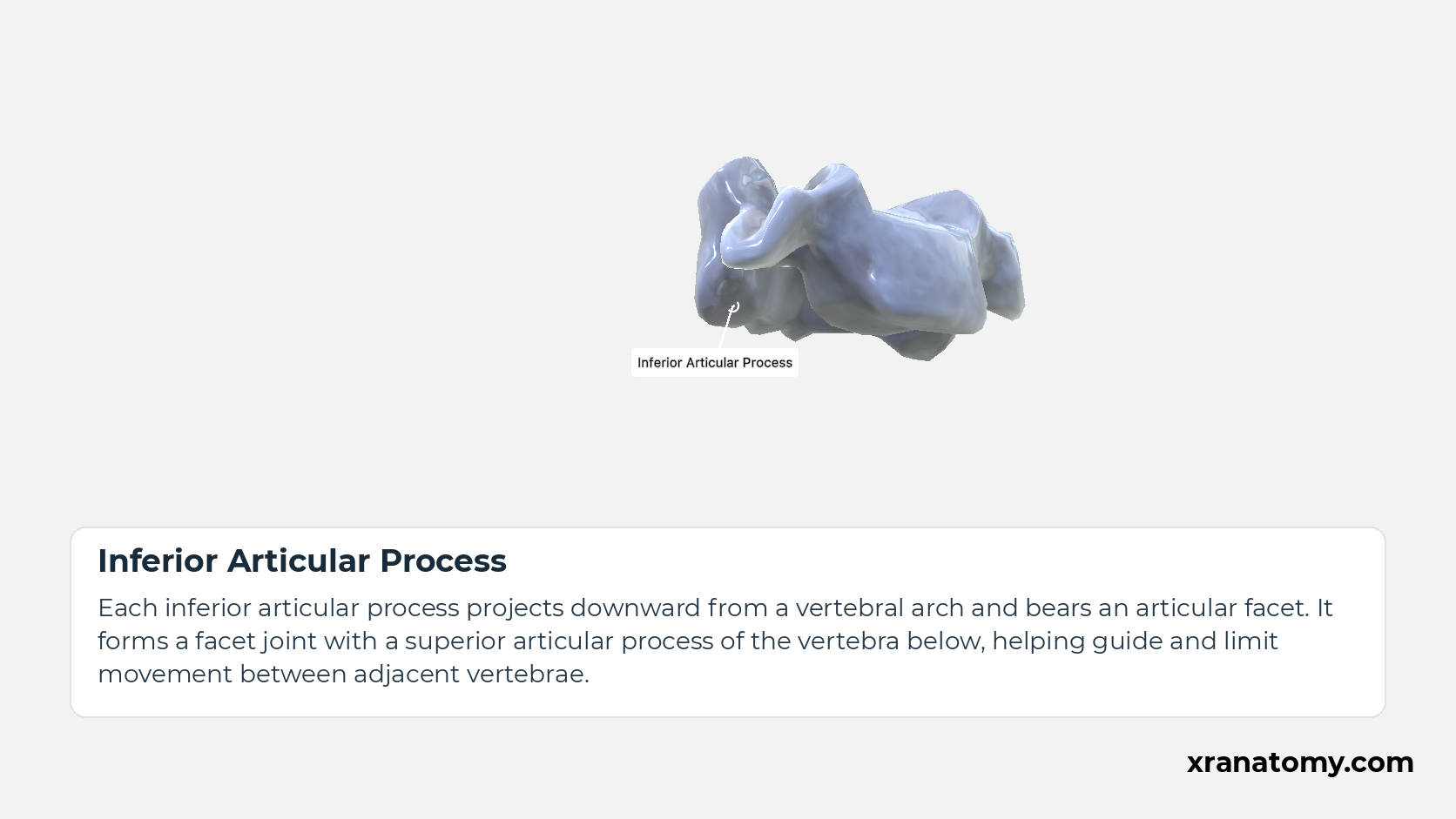
Each inferior articular process projects downward from a vertebral arch and bears an articular facet. It forms a facet joint with a superior articular process of the vertebra below, helping guide and limit movement between adjacent vertebrae.Show less
Illustration by XR Anatomy.
Uncinate Process
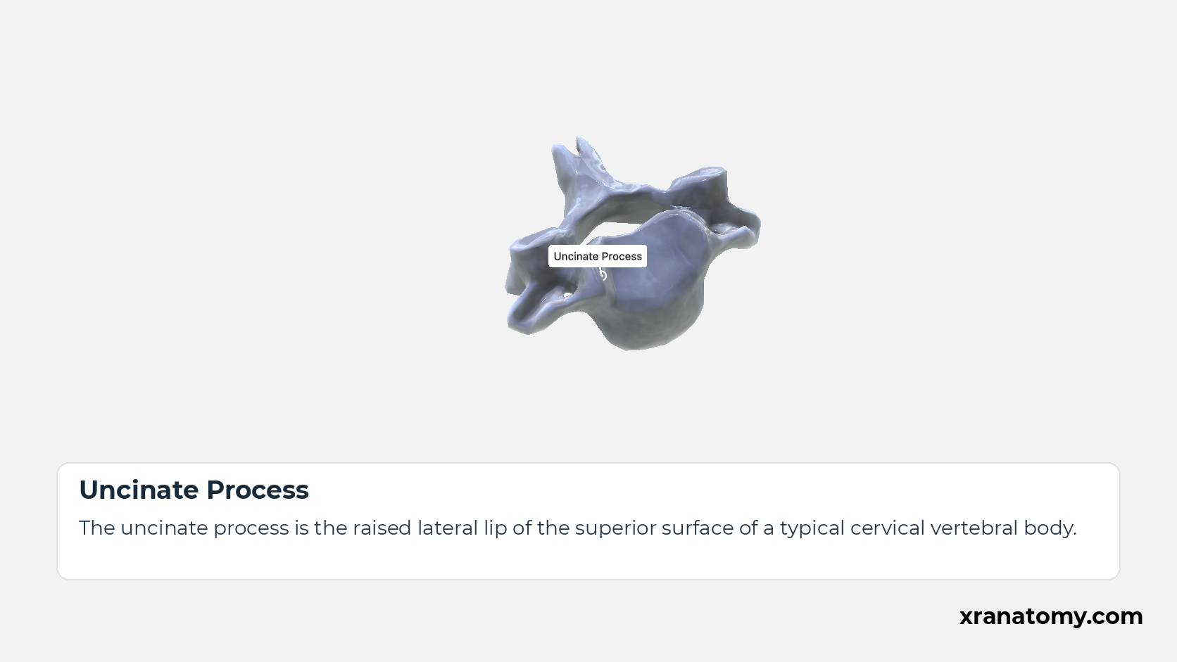
The uncinate process is the raised lateral lip of the superior surface of a typical cervical vertebral body.
Illustration by XR Anatomy.
Foramen Transversarium
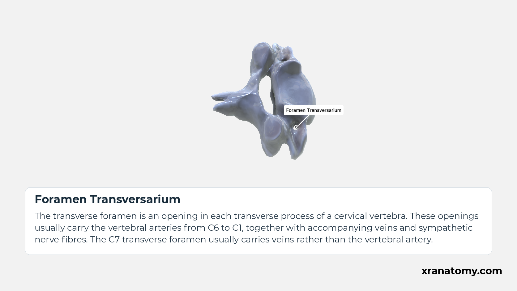
The transverse foramen is an opening in each transverse process of a cervical vertebra. These openings usually carry the vertebral arteries from C6 to C1, together with accompanying veins and sympathetic nerve fibres. The C7 transverse foramen usually carries veins rather than the vertebral artery.Show less
Illustration by XR Anatomy.
A typical cervical vertebra has a vertebral body, vertebral arch and vertebral foramen, together with spinous, articular, and transverse processes. The isolated C4 specimen provides a single typical cervical vertebra for these landmarks.
In depth · 2 sections · 1 min read2 more sections on this part2 sections open — reading In depth- Cervical Vertebra
- Vertebra Prominens (C7)
Cervical Vertebra
A cervical vertebra (vertebra cervicalis) is part of the C1–C7 regional series. The typical pattern mainly applies to C3–C6; the atlas, axis, and C7 differ from it in recognizable ways.
Vertebra Prominens (C7)
The seventh cervical vertebra (C7), or vertebra prominens, usually has a longer, non-bifid spinous process. This distinguishes it from the typical C3–C6 pattern.
Practice · Active recall · 3 questions Recall: General Features
Why this helps
Testing yourself before revealing the answer (active recall) strengthens memory far more than re-reading — so each prompt stays hidden until you choose to reveal it.
What usual spinous-process feature distinguishes C7 from typical C3–C6 vertebrae?
a longer, non-bifid spinous process
The seventh cervical vertebra (C7), or vertebra prominens, usually has a longer, non-bifid spinous process. This distinguishes it from the typical C3–C6 pattern.
Review this sectionHow well did you know it? · press 1–4
To which cervical levels does the typical vertebral pattern mainly apply?
C3–C6
A cervical vertebra (vertebra cervicalis) is part of the C1–C7 regional series. The typical pattern mainly applies to C3–C6; the atlas, axis, and C7 differ from it in recognizable ways.
Review this sectionHow well did you know it? · press 1–4
In depth · 1 question Recall: Cervical Vertebra
The typical cervical pattern mainly applies to .
C3–C6
A cervical vertebra (vertebra cervicalis) is part of the C1–C7 regional series. The typical pattern mainly applies to C3–C6; the atlas, axis, and C7 differ from it in recognizable ways.
Review this sectionHow well did you know it? · press 1–4
Nice work — revisit in a few days.
Vertebral Body
Cervical Vertebra
A typical cervical vertebra is one of the vertebrae forming the cervical spine, usually C3–C6. It has a small body, a large triangular vertebral foramen, transverse foramina and a commonly split spinous process. C4 illustrates this pattern; the atlas and axis are specialised for supporting and turning the head.Show less
Study Cervical Vertebra; C4 in this fixed image. Use the thumbnails to move between the 2D views; each view highlights one structure. The image does not rotate.
Illustration by XR Anatomy.
Vertebral Body
The vertebral body is the main anterior, load-bearing part of a vertebra. Its size generally increases toward the lumbar region as the load on the column increases, and its upper and lower surfaces meet adjacent intervertebral discs.
Illustration by XR Anatomy.
Vertebral Arch
The vertebral arch is formed by the paired pedicles and laminae behind the vertebral body.
Illustration by XR Anatomy.
Pedicle of Vertebral Arch
A pedicle joins the vertebral body to the posterior arch.
Illustration by XR Anatomy.
Lamina of Vertebral Arch
A lamina forms the posterior part of the vertebral arch and meets the opposite lamina near the spinous process.
Illustration by XR Anatomy.
Vertebral Foramen
The vertebral foramen is the opening in a vertebra bounded by its body anteriorly and its arch posteriorly and laterally. The foramina of successive vertebrae align to form the vertebral canal, which encloses the spinal cord, nerve roots and their coverings.Show less
Illustration by XR Anatomy.
Spinous Process
The spinous process projects posteriorly from the junction of the laminae of a vertebra. Its shape and direction vary by region. It provides attachment for muscles and ligaments that move and stabilize the vertebral column.
Illustration by XR Anatomy.
Transverse Process
The transverse process of the axis is a lateral projection from the vertebral arch of C2. It contains a transverse foramen for the vertebral vessels and provides attachment for muscles that help move and stabilise the neck.
Illustration by XR Anatomy.
Superior Articular Process
Each superior articular process projects upward from a vertebral arch and bears an articular facet. It forms a facet joint with an inferior articular process of the vertebra above, helping stabilize the column and guide its movements.
Illustration by XR Anatomy.
Inferior Articular Process
Each inferior articular process projects downward from a vertebral arch and bears an articular facet. It forms a facet joint with a superior articular process of the vertebra below, helping guide and limit movement between adjacent vertebrae.Show less
Illustration by XR Anatomy.
Uncinate Process
The uncinate process is the raised lateral lip of the superior surface of a typical cervical vertebral body.
Illustration by XR Anatomy.
Foramen Transversarium
The transverse foramen is an opening in each transverse process of a cervical vertebra. These openings usually carry the vertebral arteries from C6 to C1, together with accompanying veins and sympathetic nerve fibres. The C7 transverse foramen usually carries veins rather than the vertebral artery.Show less
Illustration by XR Anatomy.
In a typical cervical vertebra, the vertebral body (corpus vertebrae) is small and wider from side to side than from front to back. Its superior surface is concave from side to side, with raised lateral lips called uncinate processes.
In depth · 1 min read Uncinate Process of Vertebra
An uncinate process (processus uncinatus vertebrae) is the raised lateral lip on the superior surface of a typical cervical vertebral body. It articulates with the shallow lateral depression on the inferior surface of the vertebra above.
Practice · Active recall · 2 questions Recall: Vertebral Body
A typical cervical vertebral body is wider from than from front to back.
side to side
In a typical cervical vertebra, the vertebral body (corpus vertebrae) is small and wider from side to side than from front to back. Its superior surface is concave from side to side, with raised lateral lips called uncinate processes.
Review this sectionHow well did you know it? · press 1–4
Nice work — revisit in a few days.
In depth · 1 question Recall: Uncinate Process of Vertebra
An uncinate process is a raised lateral lip on the surface of a typical cervical vertebral body.
superior
An uncinate process (processus uncinatus vertebrae) is the raised lateral lip on the superior surface of a typical cervical vertebral body. It lies beside the shallow lateral depression on the inferior surface of the vertebra above.
Review this sectionHow well did you know it? · press 1–4
Nice work — revisit in a few days.
Pedicles and Laminae
Cervical Vertebra
A typical cervical vertebra is one of the vertebrae forming the cervical spine, usually C3–C6. It has a small body, a large triangular vertebral foramen, transverse foramina and a commonly split spinous process. C4 illustrates this pattern; the atlas and axis are specialised for supporting and turning the head.Show less
Study Cervical Vertebra; C4 in this fixed image. Use the thumbnails to move between the 2D views; each view highlights one structure. The image does not rotate.
Illustration by XR Anatomy.
Vertebral Body
The vertebral body is the main anterior, load-bearing part of a vertebra. Its size generally increases toward the lumbar region as the load on the column increases, and its upper and lower surfaces meet adjacent intervertebral discs.
Illustration by XR Anatomy.
Vertebral Arch
The vertebral arch is formed by the paired pedicles and laminae behind the vertebral body.
Illustration by XR Anatomy.
Pedicle of Vertebral Arch
A pedicle joins the vertebral body to the posterior arch.
Illustration by XR Anatomy.
Lamina of Vertebral Arch
A lamina forms the posterior part of the vertebral arch and meets the opposite lamina near the spinous process.
Illustration by XR Anatomy.
Vertebral Foramen
The vertebral foramen is the opening in a vertebra bounded by its body anteriorly and its arch posteriorly and laterally. The foramina of successive vertebrae align to form the vertebral canal, which encloses the spinal cord, nerve roots and their coverings.Show less
Illustration by XR Anatomy.
Spinous Process
The spinous process projects posteriorly from the junction of the laminae of a vertebra. Its shape and direction vary by region. It provides attachment for muscles and ligaments that move and stabilize the vertebral column.
Illustration by XR Anatomy.
Transverse Process
The transverse process of the axis is a lateral projection from the vertebral arch of C2. It contains a transverse foramen for the vertebral vessels and provides attachment for muscles that help move and stabilise the neck.
Illustration by XR Anatomy.
Superior Articular Process
Each superior articular process projects upward from a vertebral arch and bears an articular facet. It forms a facet joint with an inferior articular process of the vertebra above, helping stabilize the column and guide its movements.
Illustration by XR Anatomy.
Inferior Articular Process
Each inferior articular process projects downward from a vertebral arch and bears an articular facet. It forms a facet joint with a superior articular process of the vertebra below, helping guide and limit movement between adjacent vertebrae.Show less
Illustration by XR Anatomy.
Uncinate Process
The uncinate process is the raised lateral lip of the superior surface of a typical cervical vertebral body.
Illustration by XR Anatomy.
Foramen Transversarium
The transverse foramen is an opening in each transverse process of a cervical vertebra. These openings usually carry the vertebral arteries from C6 to C1, together with accompanying veins and sympathetic nerve fibres. The C7 transverse foramen usually carries veins rather than the vertebral artery.Show less
Illustration by XR Anatomy.
Atlas (C1)
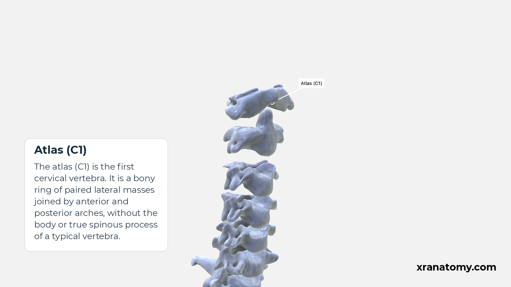
The atlas (C1) is the first cervical vertebra. It is a bony ring of paired lateral masses joined by anterior and posterior arches, without the body or true spinous process of a typical vertebra.
Study Cervical Spine; Articulation Context in this fixed image. Use the thumbnails to move between the 2D views; each view highlights one structure. The image does not rotate.
Illustration by XR Anatomy.
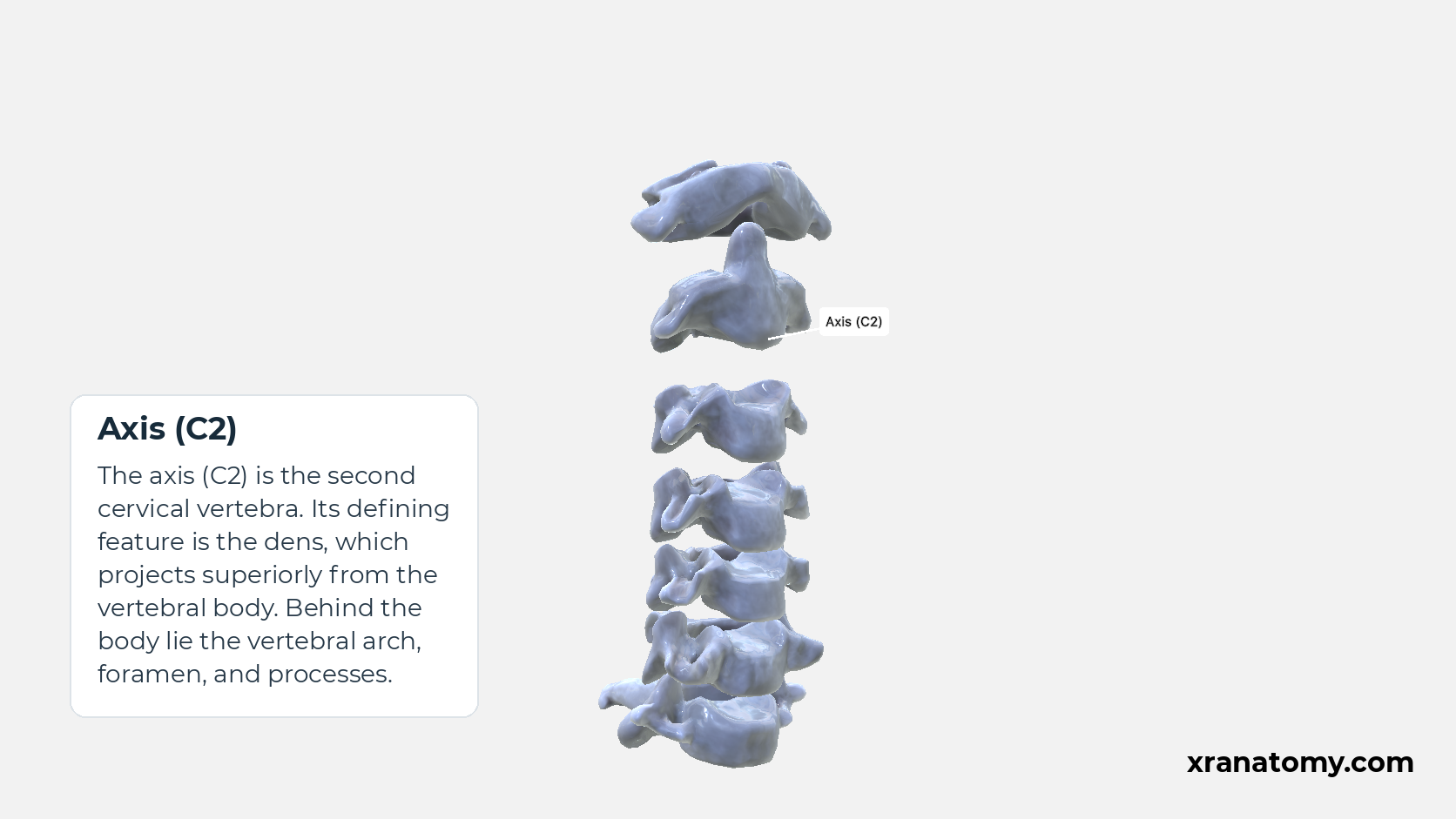
Axis (C2)
The axis (C2) is the second cervical vertebra. Its defining feature is the dens, which projects superiorly from the vertebral body. Behind the body lie the vertebral arch, foramen, and processes.
Illustration by XR Anatomy.
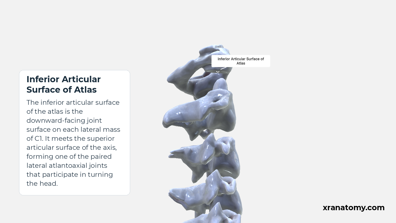
Inferior Articular Surface of Atlas
The inferior articular surface of the atlas is the downward-facing joint surface on each lateral mass of C1. It meets the superior articular surface of the axis, forming one of the paired lateral atlantoaxial joints that participate in turning the head.Show less
Illustration by XR Anatomy.
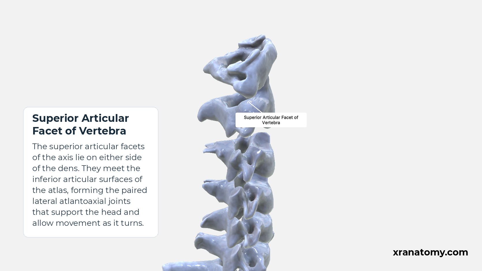
Superior Articular Facet of Vertebra
The superior articular facets of the axis lie on either side of the dens. They meet the inferior articular surfaces of the atlas, forming the paired lateral atlantoaxial joints that support the head and allow movement as it turns.
Illustration by XR Anatomy.
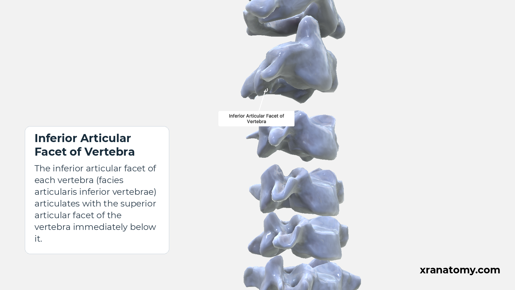
Inferior Articular Facet of Vertebra
The inferior articular facet of each vertebra (facies articularis inferior vertebrae) articulates with the superior articular facet of the vertebra immediately below it.
Illustration by XR Anatomy.
The paired pedicles and laminae form the vertebral arch. Along with the posterior surface of the body, they form the boundary of the large triangular vertebral foramen typical of C3–C6.
In depth · 3 sections · 1 min read3 more sections on this part3 sections open — reading In depth- Pedicle of Vertebral Arch
- Lamina of Vertebral Arch
- Vertebral Foramen
Pedicle of Vertebral Arch
Each pedicle (pediculus arcus vertebrae) extends posteriorly from the body. Its superior and inferior borders have the corresponding vertebral notches. In the assembled cervical spine, the notches of adjacent vertebrae bound an intervertebral foramen through which a spinal nerve exits.
Superior Vertebral Notch
The superior vertebral notch (incisura superior vertebrae) is an indentation in the superior border of a pedicle and helps form the intervertebral foramen above.
Inferior Vertebral Notch
The inferior vertebral notch (incisura inferior vertebrae) is an indentation in the inferior border of a pedicle and helps form the intervertebral foramen below.
Lamina of Vertebral Arch
Each lamina (lamina arcus vertebrae) runs posteromedially from a pedicle and joins the other lamina near the base of the spinous process. Together, the laminae form the posterior boundary of the vertebral foramen.
Vertebral Foramen
The vertebral foramen (foramen vertebrale) is large and triangular in a typical cervical vertebra. When vertebral foramina line up, they form the cervical part of the vertebral canal.
Cervical Intervertebral Foramen
The cervical intervertebral foramen is the lateral opening bounded by the vertebral notches of adjacent cervical vertebrae. A spinal nerve exits through it.
Practice · Active recall · 5 questions Recall: Pedicles and Laminae
The paired pedicles and laminae form the .
The paired pedicles and laminae form the vertebral arch. Along with the posterior surface of the body, they form the boundary of the large triangular vertebral foramen typical of C3–C6.
Review this sectionHow well did you know it? · press 1–4
Nice work — revisit in a few days.
In depth · 1 question Recall: Pedicle of Vertebral Arch
Each pedicle extends from the vertebral body.
posteriorly
Each pedicle (pediculus arcus vertebrae) extends posteriorly from the body. Its superior and inferior borders have the corresponding vertebral notches. In the assembled cervical spine, the notches of adjacent vertebrae bound an intervertebral foramen through which a spinal nerve exits.
Review this sectionHow well did you know it? · press 1–4
Nice work — revisit in a few days.
In depth · 1 question Recall: Lamina of Vertebral Arch
Each lamina joins the opposite lamina near the base of the .
Each lamina (lamina arcus vertebrae) runs posteromedially from a pedicle and joins the other lamina near the base of the spinous process. Together, the laminae form the posterior boundary of the vertebral foramen.
Review this sectionHow well did you know it? · press 1–4
Nice work — revisit in a few days.
What exits through a cervical intervertebral foramen?
spinal nerve
The cervical intervertebral foramen is the lateral opening bounded by the vertebral notches of adjacent cervical vertebrae. A spinal nerve exits through it.
Review this sectionHow well did you know it? · press 1–4
In depth · 1 question Recall: Vertebral Foramen
Aligned vertebral foramina form the cervical part of the .
The vertebral foramen (foramen vertebrale) is large and triangular in a typical cervical vertebra. When vertebral foramina line up, they form the cervical part of the vertebral canal.
Review this sectionHow well did you know it? · press 1–4
Nice work — revisit in a few days.
Spinous Process
The spinous process (processus spinosus) is short and commonly bifid in C3–C6. C7's spinous process is usually longer and non-bifid, helping distinguish the vertebra prominens from the typical pattern.
Practice · Active recall · 1 question Recall: Spinous Process
The spinous process is short and commonly in C3–C6.
bifid
The spinous process (processus spinosus) is short and commonly bifid in C3–C6. C7's spinous process is usually longer and non-bifid, helping distinguish the vertebra prominens from the typical pattern.
Review this sectionHow well did you know it? · press 1–4
Nice work — revisit in a few days.
Articular Processes
Atlas (C1)
The atlas (C1) is the first cervical vertebra. It is a bony ring of paired lateral masses joined by anterior and posterior arches, without the body or true spinous process of a typical vertebra.
Study Cervical Spine; Articulation Context in this fixed image. Use the thumbnails to move between the 2D views; each view highlights one structure. The image does not rotate.
Illustration by XR Anatomy.
Axis (C2)
The axis (C2) is the second cervical vertebra. Its defining feature is the dens, which projects superiorly from the vertebral body. Behind the body lie the vertebral arch, foramen, and processes.
Illustration by XR Anatomy.
Inferior Articular Surface of Atlas
The inferior articular surface of the atlas is the downward-facing joint surface on each lateral mass of C1. It meets the superior articular surface of the axis, forming one of the paired lateral atlantoaxial joints that participate in turning the head.Show less
Illustration by XR Anatomy.
Superior Articular Facet of Vertebra
The superior articular facets of the axis lie on either side of the dens. They meet the inferior articular surfaces of the atlas, forming the paired lateral atlantoaxial joints that support the head and allow movement as it turns.
Illustration by XR Anatomy.
Inferior Articular Facet of Vertebra
The inferior articular facet of each vertebra (facies articularis inferior vertebrae) articulates with the superior articular facet of the vertebra immediately below it.
Illustration by XR Anatomy.
On each side, the superior and inferior articular processes form an articular pillar. In typical cervical vertebrae, the superior articular facets face mainly upward and backward, while the inferior facets face mainly downward and forward.
In depth · 2 sections · 1 min read2 more sections on this part2 sections open — reading In depth- Superior Articular Process of Vertebra
- Inferior Articular Process of Vertebra
Superior Articular Process of Vertebra
Each superior articular process (processus articularis superior vertebrae) arises from the vertebral arch and carries a superior articular facet.
Superior Articular Facet of Vertebra
The superior articular facet (facies articularis superior vertebrae) lies on the superior articular process and faces mainly upward and backward.
Inferior Articular Process of Vertebra
Each inferior articular process (processus articularis inferior vertebrae) extends from the vertebral arch and carries an inferior articular facet.
Inferior Articular Facet of Vertebra
The inferior articular facet (facies articularis inferior vertebrae) lies on the inferior articular process and faces mainly downward and forward.
Practice · Active recall · 3 questions Recall: Articular Processes
In typical cervical vertebrae, the inferior articular facets face mainly .
downward and forward
On each side, the superior and inferior articular processes form an articular pillar. In typical cervical vertebrae, the superior articular facets face mainly upward and backward, while the inferior facets face mainly downward and forward.
Review this sectionHow well did you know it? · press 1–4
Nice work — revisit in a few days.
In depth · 1 question Recall: Superior Articular Process of Vertebra
Each superior articular process carries a .
superior articular facet
Each superior articular process (processus articularis superior vertebrae) arises from the vertebral arch and carries a superior articular facet.
Review this sectionHow well did you know it? · press 1–4
Nice work — revisit in a few days.
In depth · 1 question Recall: Inferior Articular Process of Vertebra
Each inferior articular process carries an .
inferior articular facet
Each inferior articular process (processus articularis inferior vertebrae) extends from the vertebral arch and carries an inferior articular facet.
Review this sectionHow well did you know it? · press 1–4
Nice work — revisit in a few days.
Transverse Process
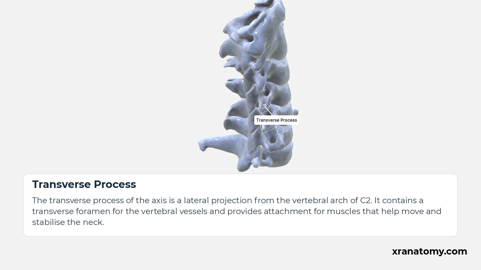
Transverse Process
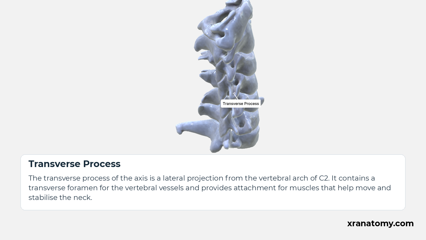
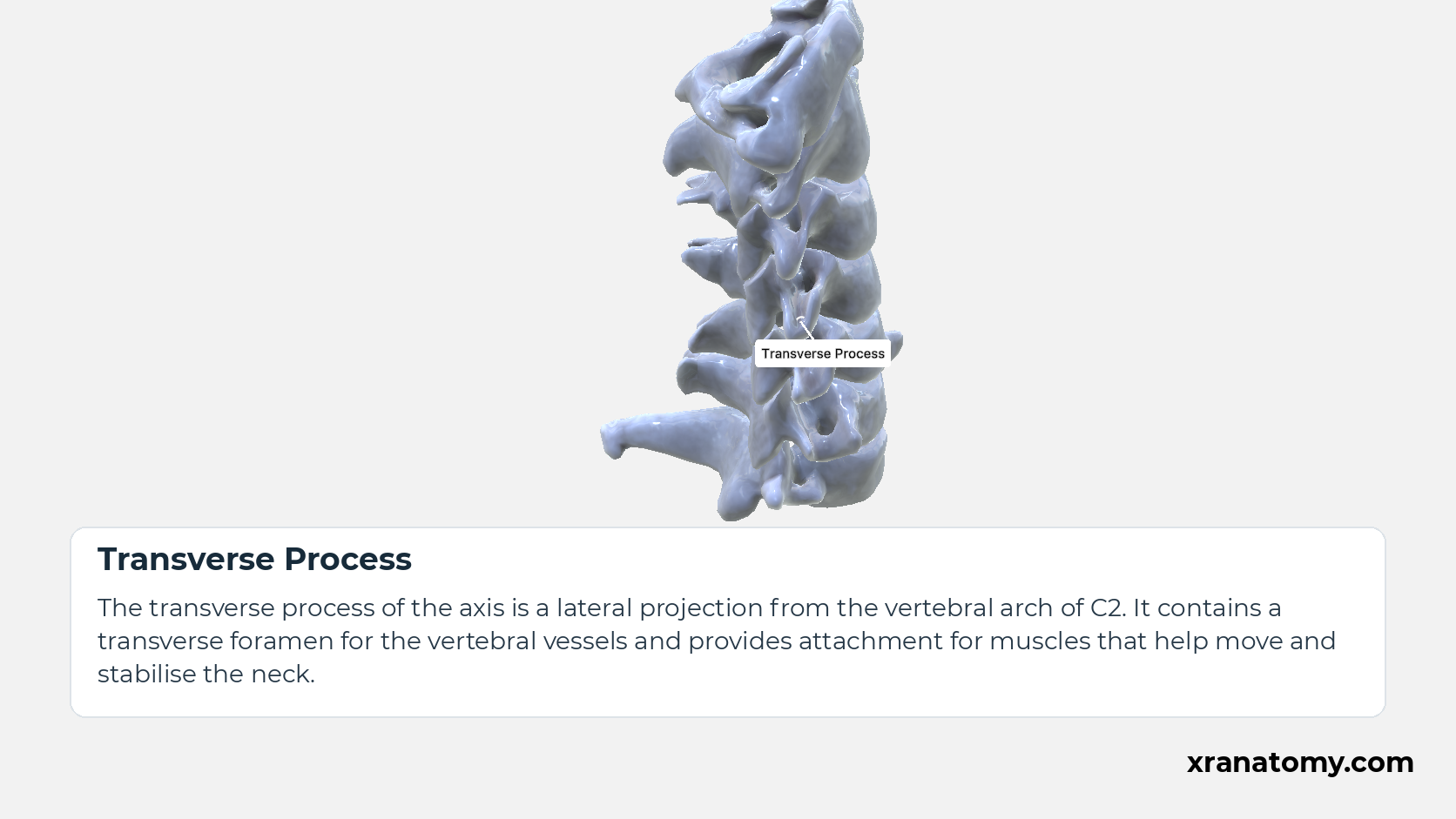
The transverse process of the axis is a lateral projection from the vertebral arch of C2. It contains a transverse foramen for the vertebral vessels and provides attachment for muscles that help move and stabilise the neck.
Study Anterior Tubercle of Transverse Process in this fixed image. Use the thumbnails to move between the 2D views; each view highlights one structure. The image does not rotate.
Illustration by XR Anatomy.
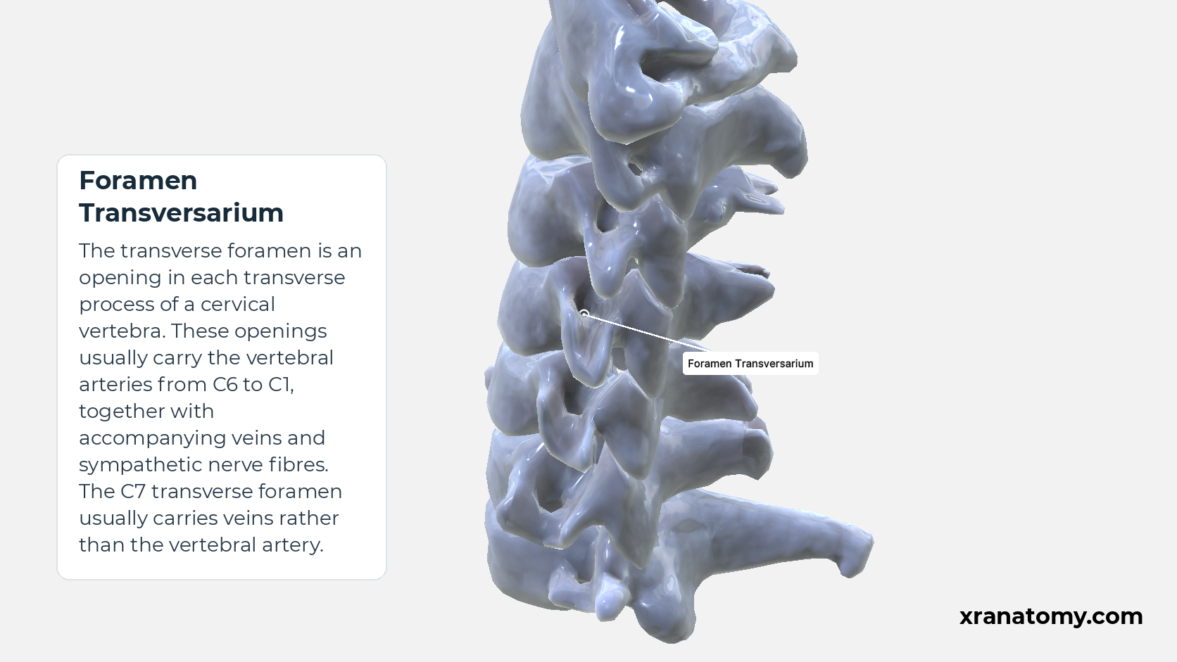
Foramen Transversarium
The transverse foramen is an opening in each transverse process of a cervical vertebra. These openings usually carry the vertebral arteries from C6 to C1, together with accompanying veins and sympathetic nerve fibres. The C7 transverse foramen usually carries veins rather than the vertebral artery.Show less
Illustration by XR Anatomy.
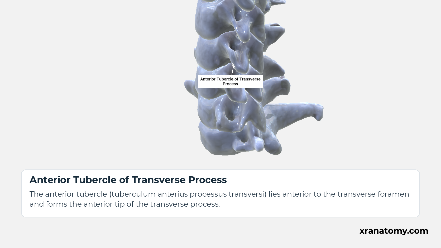
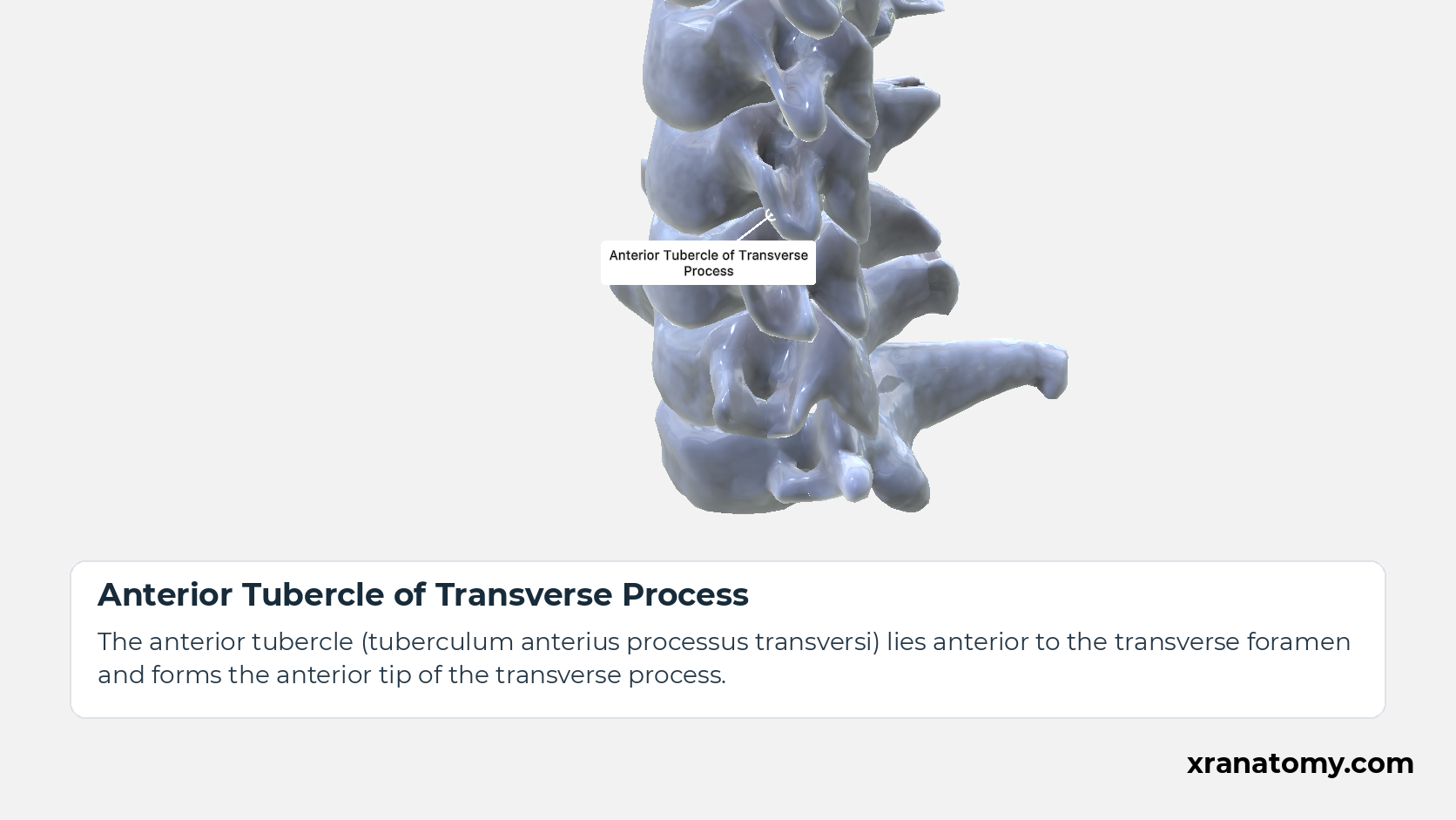
Anterior Tubercle of Transverse Process
The anterior tubercle (tuberculum anterius processus transversi) lies anterior to the transverse foramen and forms the anterior tip of the transverse process.
Illustration by XR Anatomy.
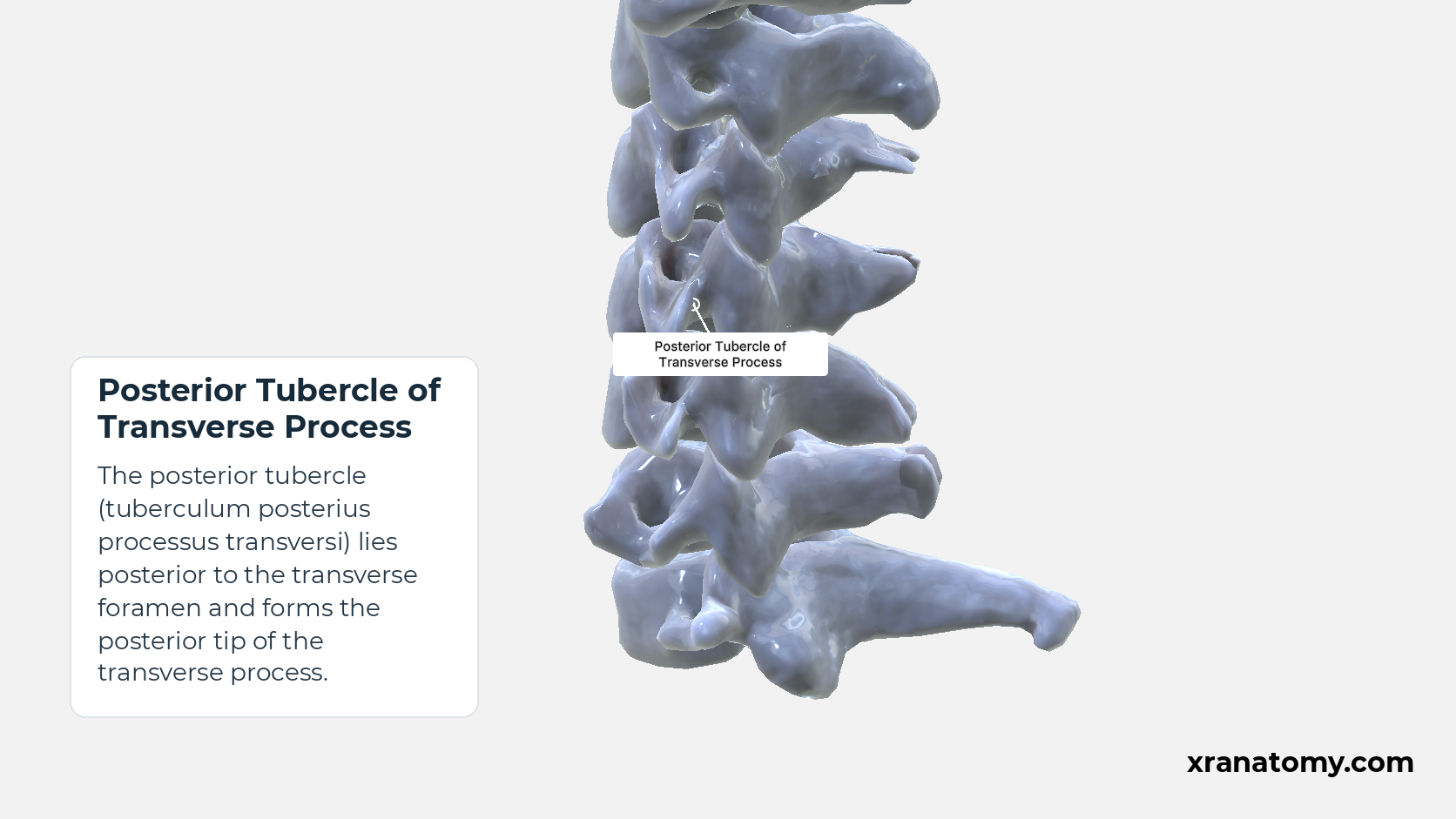
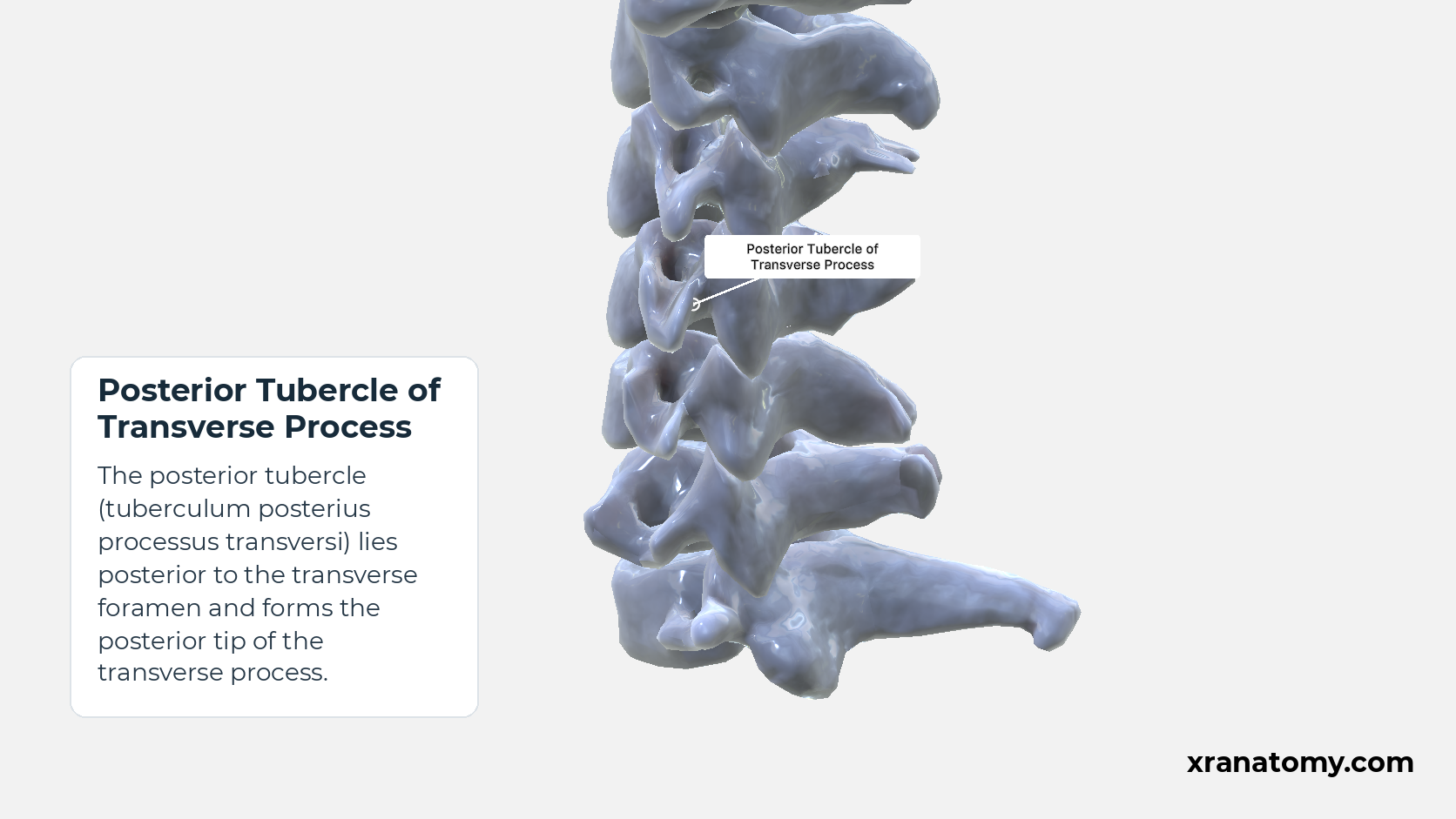
Posterior Tubercle of Transverse Process
The posterior tubercle (tuberculum posterius processus transversi) lies posterior to the transverse foramen and forms the posterior tip of the transverse process.
Illustration by XR Anatomy.
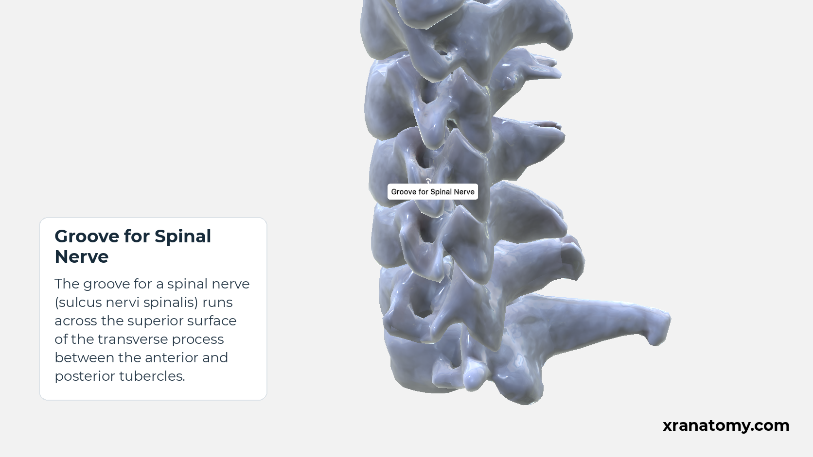
Groove for Spinal Nerve
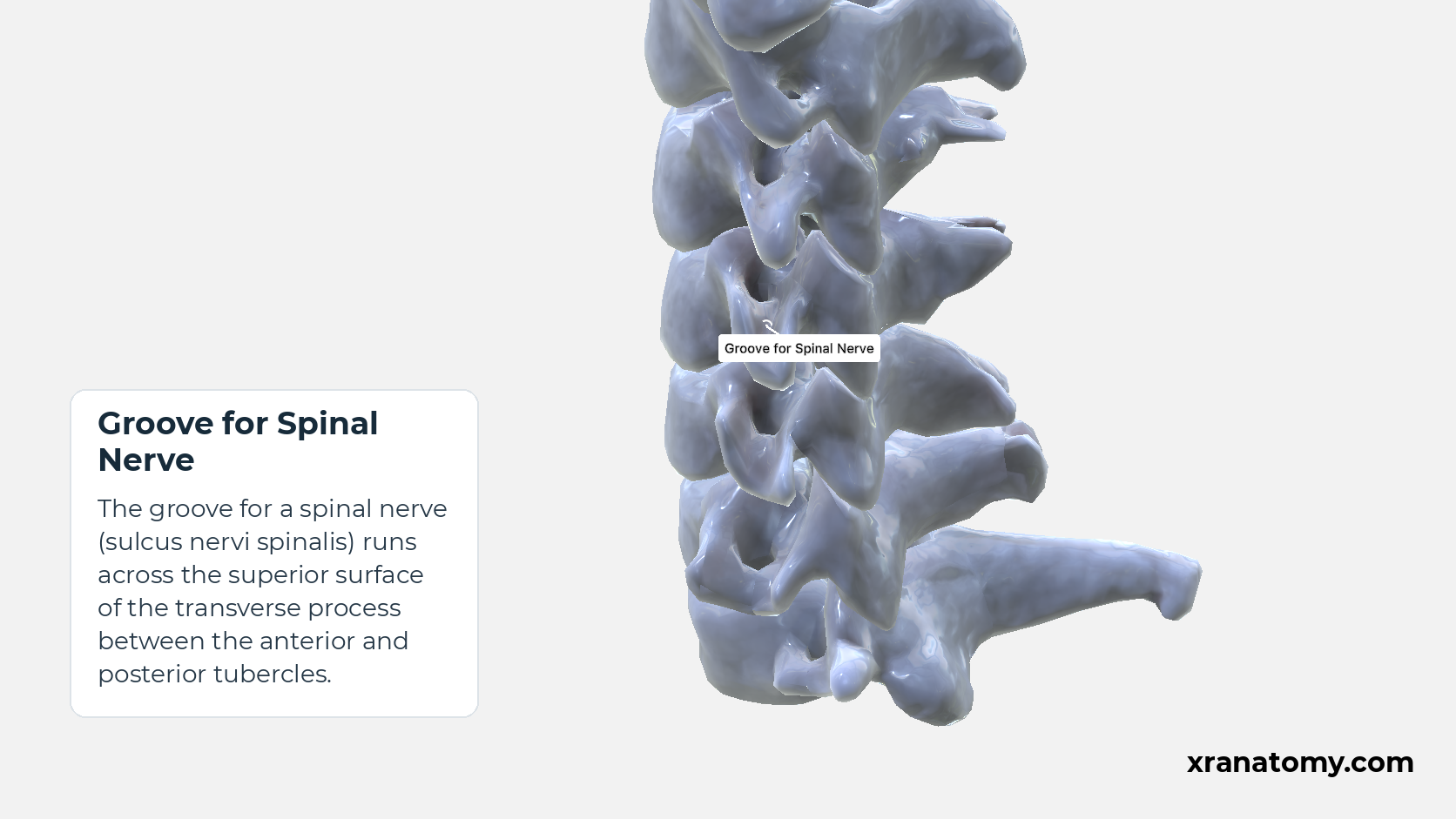
The groove for a spinal nerve (sulcus nervi spinalis) runs across the superior surface of the transverse process between the anterior and posterior tubercles.
Illustration by XR Anatomy.
Cervical Vertebra
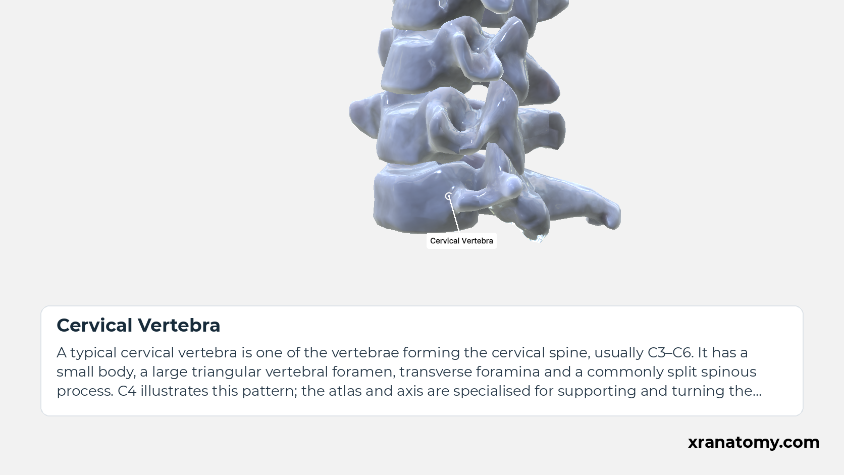
A typical cervical vertebra is one of the vertebrae forming the cervical spine, usually C3–C6. It has a small body, a large triangular vertebral foramen, transverse foramina and a commonly split spinous process. C4 illustrates this pattern; the atlas and axis are specialised for supporting and turning the head.Show less
Illustration by XR Anatomy.
Uncinate Process of Vertebra
The uncinate process is the raised lateral lip of the superior surface of a typical cervical vertebral body.
Illustration by XR Anatomy.
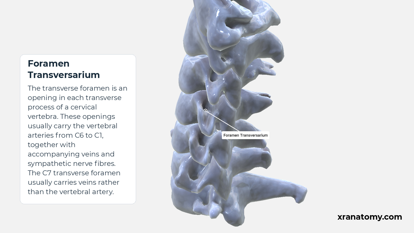
Each transverse process (processus transversus) of a typical cervical vertebra contains a transverse foramen and ends in anterior and posterior tubercles. A groove for a spinal nerve crosses its superior surface between the tubercles.
Foramen Transversarium
The transverse foramen is an opening in each transverse process of a cervical vertebra. These openings usually carry the vertebral arteries from C6 to C1, together with accompanying veins and sympathetic nerve fibres. The C7 transverse foramen usually carries veins rather than the vertebral artery.Show less
Study Foramen Transversarium in this fixed image. Use the thumbnails to move between the 2D views; each view highlights one structure. The image does not rotate.
Illustration by XR Anatomy.
Transverse Process
The transverse process of the axis is a lateral projection from the vertebral arch of C2. It contains a transverse foramen for the vertebral vessels and provides attachment for muscles that help move and stabilise the neck.
Illustration by XR Anatomy.
Cervical Vertebra
A typical cervical vertebra is one of the vertebrae forming the cervical spine, usually C3–C6. It has a small body, a large triangular vertebral foramen, transverse foramina and a commonly split spinous process. C4 illustrates this pattern; the atlas and axis are specialised for supporting and turning the head.Show less
Illustration by XR Anatomy.
Foramen Transversarium
The transverse foramen is an opening in each transverse process of a cervical vertebra. These openings usually carry the vertebral arteries from C6 to C1, together with accompanying veins and sympathetic nerve fibres. The C7 transverse foramen usually carries veins rather than the vertebral artery.Show less
Study Foramen Transversarium and Transverse-Process Landmarks in this fixed image. Use the thumbnails to move between the 2D views; each view highlights one structure. The image does not rotate.
Illustration by XR Anatomy.
Transverse Process
The transverse process of the axis is a lateral projection from the vertebral arch of C2. It contains a transverse foramen for the vertebral vessels and provides attachment for muscles that help move and stabilise the neck.
Illustration by XR Anatomy.
Anterior Tubercle of Transverse Process
The anterior tubercle (tuberculum anterius processus transversi) lies anterior to the transverse foramen and forms the anterior tip of the transverse process.
Illustration by XR Anatomy.
Posterior Tubercle of Transverse Process
The posterior tubercle (tuberculum posterius processus transversi) lies posterior to the transverse foramen and forms the posterior tip of the transverse process.
Illustration by XR Anatomy.
Groove for Spinal Nerve
The groove for a spinal nerve (sulcus nervi spinalis) runs across the superior surface of the transverse process between the anterior and posterior tubercles.
Illustration by XR Anatomy.
Uncinate Process of Vertebra
The uncinate process is the raised lateral lip of the superior surface of a typical cervical vertebral body.
Illustration by XR Anatomy.
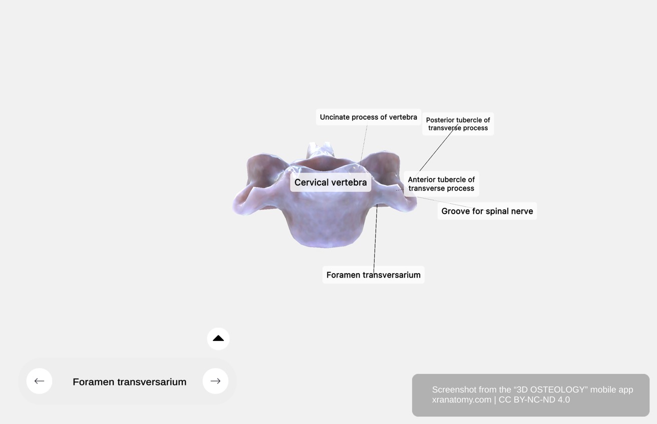 ↺ drag to rotate · scroll to zoom
Foramen Transversarium and Transverse-Process Landmarks
Foramen Transversarium and Transverse-Process Landmarks
Drag or use the controls to bring each labelled transverse-process landmark into a clear profile.
Sourced from 3D Osteology.
In depth · 5 sections · 1 min read5 more sections on this part5 sections open — reading In depth- Foramen Transversarium
- Anterior Tubercle of Transverse Process
- Posterior Tubercle of Transverse Process
- Groove for Spinal Nerve
- Check Your Understanding
Foramen Transversarium
The transverse foramen (foramen transversarium) pierces the transverse process.
Anterior Tubercle of Transverse Process
The anterior tubercle (tuberculum anterius processus transversi) lies anterior to the transverse foramen and forms the anterior tip of the transverse process.
Posterior Tubercle of Transverse Process
The posterior tubercle (tuberculum posterius processus transversi) lies posterior to the transverse foramen and forms the posterior tip of the transverse process.
Groove for Spinal Nerve
The groove for a spinal nerve (sulcus nervi spinalis) runs across the superior surface of the transverse process between the anterior and posterior tubercles.
Practice · Active recall · 5 questions Recall: Transverse Process
A typical cervical transverse process ends in anterior and posterior .
tubercles
Each transverse process (processus transversus) of a typical cervical vertebra contains a transverse foramen and ends in anterior and posterior tubercles. A groove for a spinal nerve crosses its superior surface between the tubercles.
Review this sectionHow well did you know it? · press 1–4
Nice work — revisit in a few days.
In depth · 1 question Recall: Foramen Transversarium
The transverse foramen pierces the .
transverse process
The transverse foramen (foramen transversarium) pierces the transverse process.
Review this sectionHow well did you know it? · press 1–4
Nice work — revisit in a few days.
In depth · 1 question Recall: Anterior Tubercle of Transverse Process
The tubercle in front of the transverse foramen is the tubercle.
anterior
The anterior tubercle (tuberculum anterius processus transversi) lies anterior to the transverse foramen and forms the anterior tip of the transverse process.
Review this sectionHow well did you know it? · press 1–4
Nice work — revisit in a few days.
In depth · 1 question Recall: Posterior Tubercle of Transverse Process
The tubercle behind the transverse foramen is the tubercle.
posterior
The posterior tubercle (tuberculum posterius processus transversi) lies posterior to the transverse foramen and forms the posterior tip of the transverse process.
Review this sectionHow well did you know it? · press 1–4
Nice work — revisit in a few days.
In depth · 1 question Recall: Groove for Spinal Nerve
The spinal-nerve groove crosses the surface of the transverse process.
superior
The groove for a spinal nerve (sulcus nervi spinalis) runs across the superior surface of the transverse process between the anterior and posterior tubercles.
Review this sectionHow well did you know it? · press 1–4
Nice work — revisit in a few days.
In depth · continued · 1 more section- Check Your Understanding
Check Your Understanding
- Which opening identifies a vertebra as cervical?
A transverse foramen in each transverse process.
- Which structures form the raised lateral margins of a typical cervical vertebral body?
The uncinate processes.
- Through which cervical levels does the vertebral artery usually ascend in the transverse foramina?
C6 through C1.
Practice · Check your understanding · 1 question Recall: Check Your Understanding
Which opening identifies a vertebra as cervical?
A transverse foramen in each transverse process.
1. Which opening identifies a vertebra as cervical? A transverse foramen in each transverse process.
Review this sectionHow well did you know it? · press 1–4
Bibliography
Every claim on this page is sourced against the standard anatomical references.
- FIPAT. Terminologia Anatomica, 2nd edition, Part 2: Bones. https://cdn.dal.ca/content/dam/dalhousie/pdf/library/FIPAT/TA2/FIPAT-TA2-Part-2.pdf
- Gray H. Anatomy of the Human Body. 21st ed. Philadelphia: Lea & Febiger; 1924.
- Drake RL, Vogl AW, Mitchell AWM. Gray's Anatomy for Students. Elsevier.
- Betts JG, et al. Anatomy and Physiology 2e. OpenStax; Rice University.
- Waxenbaum JA, Reddy V, Margetis K. Anatomy, Back, Cervical Vertebrae. [Updated 2026 Jan 31]. In: StatPearls [Internet]. Treasure Island (FL): StatPearls Publishing; 2026 Jan-. https://www.ncbi.nlm.nih.gov/books/NBK459200/
- Cho W, Maeda T, Park Y, Buchowski JM, Nabb CE, Riew KD. The Incidence of Bifid C7 Spinous Processes. Global Spine J. 2012;2(2):99-104. https://pmc.ncbi.nlm.nih.gov/articles/PMC3864495/
- Winegar BA, Kay MD, Taljanovic M. Magnetic resonance imaging of the spine. Pol J Radiol. 2020;85:e550-e574. doi:10.5114/pjr.2020.99887. https://pmc.ncbi.nlm.nih.gov/articles/PMC7571515/
