Anatomy
Shared valve principles
The heart's four valves keep blood moving one way: two atrioventricular valves between atria and ventricles, and two semilunar valves at the ventricular outlets.
As blood moves through the heart, four valves help keep it travelling forwards. Their positions divide them into two families: valves between atria and ventricles, and valves at the ventricular outlets. Each family has a different arrangement of supporting structures.
Four valves, two families
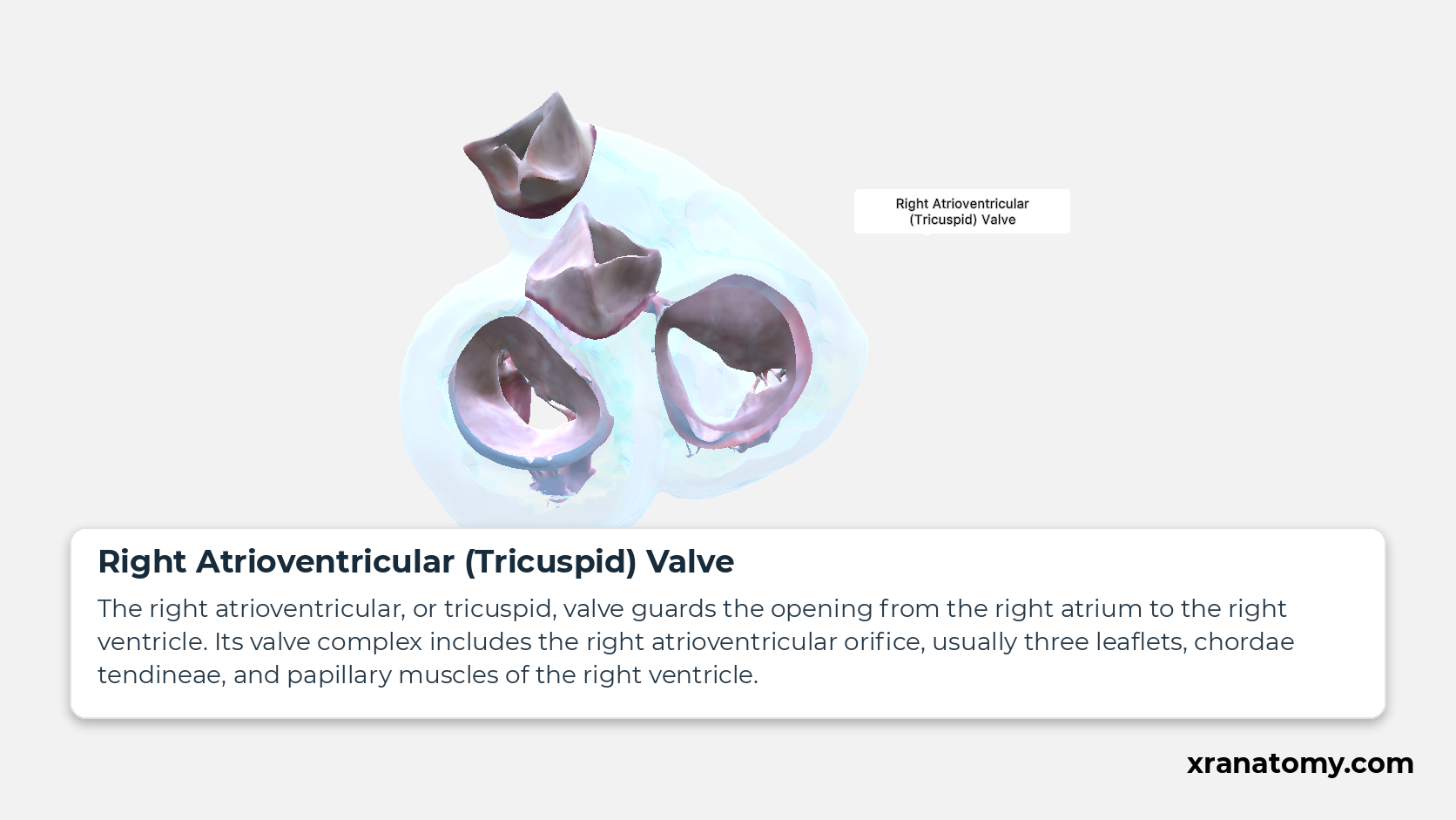
Right Atrioventricular (Tricuspid) Valve
The right atrioventricular, or tricuspid, valve guards the opening from the right atrium to the right ventricle. Its valve complex includes the right atrioventricular orifice, usually three leaflets, chordae tendineae, and papillary muscles of the right ventricle.Show less
Study Four valves, two families in this fixed image. Use the thumbnails to move between the 2D views; each view highlights one structure. The image does not rotate.
Illustration by XR Anatomy.
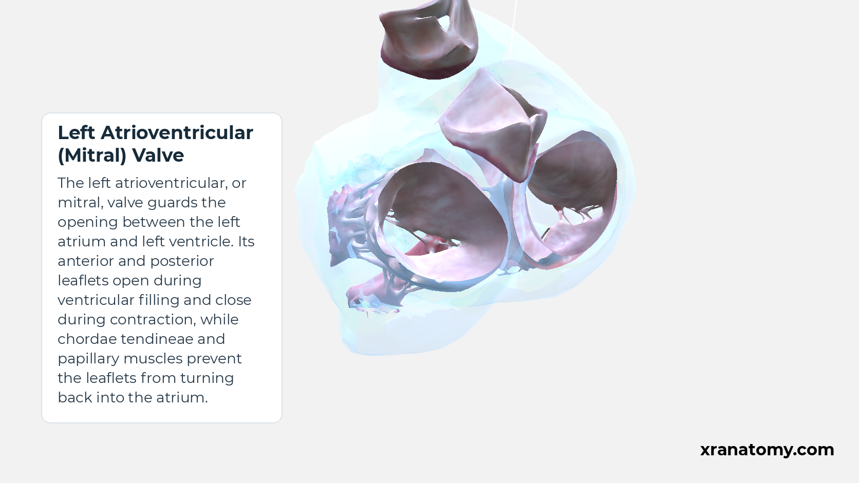
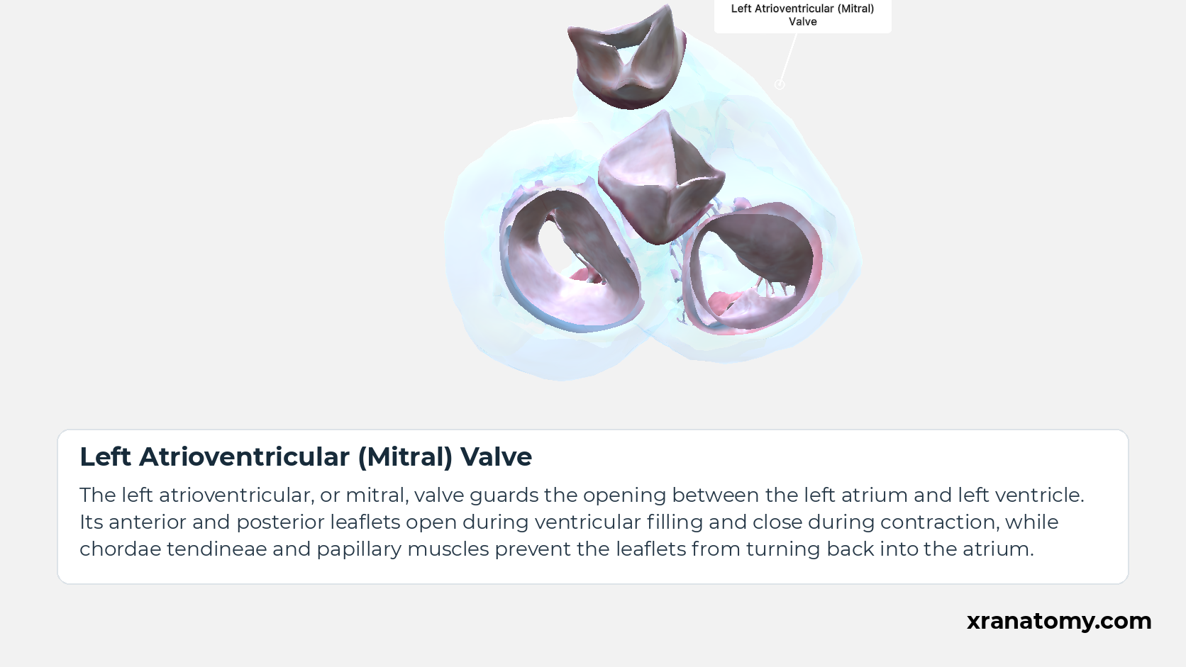
Left Atrioventricular (Mitral) Valve
The left atrioventricular, or mitral, valve guards the opening between the left atrium and left ventricle. Its anterior and posterior leaflets open during ventricular filling and close during contraction, while chordae tendineae and papillary muscles prevent the leaflets from turning back into the atrium.Show less
Illustration by XR Anatomy.
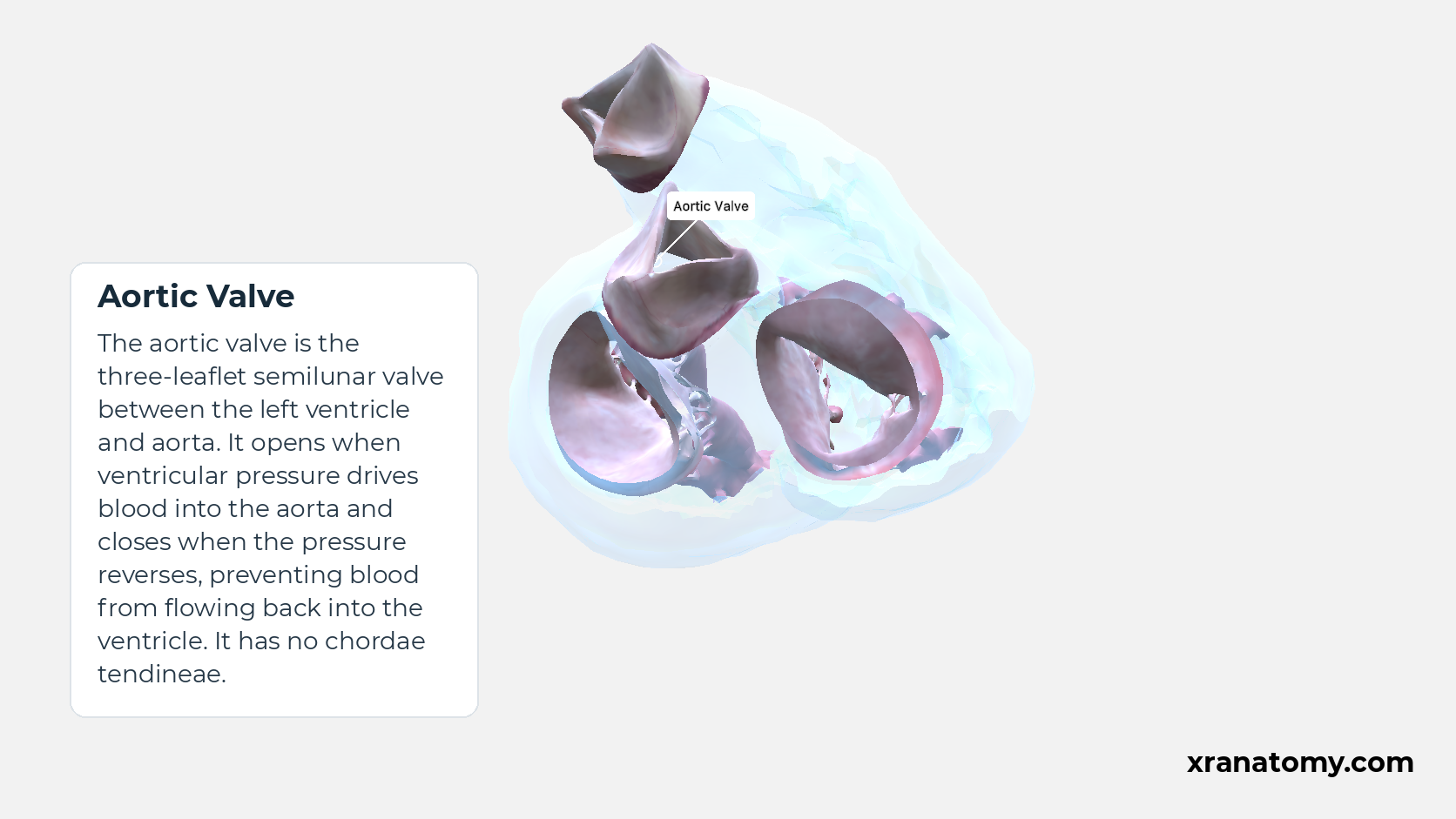
Aortic Valve
The aortic valve is the three-leaflet semilunar valve between the left ventricle and aorta. It opens when ventricular pressure drives blood into the aorta and closes when the pressure reverses, preventing blood from flowing back into the ventricle. It has no chordae tendineae.Show less
Illustration by XR Anatomy.
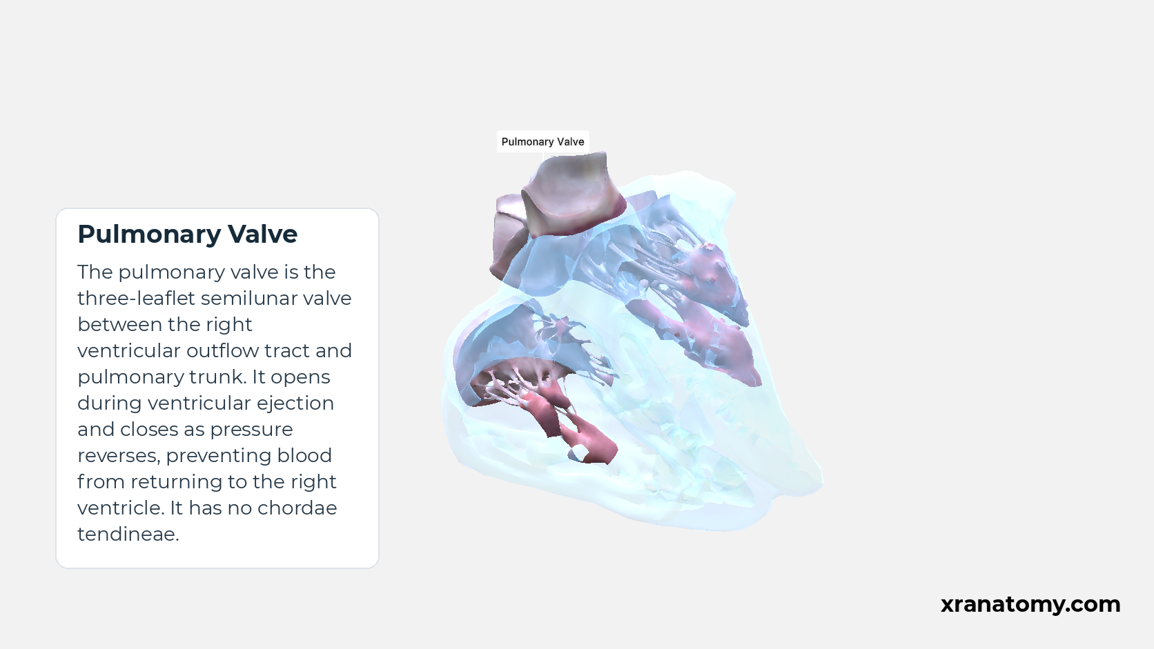
Pulmonary Valve
The pulmonary valve is the three-leaflet semilunar valve between the right ventricular outflow tract and pulmonary trunk. It opens during ventricular ejection and closes as pressure reverses, preventing blood from returning to the right ventricle. It has no chordae tendineae.Show less
Illustration by XR Anatomy.
The two atrioventricular valves
Between the right atrium and right ventricle, the tricuspid valve controls the opening into the ventricle. The corresponding valve on the left is the mitral valve. Both are called atrioventricular valves because they lie at the junction of an atrium and a ventricle. The tricuspid valve usually has three main leaflets, while the mitral valve has two principal leaflets whose margins meet at commissural regions. In both valves, chordae tendineae connect the leaflets to supporting papillary muscles.
The two semilunar valves
At the ventricular exits, the aortic valve controls the left ventricular outlet into the aorta, while the pulmonary valve controls the right ventricular outlet into the pulmonary trunk. These are the semilunar valves. Each normally has three leaflets supported within its arterial root. Their support comes from this root arrangement, so they have no chordae tendineae or papillary-muscle apparatus.
Valve relationships
How pressure moves the leaflets and muscle supports them
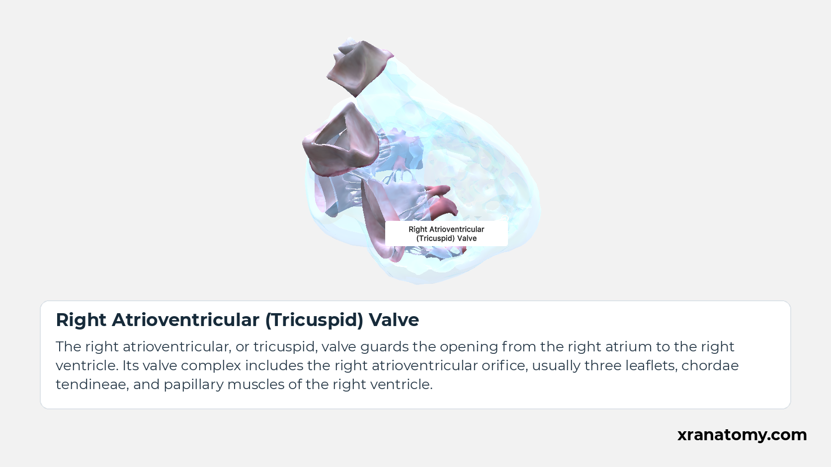
Right Atrioventricular (Tricuspid) Valve
The right atrioventricular, or tricuspid, valve guards the opening from the right atrium to the right ventricle. Its valve complex includes the right atrioventricular orifice, usually three leaflets, chordae tendineae, and papillary muscles of the right ventricle.Show less
Study Pressure, leaflets, chordae, and papillary support in this fixed image. Use the thumbnails to move between the 2D views; each view highlights one structure. The image does not rotate.
Illustration by XR Anatomy.
Left Atrioventricular (Mitral) Valve
The left atrioventricular, or mitral, valve guards the opening between the left atrium and left ventricle. Its anterior and posterior leaflets open during ventricular filling and close during contraction, while chordae tendineae and papillary muscles prevent the leaflets from turning back into the atrium.Show less
Illustration by XR Anatomy.
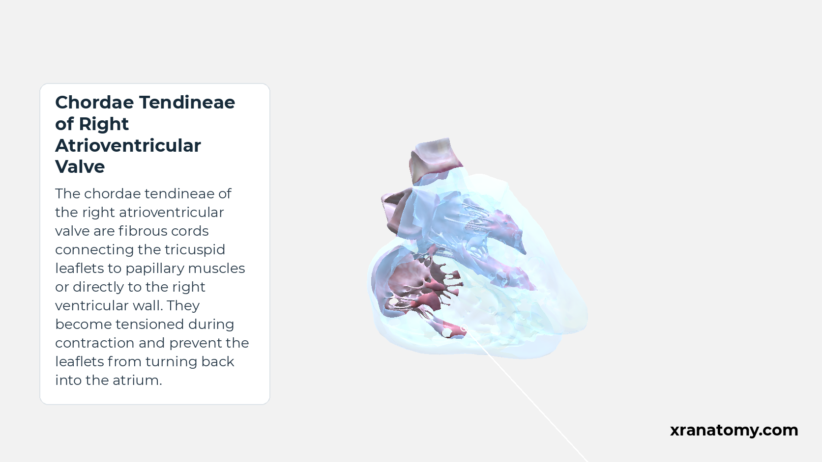
Chordae Tendineae of Right Atrioventricular Valve
The chordae tendineae of the right atrioventricular valve are fibrous cords connecting the tricuspid leaflets to papillary muscles or directly to the right ventricular wall. They become tensioned during contraction and prevent the leaflets from turning back into the atrium.Show less
Illustration by XR Anatomy.
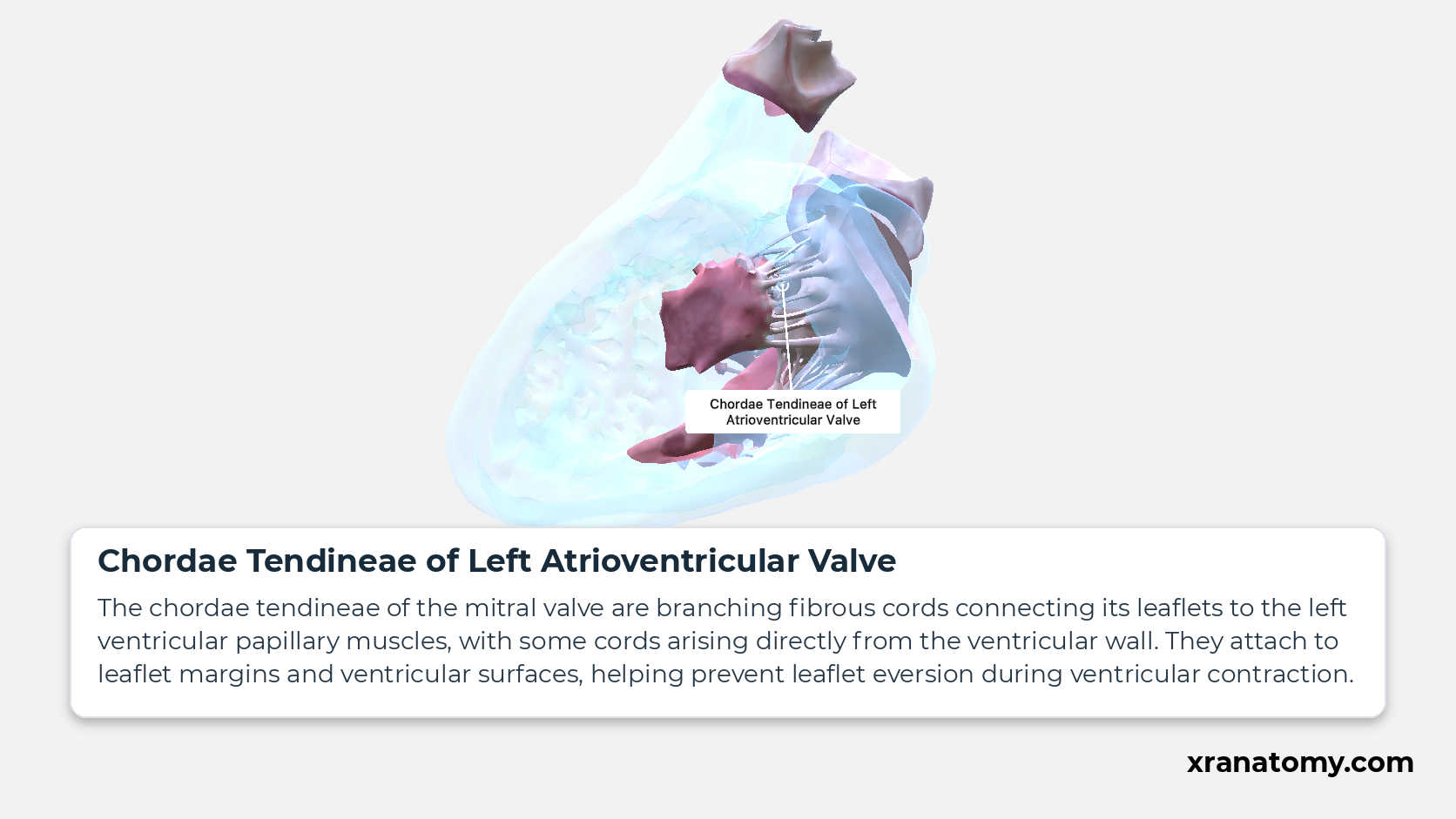
Chordae Tendineae of Left Atrioventricular Valve
The chordae tendineae of the mitral valve are branching fibrous cords connecting its leaflets to the left ventricular papillary muscles, with some cords arising directly from the ventricular wall. They attach to leaflet margins and ventricular surfaces, helping prevent leaflet eversion during ventricular contraction.Show less
Illustration by XR Anatomy.
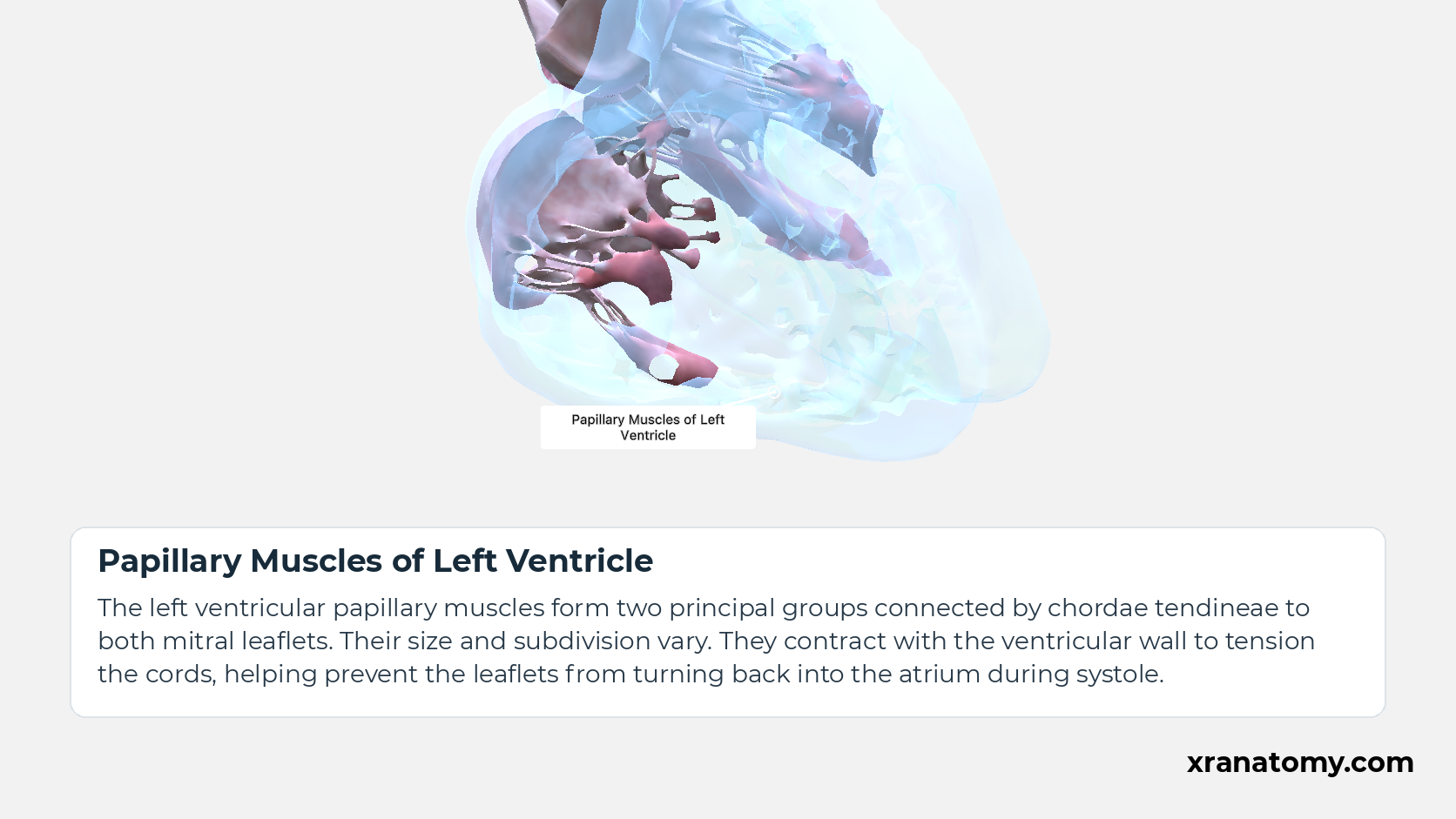
Papillary Muscles of Left Ventricle
The left ventricular papillary muscles form two principal groups connected by chordae tendineae to both mitral leaflets. Their size and subdivision vary. They contract with the ventricular wall to tension the cords, helping prevent the leaflets from turning back into the atrium during systole.Show less
Illustration by XR Anatomy.
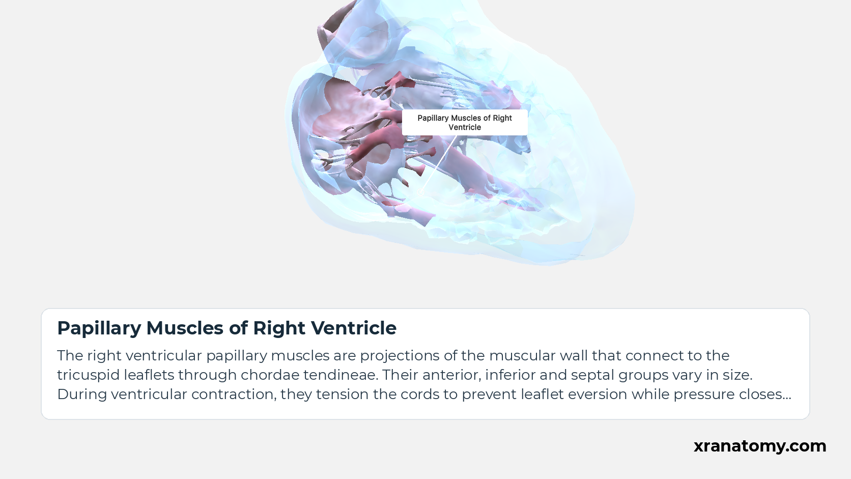
Papillary Muscles of Right Ventricle
The right ventricular papillary muscles are projections of the muscular wall that connect to the tricuspid leaflets through chordae tendineae. Their anterior, inferior and septal groups vary in size. During ventricular contraction, they tension the cords to prevent leaflet eversion while pressure closes the valve.Show less
Illustration by XR Anatomy.
Pressure opens and closes the valve
The valve leaflets move in response to pressure on their two sides. During ventricular filling, pressure in the atria exceeds pressure in the ventricles, allowing the atrioventricular valves to open. As the ventricles contract and their pressure rises above atrial pressure, the leaflets meet and close.
For blood to leave the ventricles, ventricular pressure must then exceed the pressure in the aorta or pulmonary trunk. This opens the semilunar valves. When the pressure difference reverses, their leaflets meet again, preventing blood from returning to the ventricles. In each case, the direction of the pressure difference explains the movement of the valve.
Chordae and papillary muscles support the leaflets
The papillary muscles support the atrioventricular leaflets while this pressure-driven movement takes place. They contract with the ventricular myocardium and tension the chordae tendineae. The tension restrains the closed leaflets, limiting their tendency to turn back into the atrium, a movement called eversion. The muscles keep the leaflets supported while pressure changes open and close the valve.
In depth · 2 sections · 1 min read2 more sections on this part2 sections open — reading In depth- Comparing the attachment arrangements
- Shared semilunar leaflet margins
Comparing the attachment arrangements
The tricuspid attachments and mitral attachments follow a chain from ventricular muscle through papillary muscle and chordae to the leaflets. The tricuspid valve illustrates this arrangement; the pulmonary root illustrates the support of a semilunar valve by its arterial wall.
Shared semilunar leaflet margins
Both aortic and pulmonary semilunar leaflets have a small central thickening at the free margin, the nodule, with crescent-shaped regions along the free edge, the lunules, on either side. These regions contribute to the surfaces that meet during closure. They are corresponding features of two separate valves, shown for the aortic valve and the pulmonary valve.
