Anatomy
Pericardium and heart in situ
The pericardium encloses the heart and the roots of its great vessels. It has a tough fibrous sac outside and the serous pericardium inside.
Pericardial covering and major vessels
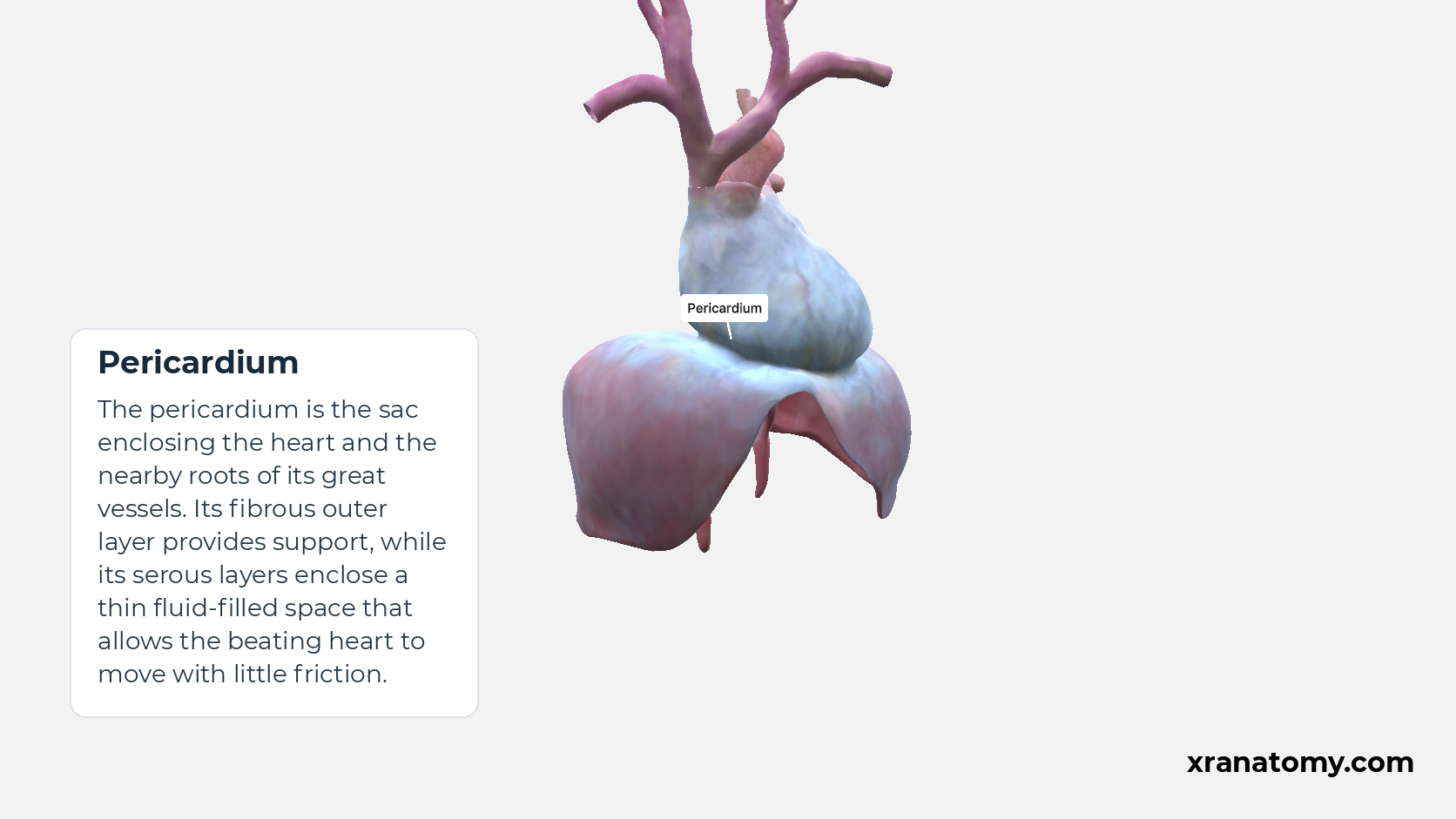
Pericardium
The pericardium is the sac enclosing the heart and the nearby roots of its great vessels. Its fibrous outer layer provides support, while its serous layers enclose a thin fluid-filled space that allows the beating heart to move with little friction.Show less
Study Pericardial covering and major vessels in this fixed image. Use the thumbnails to move between the 2D views; each view highlights one structure. The image does not rotate.
Illustration by XR Anatomy.
The heart within its covering
The pericardium forms a covering around the heart and extends onto the nearby roots of its major vessels. Looking at the organ in situ means seeing it within these surroundings. In that view, the covering can be related to the heart, the caval and pulmonary veins bringing blood in, and the aorta and pulmonary trunk carrying blood out.
From the outer sac to the heart’s surface
The covering has an outer fibrous part and an inner serous part. The fibrous pericardium is a tough connective-tissue sac. Inside it, the serous pericardium forms two layers: a parietal layer lining the sac and a visceral layer lying against the heart. At the heart wall, this visceral covering is associated with the epicardium. Following the layers from outside in therefore leads from the sac towards the surface of the organ.
Layers, reflections and sinuses
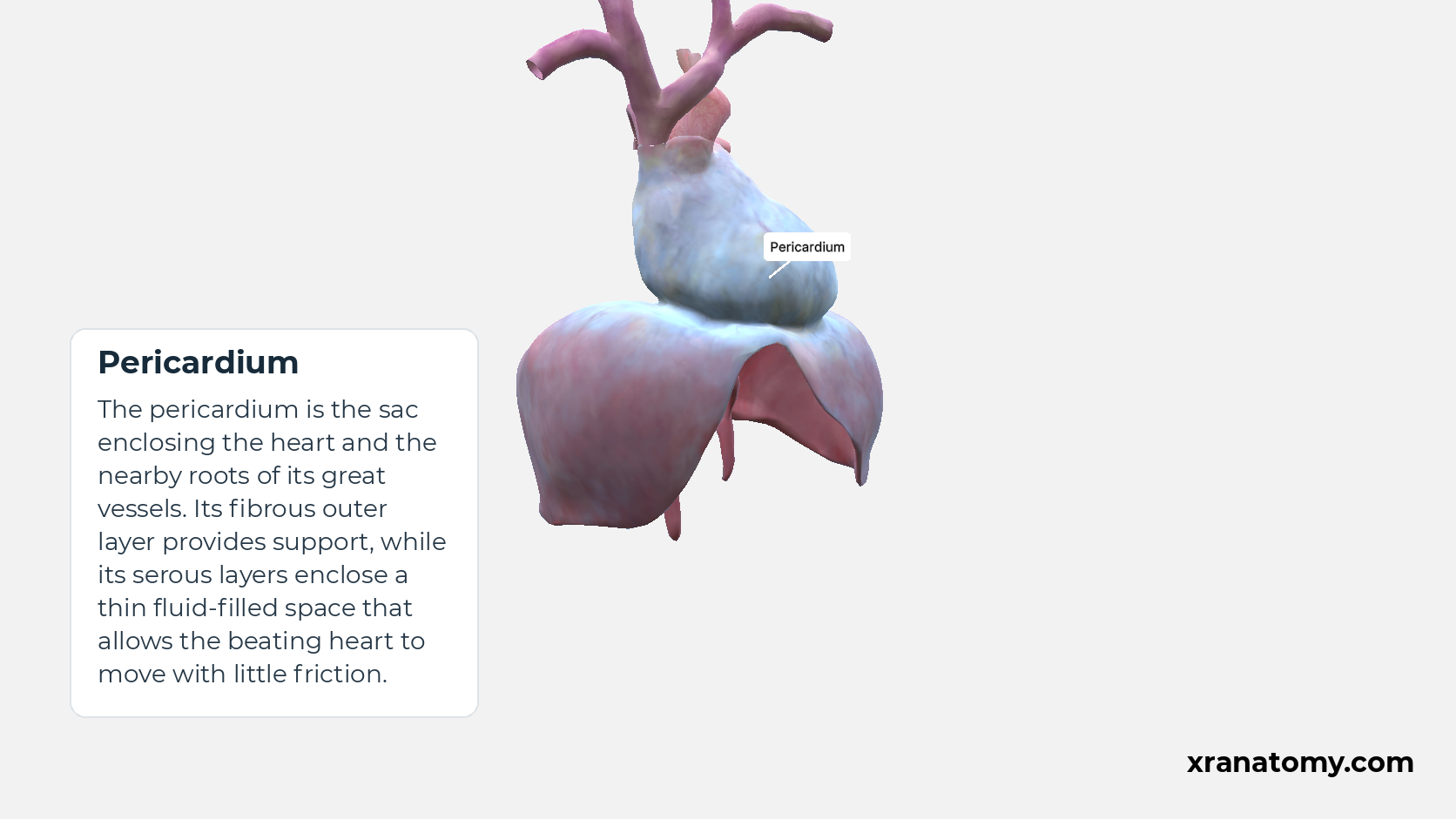
Pericardium
The pericardium is the sac enclosing the heart and the nearby roots of its great vessels. Its fibrous outer layer provides support, while its serous layers enclose a thin fluid-filled space that allows the beating heart to move with little friction.Show less
Study Pericardial covering and diaphragm in this fixed image. Use the thumbnails to move between the 2D views; each view highlights one structure. The image does not rotate.
Illustration by XR Anatomy.
The space that allows the layers to move
Between the parietal and visceral serous layers lies the pericardial cavity. It is a potential space containing a thin film of serous fluid, which lets the facing surfaces move smoothly against each other as the heart changes shape. Where the serous covering folds back on itself, these reflections form the transverse pericardial sinus and the oblique sinus. The sinuses are thus spaces created by the arrangement of the covering.
In depth · 2 sections · 1 min read2 more sections on this part2 sections open — reading In depth- Reflections around the great vessels
- Transverse and oblique sinuses
Reflections around the great vessels
The serous layers continue into one another at reflections around the great vessels. One group of reflections surrounds the ascending aorta and pulmonary trunk; another is associated with the caval and pulmonary veins. Their arrangement creates passages and recesses within the pericardial space.
Transverse and oblique sinuses
The transverse pericardial sinus passes behind the ascending aorta and pulmonary trunk, between the arterial and venous groups of reflections. The oblique sinus is a recess behind the left atrium, related to the venous reflections. These are spaces shaped by the pericardium and surrounding anatomy, not solid structures or additional heart chambers. Their detailed arrangement can vary.
