Anatomy
Surfaces, base, and apex
The heart's four named surfaces face the sternum, the diaphragm and the lungs, and its posterior base lies opposite the apex.
The outside of the heart gives useful clues to the chambers within it. Each surface faces a neighbouring structure, and the chamber beneath that surface helps give it its shape. Following these relationships makes the external anatomy easier to recognise.
Surfaces of the heart
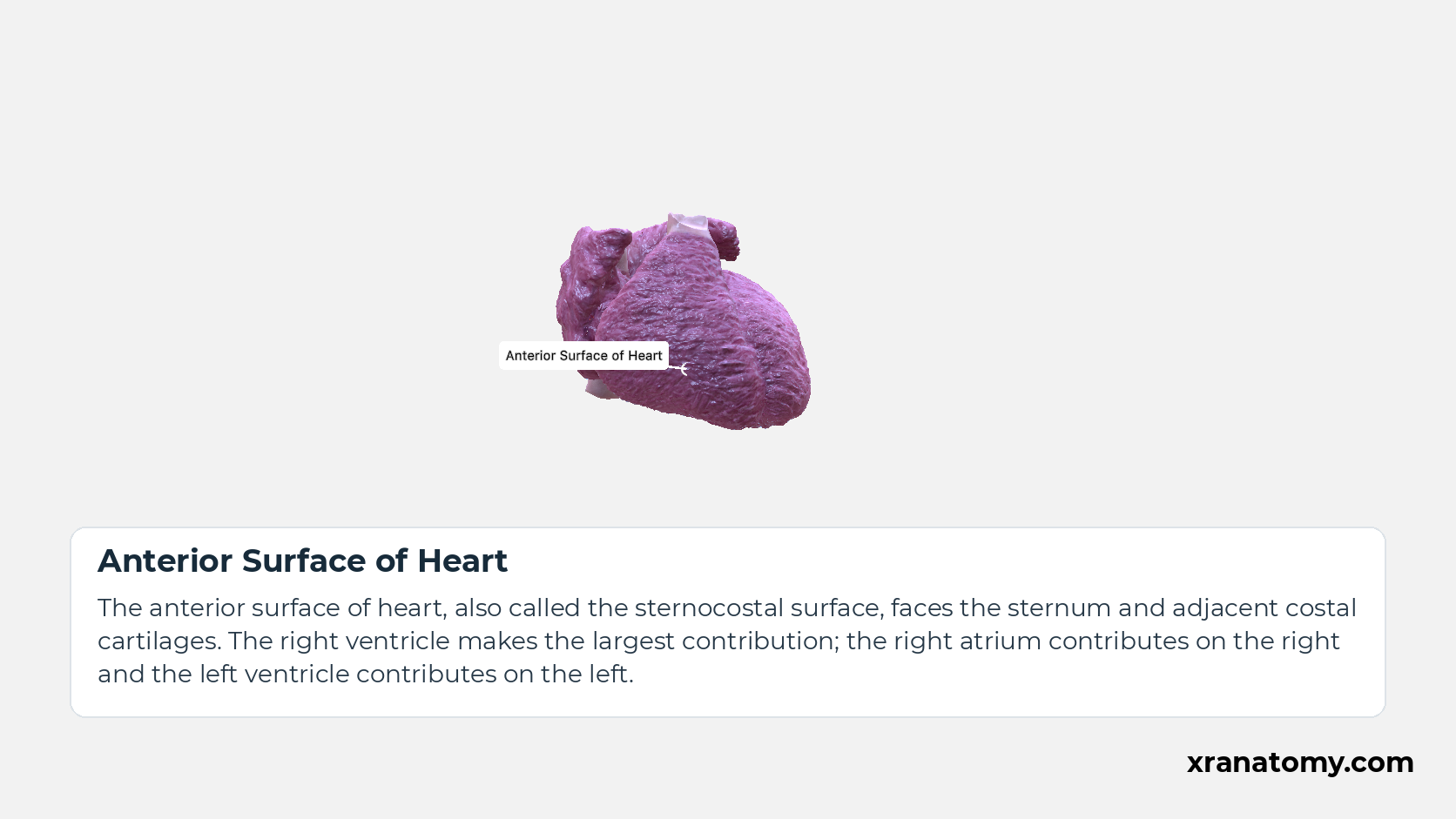
Anterior Surface of Heart
The anterior surface of heart, also called the sternocostal surface, faces the sternum and adjacent costal cartilages. The right ventricle makes the largest contribution; the right atrium contributes on the right and the left ventricle contributes on the left.Show less
Study Anterior and inferior surfaces in this fixed image. Use the thumbnails to move between the 2D views; each view highlights one structure. The image does not rotate.
Illustration by XR Anatomy.
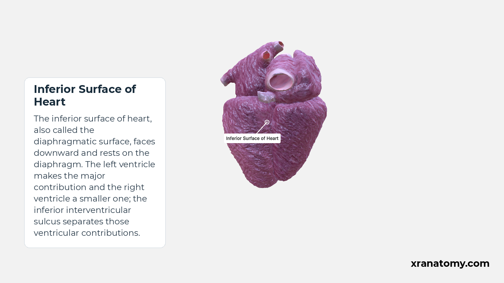
Inferior Surface of Heart
The inferior surface of heart, also called the diaphragmatic surface, faces downward and rests on the diaphragm. The left ventricle makes the major contribution and the right ventricle a smaller one; the inferior interventricular sulcus separates those ventricular contributions.Show less
Illustration by XR Anatomy.
Right Surface of Heart
The right surface of heart, also called the right pulmonary surface, faces the right lung. The right pulmonary surface is formed by the right atrium and extends between the superior vena cava and inferior vena cava.
Illustration by XR Anatomy.
Left Surface of Heart
The left surface of heart, also called the left pulmonary surface, faces the left lung. The left pulmonary surface is formed mainly by the left ventricle, with part of the left atrium contributing.
Illustration by XR Anatomy.
Base of Heart
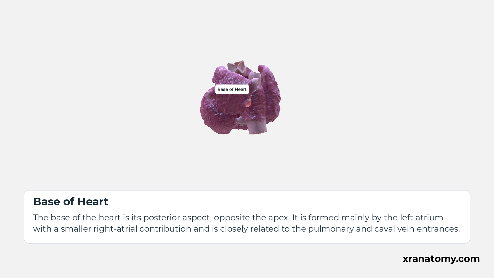
The base of the heart is its posterior aspect, opposite the apex. It is formed mainly by the left atrium with a smaller right-atrial contribution and is closely related to the pulmonary and caval vein entrances.
Illustration by XR Anatomy.
Apex of Heart
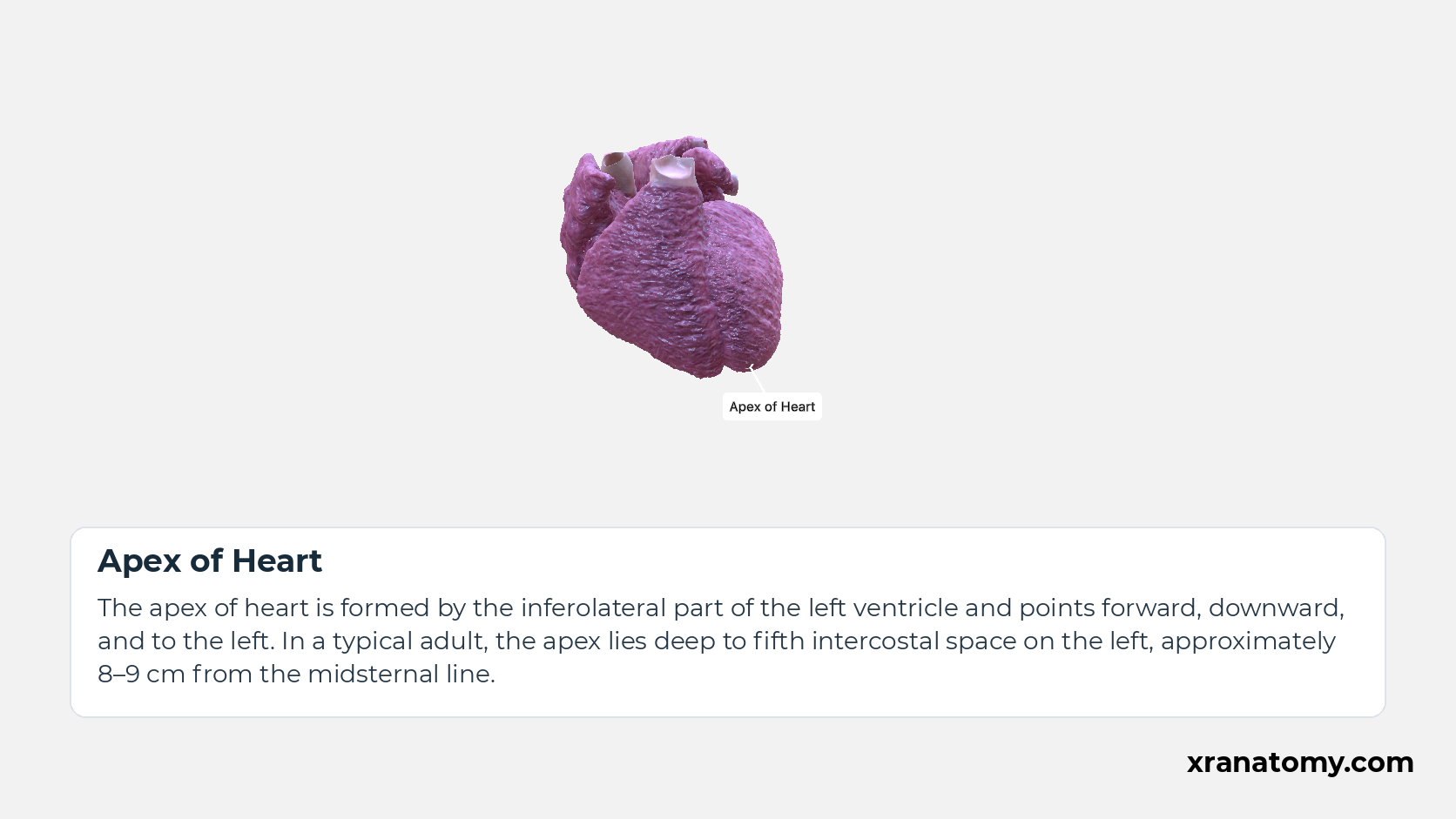
The apex of heart is formed by the inferolateral part of the left ventricle and points forward, downward, and to the left. In a typical adult, the apex lies deep to fifth intercostal space on the left, approximately 8–9 cm from the midsternal line.Show less
Illustration by XR Anatomy.
 ↺ drag to rotate · scroll to zoom
Anterior and inferior surfaces
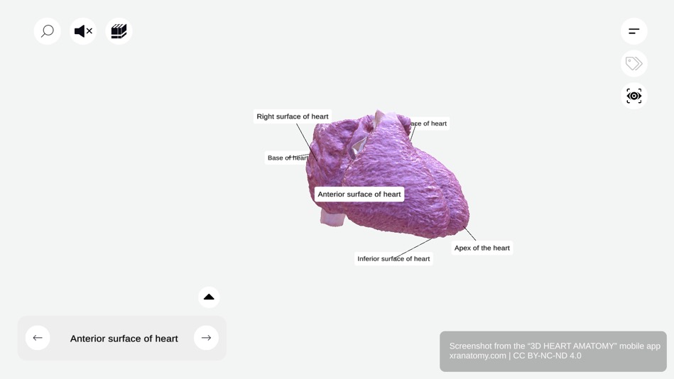
This figure shows anterior surface of heart, inferior surface of heart, right ventricle, left ventricle, right atrium, inferior interventricular sulcus.
Rotate the model to compare the heart's surface, base and apex labels.
Sourced from 3D Heart Anatomy.
Anterior and inferior surfaces
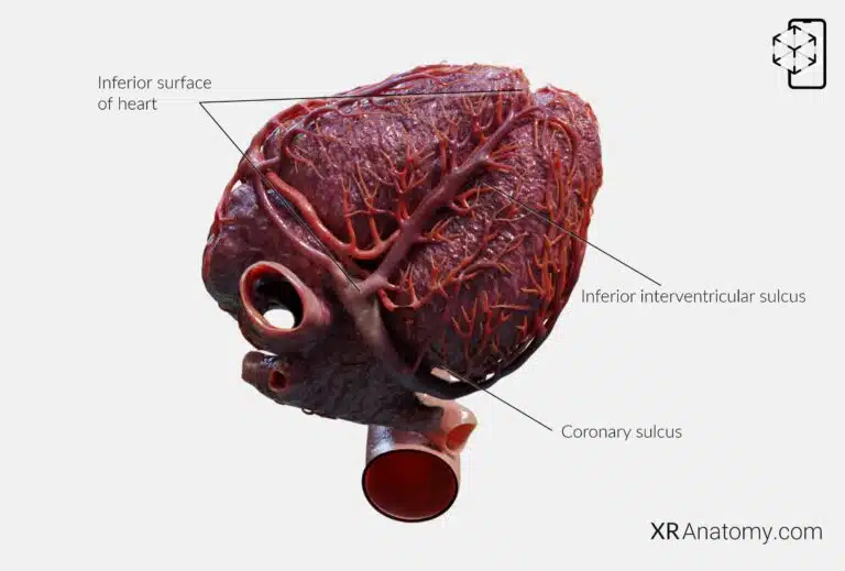
This figure shows anterior surface of heart, inferior surface of heart, right ventricle, left ventricle, right atrium, inferior interventricular sulcus.
Compare the labelled inferior surface with the grooves beside it.
Visualise this in augmented reality with XR Anatomy.
At the front, the anterior surface faces the sternum and is formed mainly by the right ventricle. It is also called the sternocostal surface. Below it, the inferior surface rests on the diaphragm and is formed mainly by the left ventricle, which is why this surface is also called diaphragmatic.
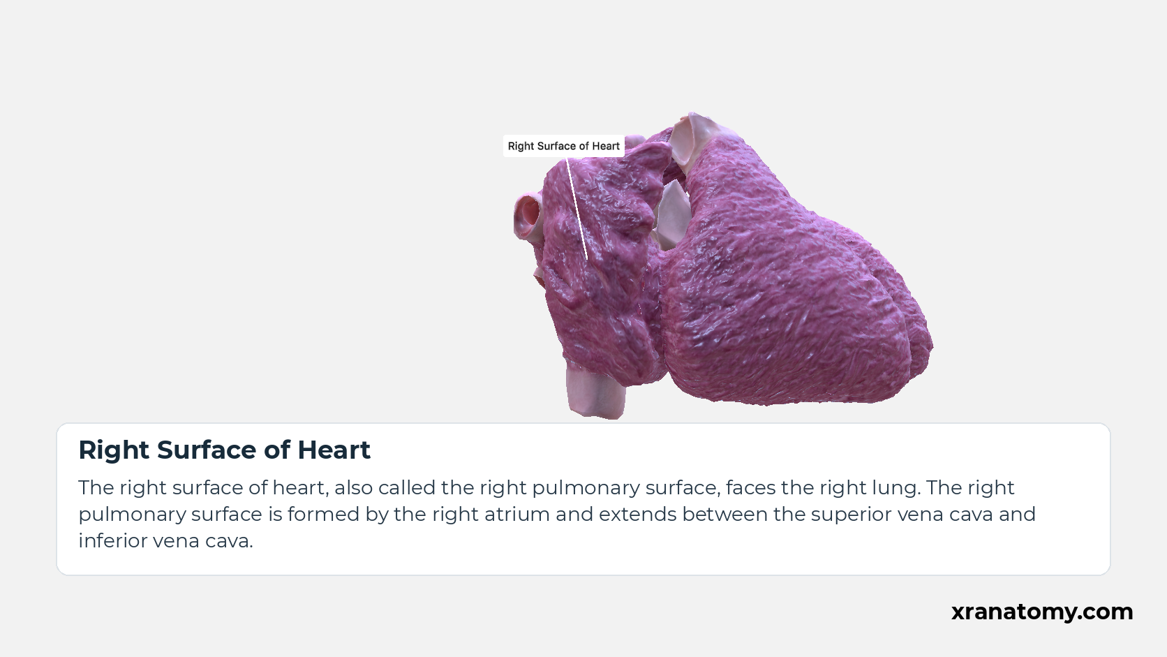
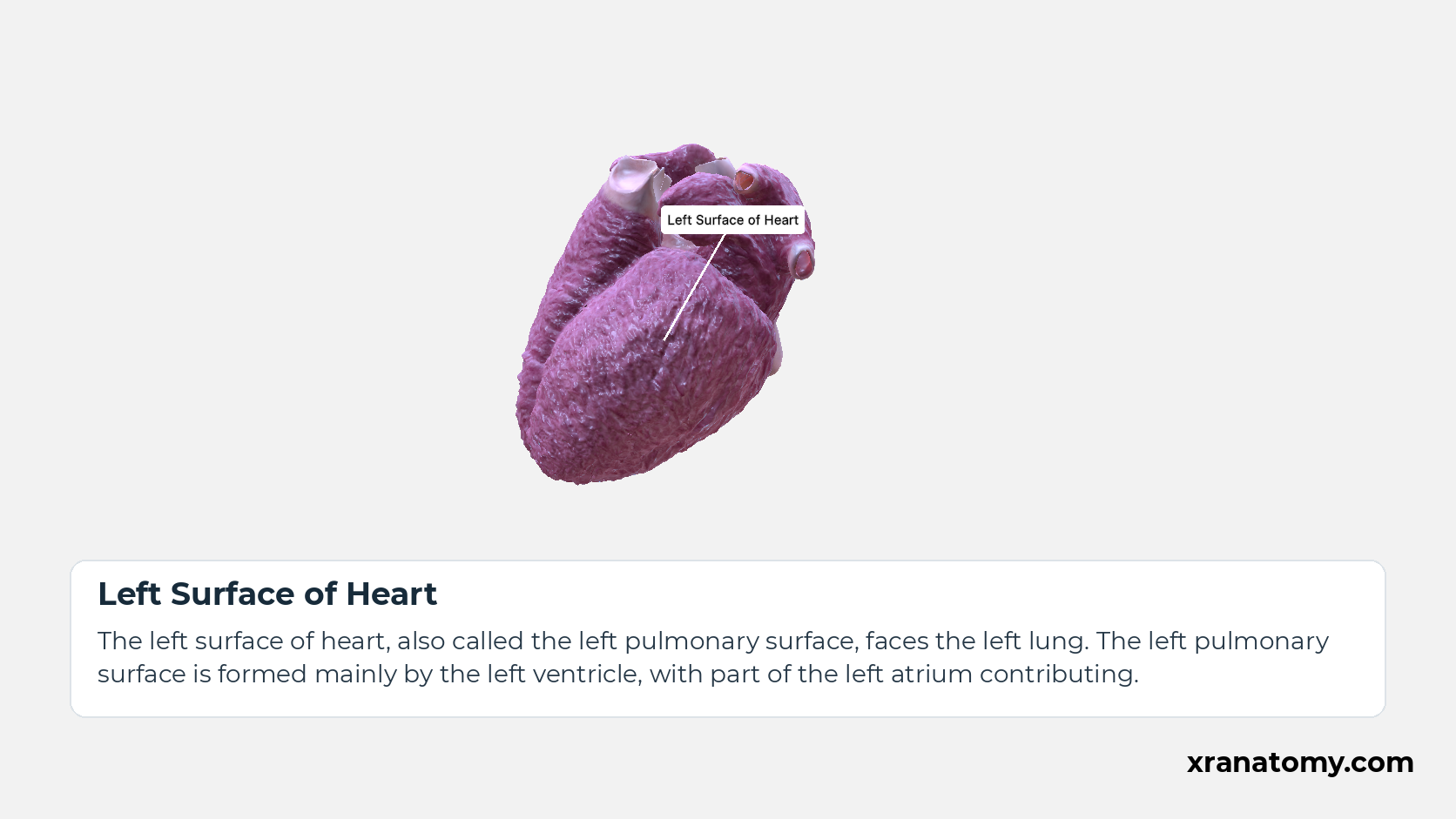
The remaining two surfaces face the lungs. The right pulmonary surface is formed by the right atrium, while the left pulmonary surface is formed mainly by the left ventricle. These relationships connect each surface to a neighbouring structure and an underlying chamber.
First Rib
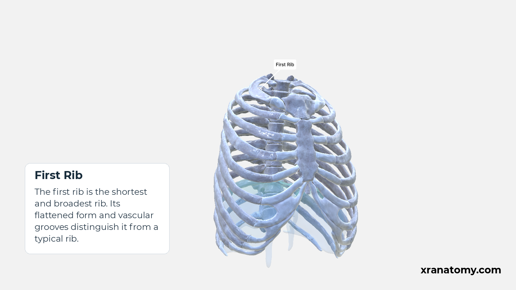
The first rib is the shortest and broadest rib. Its flattened form and vascular grooves distinguish it from a typical rib.
Study Bony Boundaries and Openings in this fixed image. Use the thumbnails to move between the 2D views; each view highlights one structure. The image does not rotate.
Illustration by XR Anatomy.
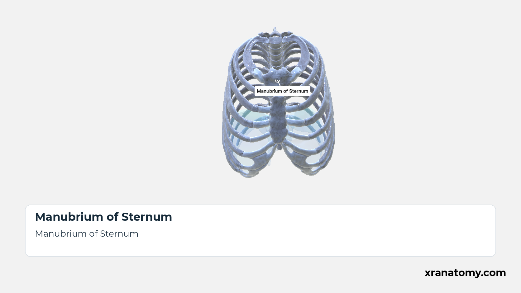
Manubrium of Sternum
The manubrium is the broad superior part of the sternum and its thickest piece. Its superior border carries a central jugular notch between paired clavicular notches, and each manubrial lateral border carries the attachment of the first costal cartilage. These features are described next. [2] [3] [4] [1]Show less
Illustration by XR Anatomy.
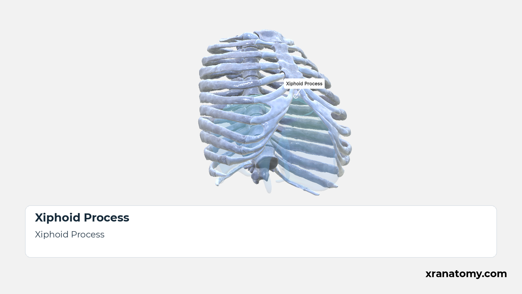
Xiphoid Process
The xiphoid process is the smallest and thinnest part, a tongue of bone that projects downwards from the body into the upper abdominal wall. It ossifies progressively from childhood and is usually bony, and often fused to the body, by middle age; its shape varies, being sometimes bifid or perforated. Its joint with the body and its soft-tissue attachments follow. [2] [3] [4] [1]Show less
Illustration by XR Anatomy.
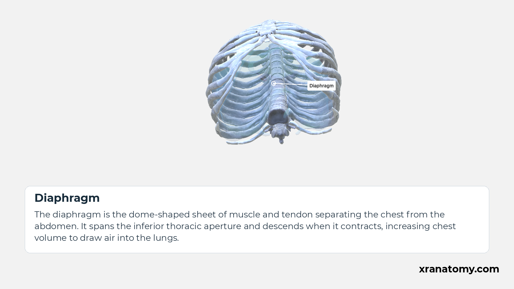
Diaphragm
The diaphragm is the dome-shaped sheet of muscle and tendon separating the chest from the abdomen. It spans the inferior thoracic aperture and descends when it contracts, increasing chest volume to draw air into the lungs.
Illustration by XR Anatomy.
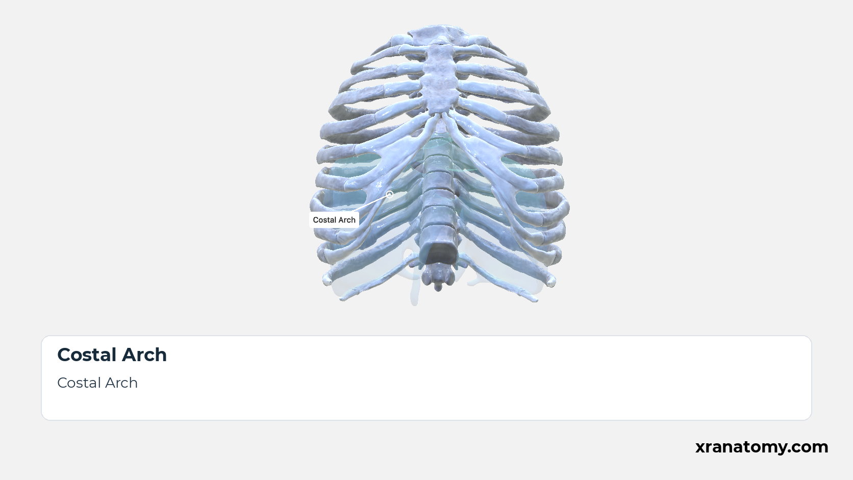
Costal Arch
The costal arch, or costal margin, is the curved lower edge of the front of the cage, formed on each side by the cartilages of ribs 7 to 10, and it outlines the anterolateral boundary of the inferior thoracic aperture. Its arrangement and variations are described with the rib cage. [5] [2] [3] [1]Show less
Illustration by XR Anatomy.
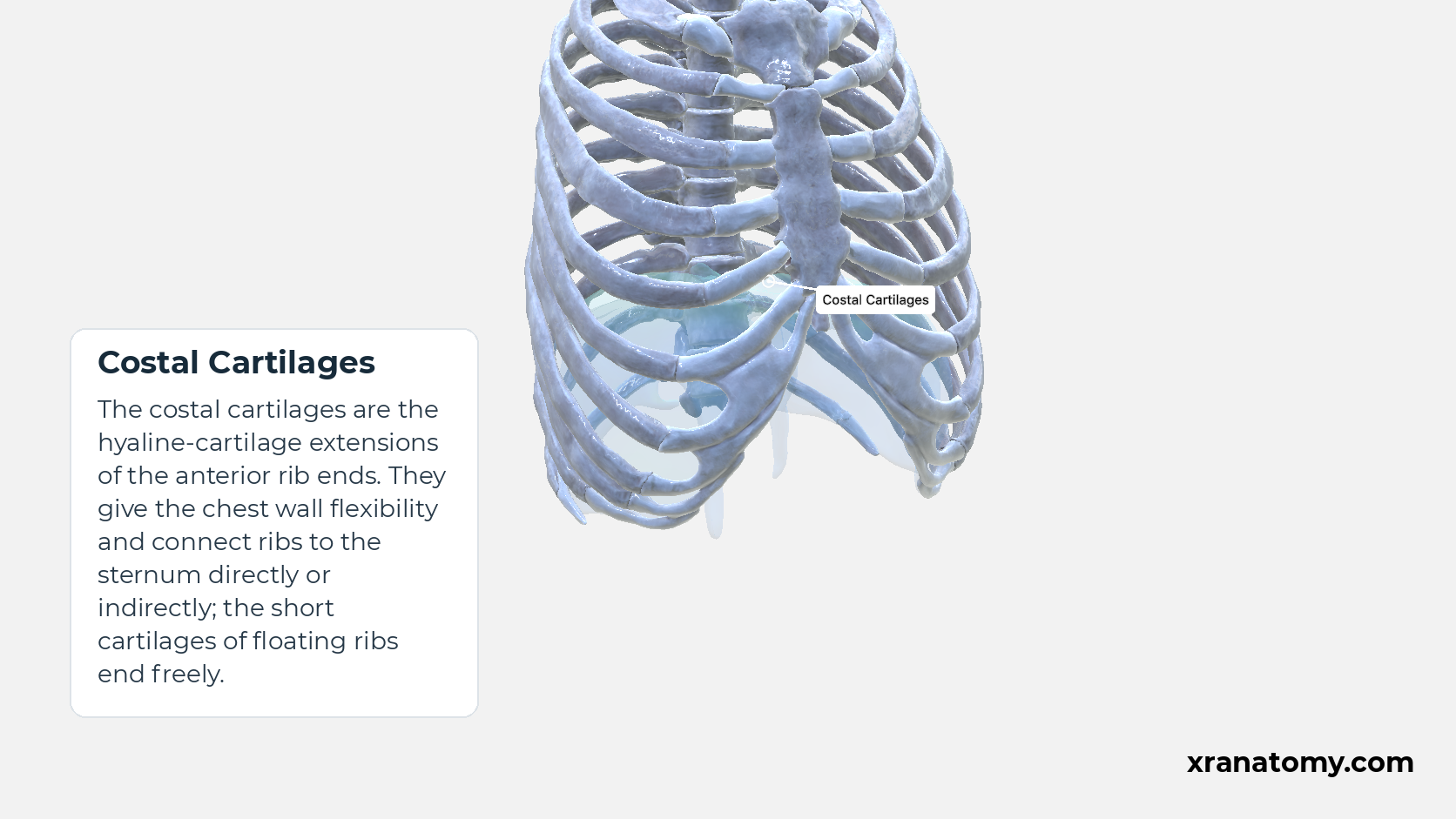
Costal Cartilages
The costal cartilages are the hyaline-cartilage extensions of the anterior rib ends. They give the chest wall flexibility and connect ribs to the sternum directly or indirectly; the short cartilages of floating ribs end freely.
Illustration by XR Anatomy.
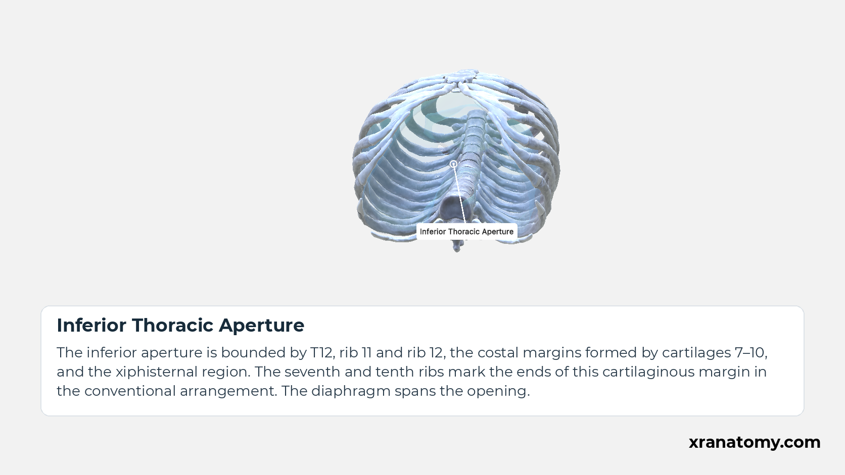
Inferior Thoracic Aperture
The inferior aperture is bounded by T12, rib 11 and rib 12, the costal margins formed by cartilages 7–10, and the xiphisternal region. The seventh and tenth ribs mark the ends of this cartilaginous margin in the conventional arrangement. The diaphragm spans the opening.Show less
Illustration by XR Anatomy.
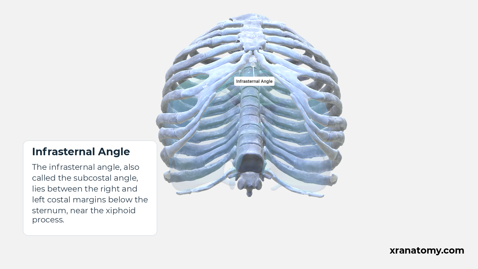
Infrasternal Angle
The infrasternal angle, also called the subcostal angle, lies between the right and left costal margins below the sternum, near the xiphoid process.
Illustration by XR Anatomy.
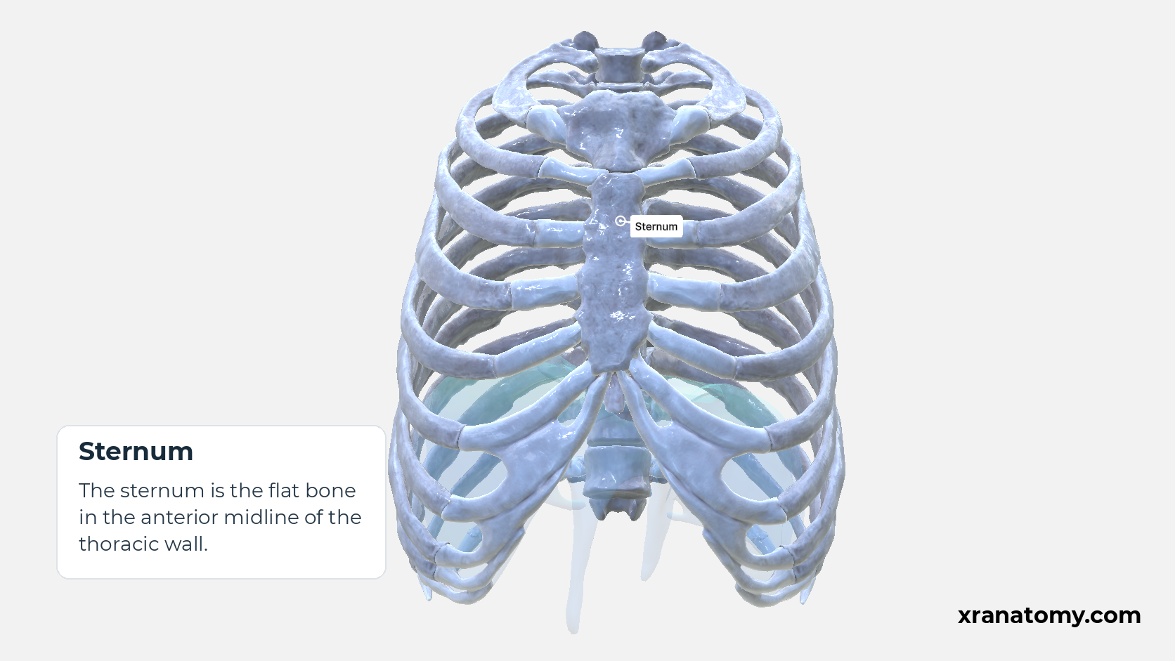
Sternum
The sternum is the flat bone in the anterior midline of the thoracic wall.
Illustration by XR Anatomy.
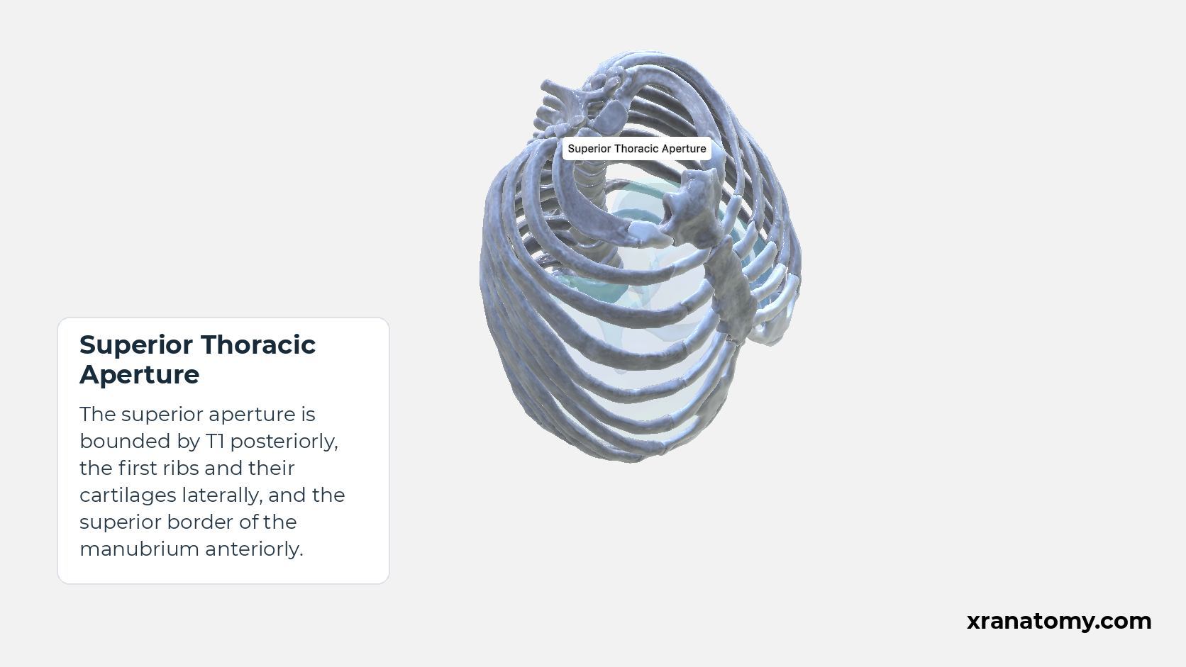
Superior Thoracic Aperture
The superior aperture is bounded by T1 posteriorly, the first ribs and their cartilages laterally, and the superior border of the manubrium anteriorly.
Illustration by XR Anatomy.
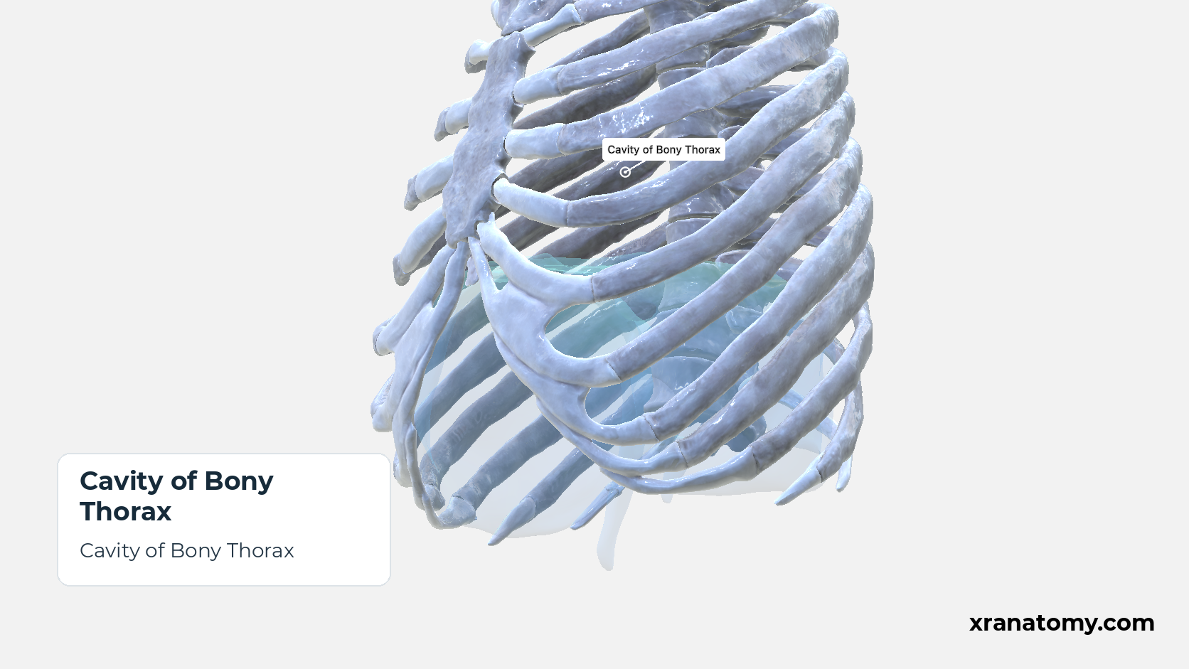
Cavity of Bony Thorax
The cavity of the bony thorax is the space enclosed by the skeletal framework alone. The thoracic cavity proper is smaller, because the complete wall also includes muscles and other soft tissues and its floor is the diaphragm, which domes up into the bony cage. [2] [3] [1]Show less
Illustration by XR Anatomy.
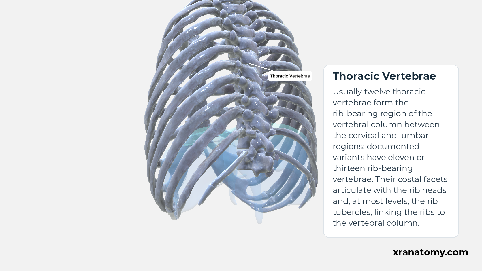
Thoracic Vertebrae
Usually twelve thoracic vertebrae form the rib-bearing region of the vertebral column between the cervical and lumbar regions; documented variants have eleven or thirteen rib-bearing vertebrae. Their costal facets articulate with the rib heads and, at most levels, the rib tubercles, linking the ribs to the vertebral column.Show less
Illustration by XR Anatomy.
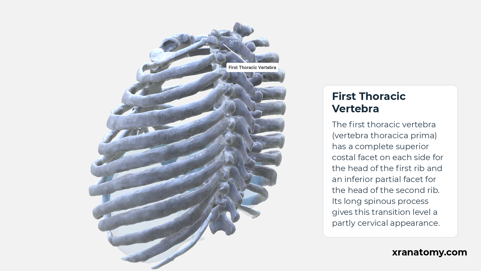
First Thoracic Vertebra
The first thoracic vertebra (vertebra thoracica prima) has a complete superior costal facet on each side for the head of the first rib and an inferior partial facet for the head of the second rib. Its long spinous process gives this transition level a partly cervical appearance.Show less
Illustration by XR Anatomy.
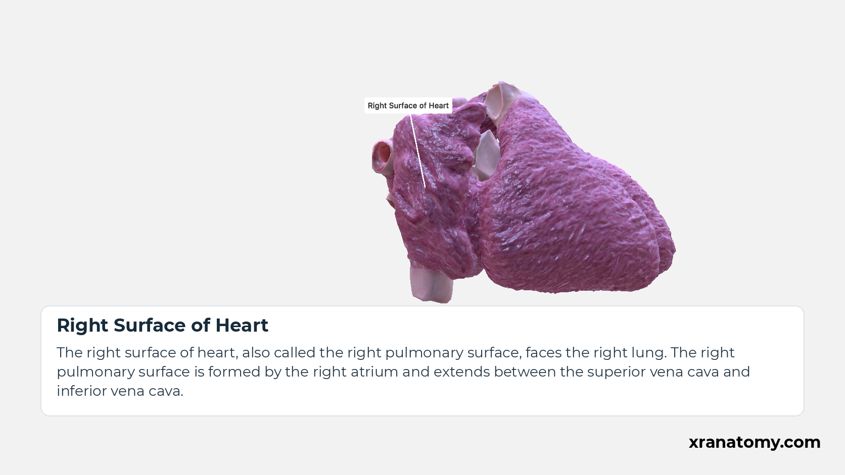
Right Surface of Heart
The right surface of heart, also called the right pulmonary surface, faces the right lung. The right pulmonary surface is formed by the right atrium and extends between the superior vena cava and inferior vena cava.
Study Right and left surfaces in this fixed image. Use the thumbnails to move between the 2D views; each view highlights one structure. The image does not rotate.
Illustration by XR Anatomy.
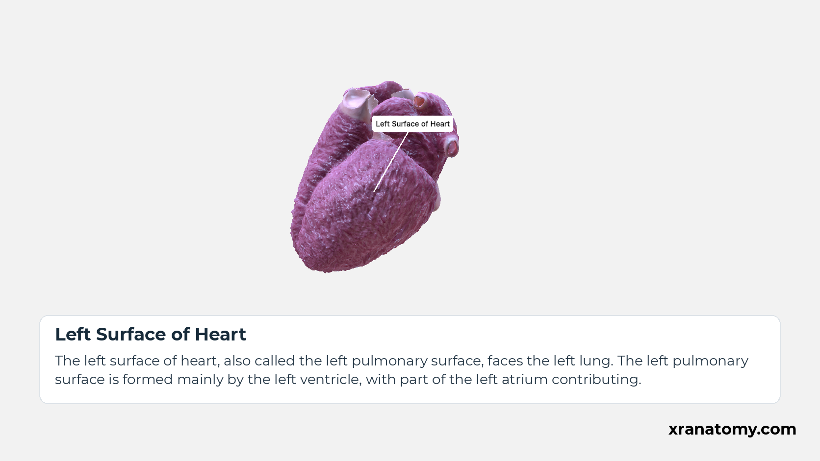
Left Surface of Heart
The left surface of heart, also called the left pulmonary surface, faces the left lung. The left pulmonary surface is formed mainly by the left ventricle, with part of the left atrium contributing.
Illustration by XR Anatomy.
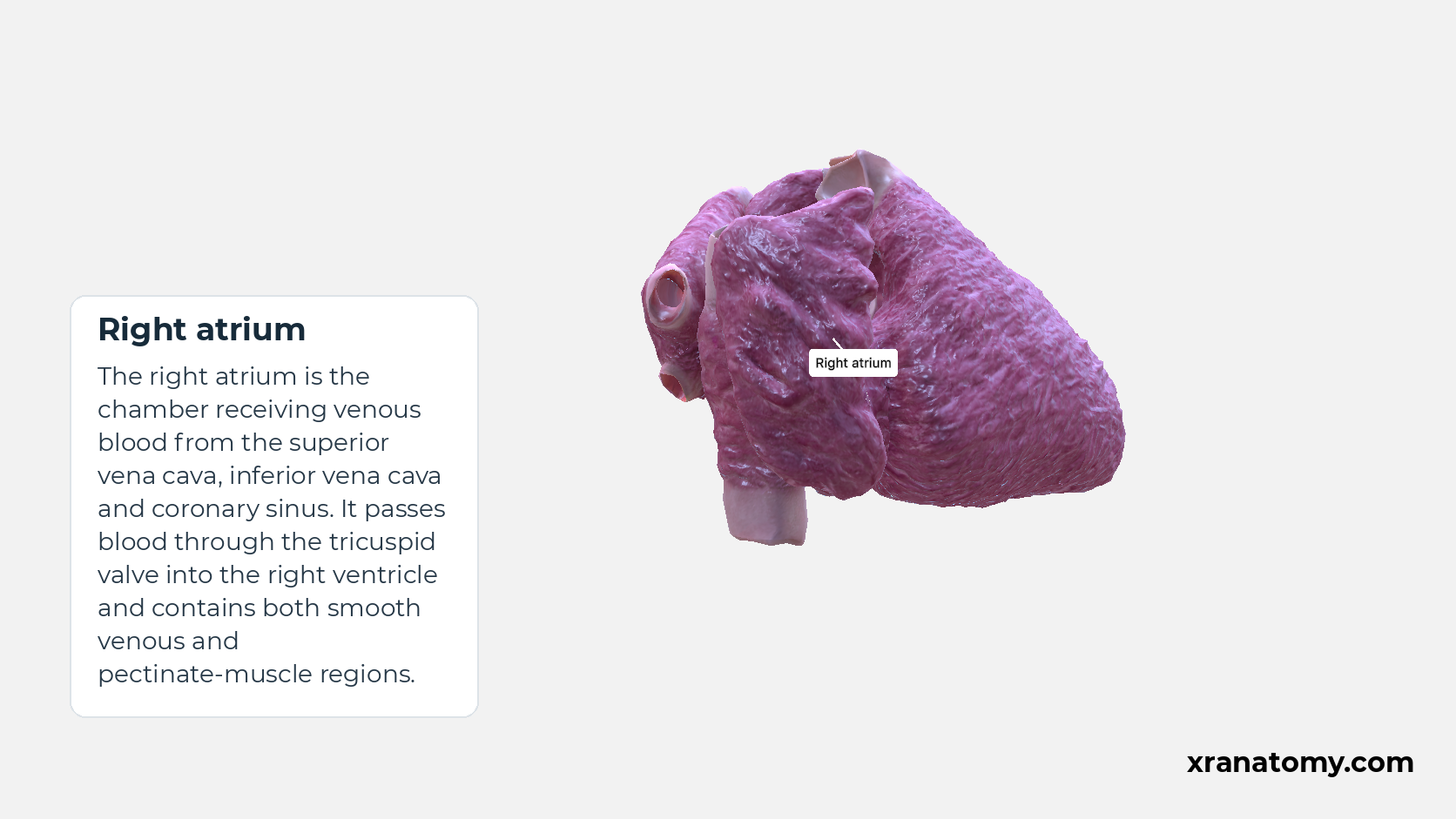
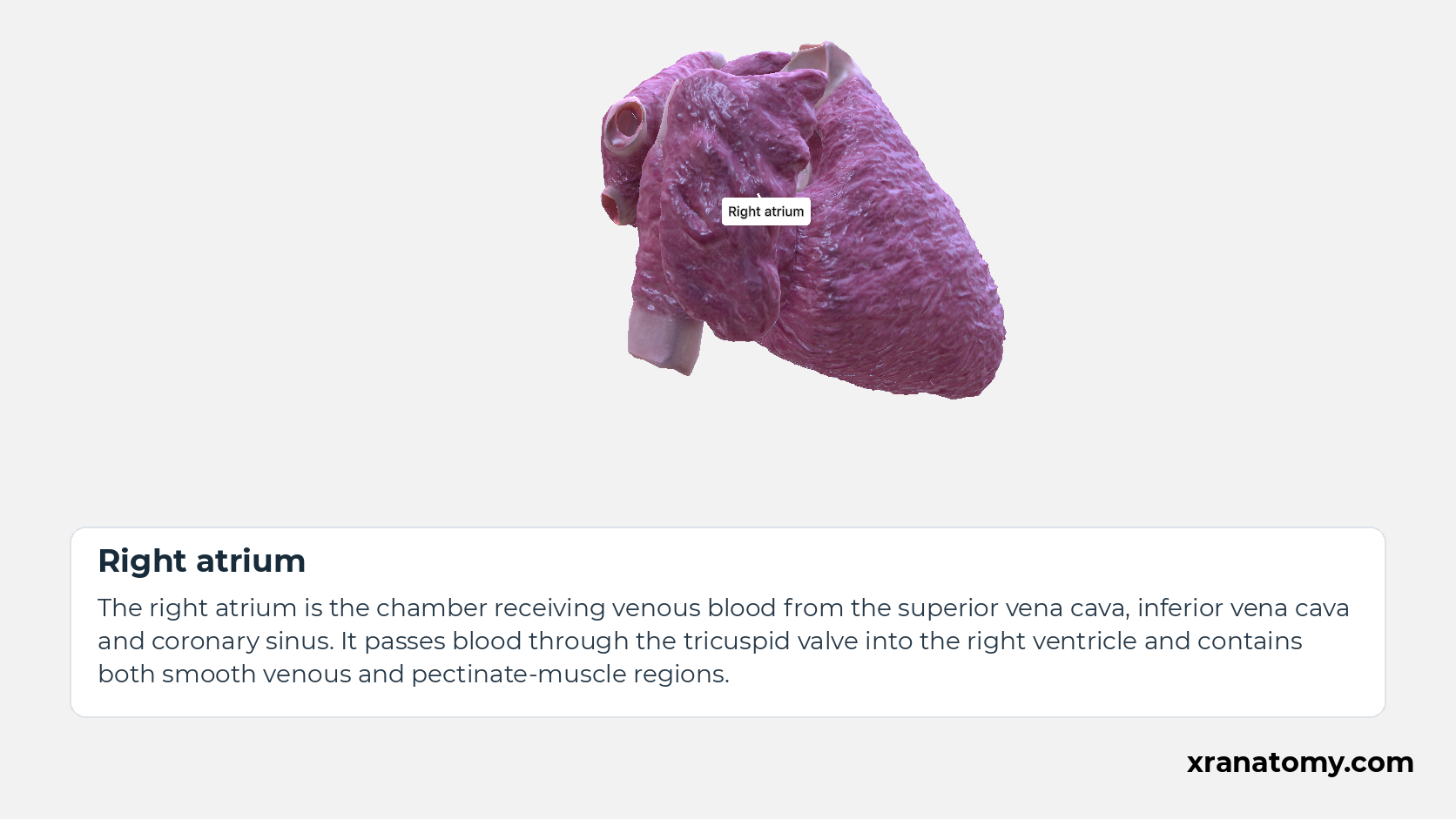
Right atrium
The right atrium is the chamber receiving venous blood from the superior vena cava, inferior vena cava and coronary sinus. It passes blood through the tricuspid valve into the right ventricle and contains both smooth venous and pectinate-muscle regions.Show less
Illustration by XR Anatomy.
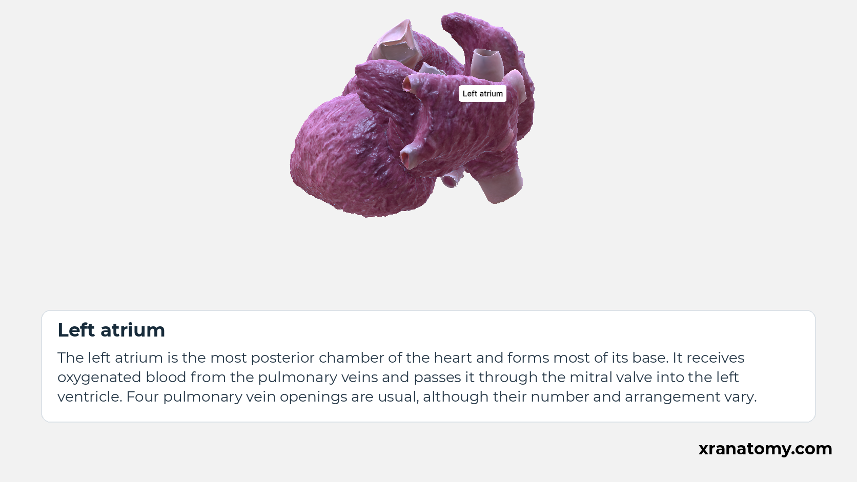
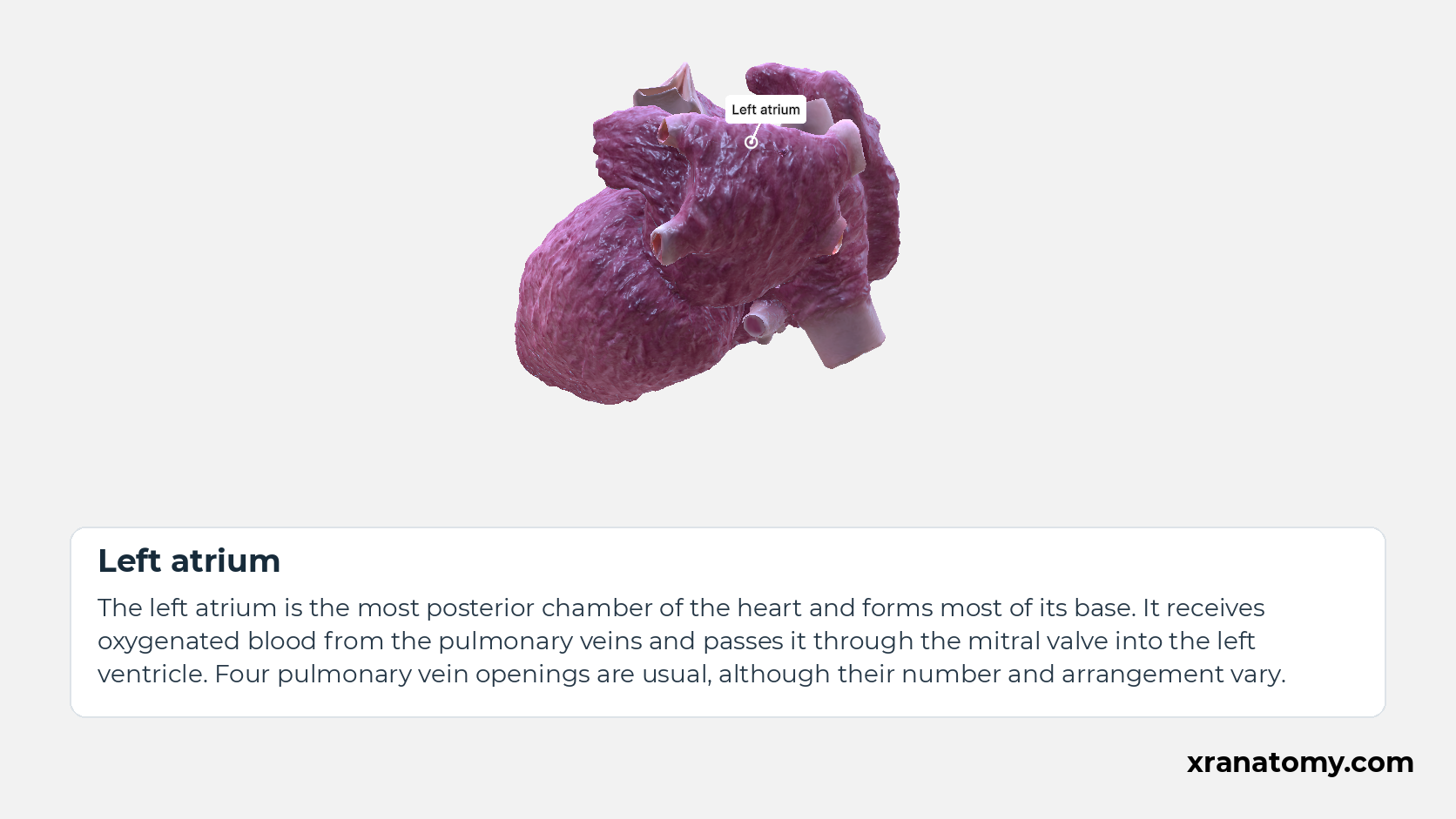
Left atrium
The left atrium is the most posterior chamber of the heart and forms most of its base. It receives oxygenated blood from the pulmonary veins and passes it through the mitral valve into the left ventricle. Four pulmonary vein openings are usual, although their number and arrangement vary.Show less
Illustration by XR Anatomy.
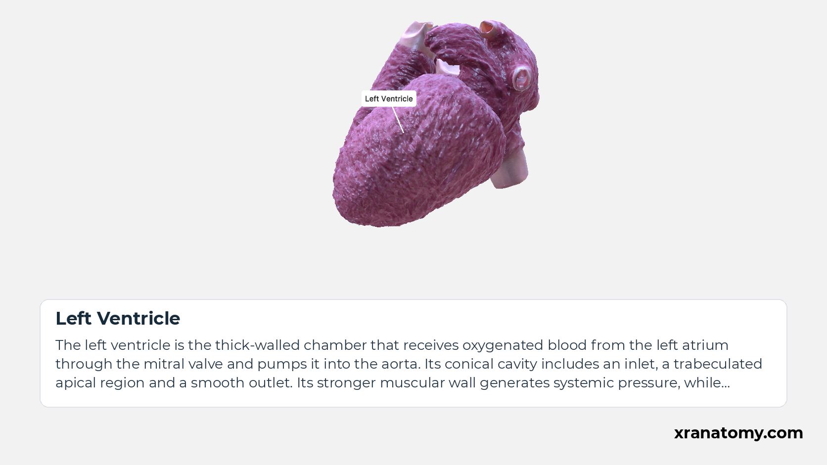
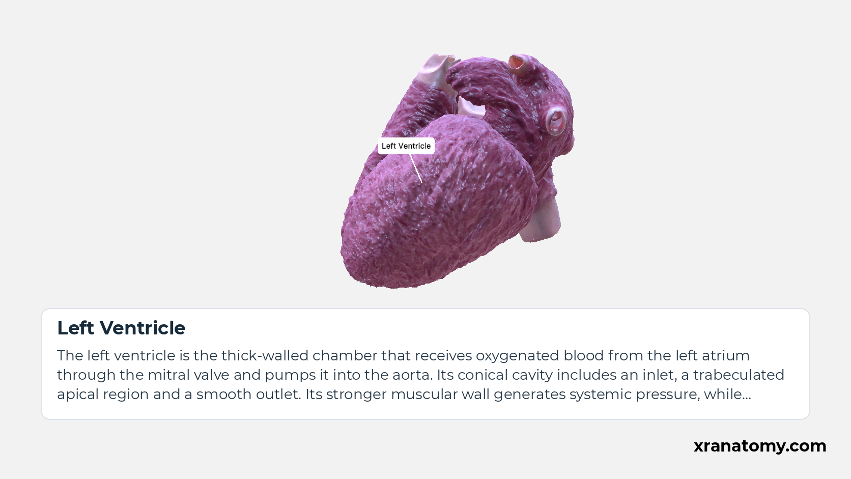
Left Ventricle
The left ventricle is the thick-walled chamber that receives oxygenated blood from the left atrium through the mitral valve and pumps it into the aorta. Its conical cavity includes an inlet, a trabeculated apical region and a smooth outlet. Its stronger muscular wall generates systemic pressure, while normally ejecting the same volume per beat as the right ventricle.Show less
Illustration by XR Anatomy.
In depth · 4 sections · 1 min read4 more sections on this part4 sections open — reading In depth- Surfaces of the Heart
- Anterior Surface of Heart
- Inferior Surface of Heart
- Right and left surfaces
Surfaces of the Heart
Anterior Surface of Heart
The anterior surface of heart, also called the sternocostal surface, faces the sternum and adjacent costal cartilages. The right ventricle makes the largest contribution; the right atrium contributes on the right and the left ventricle contributes on the left.
Inferior Surface of Heart
The inferior surface of heart, also called the diaphragmatic surface, faces downwards and rests on the diaphragm. The left ventricle makes the major contribution and the right ventricle a smaller one; the inferior interventricular sulcus separates those ventricular contributions.
Right and left surfaces
Left Surface of Heart
The left surface of heart, also called the left pulmonary surface, faces the left lung. The left pulmonary surface is formed mainly by the left ventricle, with part of the left atrium contributing.
Right Surface of Heart
The right surface of heart, also called the right pulmonary surface, faces the right lung. The right pulmonary surface is formed by the right atrium and extends between the superior vena cava and inferior vena cava.
Practice · Check your understanding · 7 questions Recall: Surfaces of the heart
Why this helps
Testing yourself before revealing the answer (active recall) strengthens memory far more than re-reading — so each prompt stays hidden until you choose to reveal it.
Which chamber makes the largest contribution to the anterior surface of the heart?
right ventricle
The anterior surface of heart, also called the sternocostal surface, faces the sternum and adjacent costal cartilages. The right ventricle makes the largest contribution; the right atrium contributes on the right and the left ventricle contributes on the left.
Review this sectionHow well did you know it? · press 1–4
Which chamber forms the pulmonary surface situated between the superior vena cava and inferior vena cava?
right atrium
The right surface of heart, also called the right pulmonary surface, faces the right lung. The right pulmonary surface is formed by the right atrium and extends between the superior vena cava and inferior vena cava.
Review this sectionHow well did you know it? · press 1–4
What are the four named surfaces of the heart?
The heart has four named surfaces: the anterior surface, inferior surface, right surface and left surface.
The heart has four named surfaces: the anterior surface, inferior surface, right surface and left surface.
Review this sectionHow well did you know it? · press 1–4
What does the anterior, or sternocostal, surface of the heart face?
The anterior surface of heart, also called the sternocostal surface, faces the sternum and adjacent costal cartilages.
The anterior surface of heart, also called the sternocostal surface, faces the sternum and adjacent costal cartilages. The right ventricle makes the largest contribution; the right atrium contributes on the right and the left ventricle contributes on the left.
Review this sectionHow well did you know it? · press 1–4
Which chambers form the left pulmonary surface of the heart?
mainly by the left ventricle, with part of the left atrium contributing
The left surface of heart, also called the left pulmonary surface, faces the left lung. The left pulmonary surface is formed mainly by the left ventricle, with part of the left atrium contributing.
Review this sectionHow well did you know it? · press 1–4
Which chamber forms the pulmonary surface that extends between the superior vena cava and inferior vena cava?
the right atrium
The right surface of heart, also called the right pulmonary surface, faces the right lung. The right pulmonary surface is formed by the right atrium and extends between the superior vena cava and inferior vena cava.
Review this sectionHow well did you know it? · press 1–4
Where does the inferior, or diaphragmatic, surface of the heart face and rest?
faces downwards and rests on the diaphragm
The inferior surface of heart, also called the diaphragmatic surface, faces downward and rests on the diaphragm. The left ventricle makes the major contribution and the right ventricle a smaller one; the inferior interventricular sulcus separates those ventricular contributions.
Review this sectionHow well did you know it? · press 1–4
Posterior base and ventricular apex
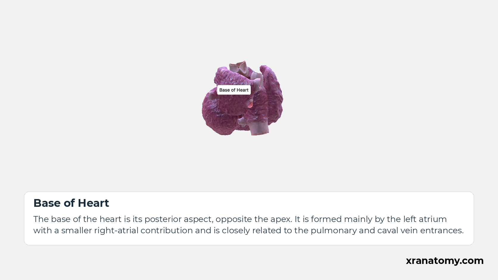
Base of Heart
The base of the heart is its posterior aspect, opposite the apex. It is formed mainly by the left atrium with a smaller right-atrial contribution and is closely related to the pulmonary and caval vein entrances.
Study Posterior base and ventricular apex in this fixed image. Use the thumbnails to move between the 2D views; each view highlights one structure. The image does not rotate.
Illustration by XR Anatomy.
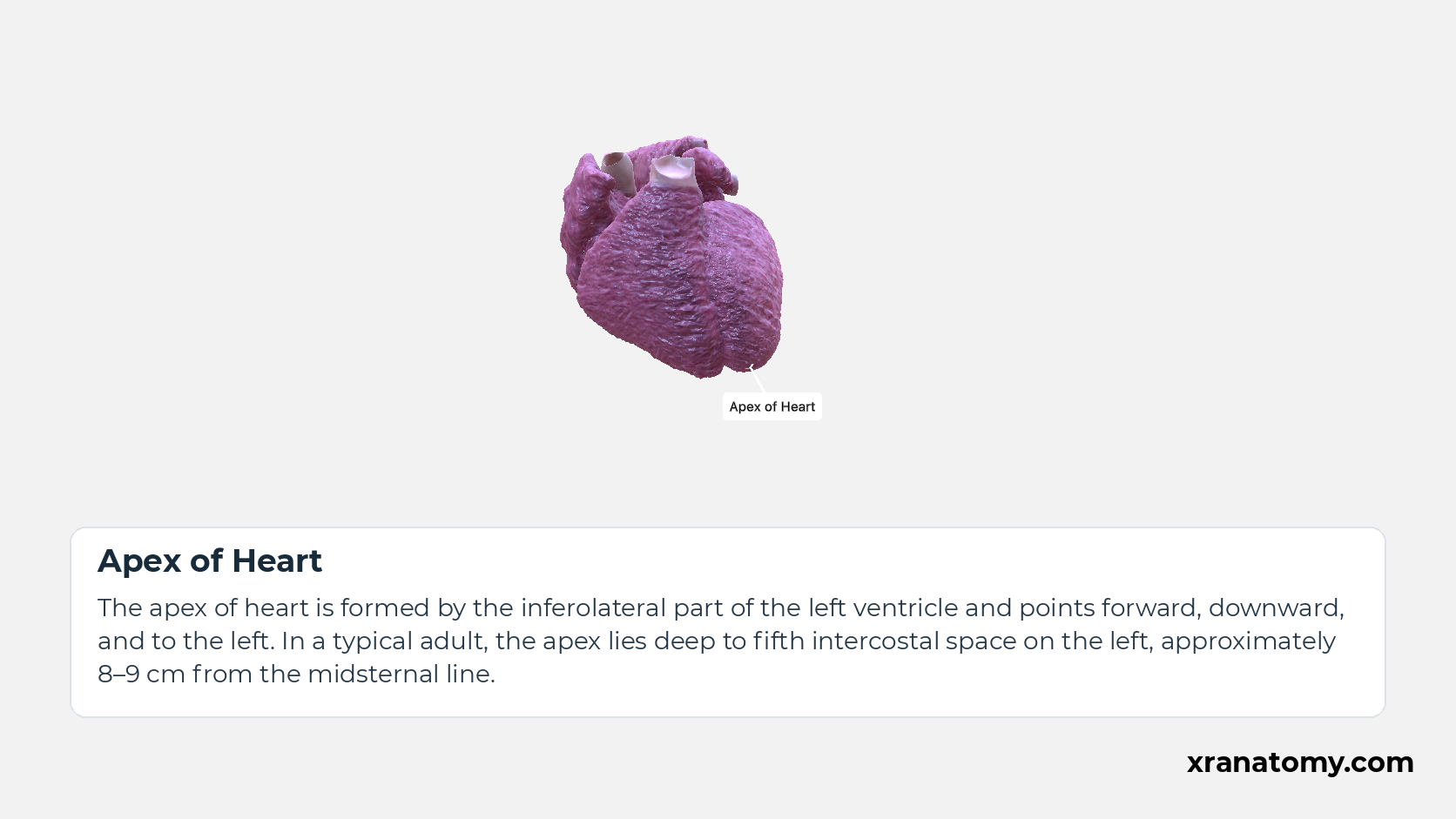
Apex of Heart
The apex of heart is formed by the inferolateral part of the left ventricle and points forward, downward, and to the left. In a typical adult, the apex lies deep to fifth intercostal space on the left, approximately 8–9 cm from the midsternal line.Show less
Illustration by XR Anatomy.
Right atrium
The right atrium is the chamber receiving venous blood from the superior vena cava, inferior vena cava and coronary sinus. It passes blood through the tricuspid valve into the right ventricle and contains both smooth venous and pectinate-muscle regions.Show less
Illustration by XR Anatomy.
Left atrium
The left atrium is the most posterior chamber of the heart and forms most of its base. It receives oxygenated blood from the pulmonary veins and passes it through the mitral valve into the left ventricle. Four pulmonary vein openings are usual, although their number and arrangement vary.Show less
Illustration by XR Anatomy.
Left Ventricle
The left ventricle is the thick-walled chamber that receives oxygenated blood from the left atrium through the mitral valve and pumps it into the aorta. Its conical cavity includes an inlet, a trabeculated apical region and a smooth outlet. Its stronger muscular wall generates systemic pressure, while normally ejecting the same volume per beat as the right ventricle.Show less
Illustration by XR Anatomy.
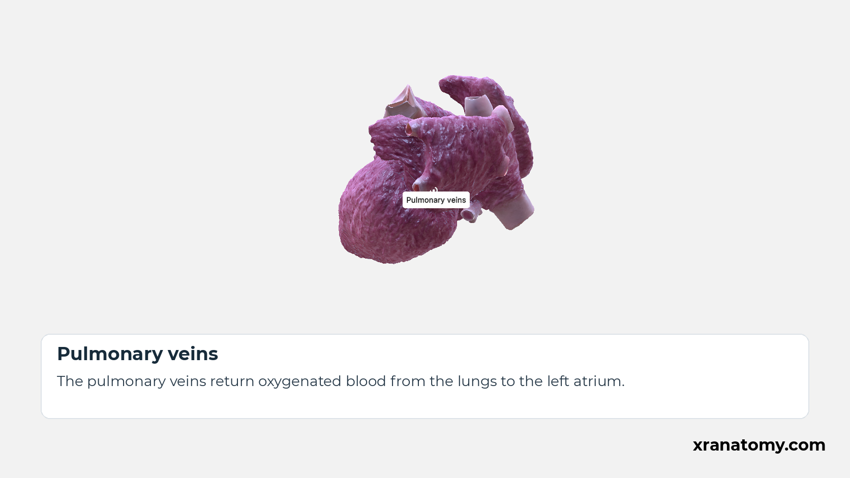
Pulmonary veins
The pulmonary veins return oxygenated blood from the lungs to the left atrium.
Illustration by XR Anatomy.
The base and apex provide two further points of orientation. The base is the broad posterior aspect of the heart and is formed mainly by the left atrium. Opposite it lies the apex, the pointed tip formed by the left ventricle. Because the heart sits at an angle, this tip points forwards, downwards and to the left.
In depth · 2 sections · 1 min read2 more sections on this part2 sections open — reading In depth- Base of Heart
- Apex of Heart
Base of Heart
The base of heart is quadrilateral and lies opposite the apex. In addition to the main contribution from the left atrium, it includes a smaller right-atrial part. The pulmonary veins enter posteriorly; the superior vena cava and inferior vena cava enter the right atrium at its right side.
Apex of Heart
The apex of heart is formed by the inferolateral part of the left ventricle. In a typical adult, the apex lies deep to the left fifth intercostal space, near the midclavicular line.
Practice · Check your understanding · 3 questions Recall: Posterior base and ventricular apex
Which chamber forms most of the posterior base of the heart?
left atrium
The base of heart is its quadrilateral posterior aspect, opposite the apex. It is formed mainly by the left atrium, with a smaller right-atrial contribution, and receives the pulmonary veins; the superior vena cava and inferior vena cava enter the right atrium at its right side.
Review this sectionHow well did you know it? · press 1–4
What is the base of the heart in shape and position relative to the apex?
The base of heart is its quadrilateral posterior aspect, opposite the apex.
The base of heart is its quadrilateral posterior aspect, opposite the apex. It is formed mainly by the left atrium, with a smaller right-atrial contribution, and receives the pulmonary veins; the superior vena cava and inferior vena cava enter the right atrium at its right side.
Review this sectionHow well did you know it? · press 1–4
Which chamber forms the apex of the heart, and in which directions does it point?
formed by the inferolateral part of the left ventricle and points forwards, downwards, and to the left
The apex of heart is formed by the inferolateral part of the left ventricle and points forward, downward, and to the left. In a typical adult, the apex lies deep to fifth intercostal space on the left, approximately 8–9 cm from the midsternal line.
Review this sectionHow well did you know it? · press 1–4
Bibliography
Every claim on this page is sourced against the standard anatomical references.
- FIPAT. Terminologia Anatomica, 2nd edition, Part 4: Systemata integrantia I. Version 2.07. 2021. https://cdn.dal.ca/content/dam/dalhousie/pdf/library/FIPAT/TA2/FIPAT-TA2-Part-4.pdf
- Gray H. Anatomy of the Human Body. 21st ed. Philadelphia: Lea & Febiger; 1924.
- Drake RL, Vogl AW, Mitchell AWM. Gray's Anatomy for Students. Elsevier.
- Betts JG, et al. Anatomy and Physiology 2e. OpenStax; Rice University.
