Anatomy
Left atrium
The left atrium is the heart's most posterior chamber and forms most of its base. It receives the pulmonary veins and passes blood through the mitral opening.
The left atrium is the chamber that receives oxygenated blood from the lungs through the pulmonary veins and passes it to the left ventricle. Most of its cavity has a smooth wall. A small muscular pouch, the left auricle, projects from it, while the smooth vestibule surrounds the opening towards the mitral valve. These regions provide the framework for the descriptions that follow.
Pulmonary venous component and smooth body
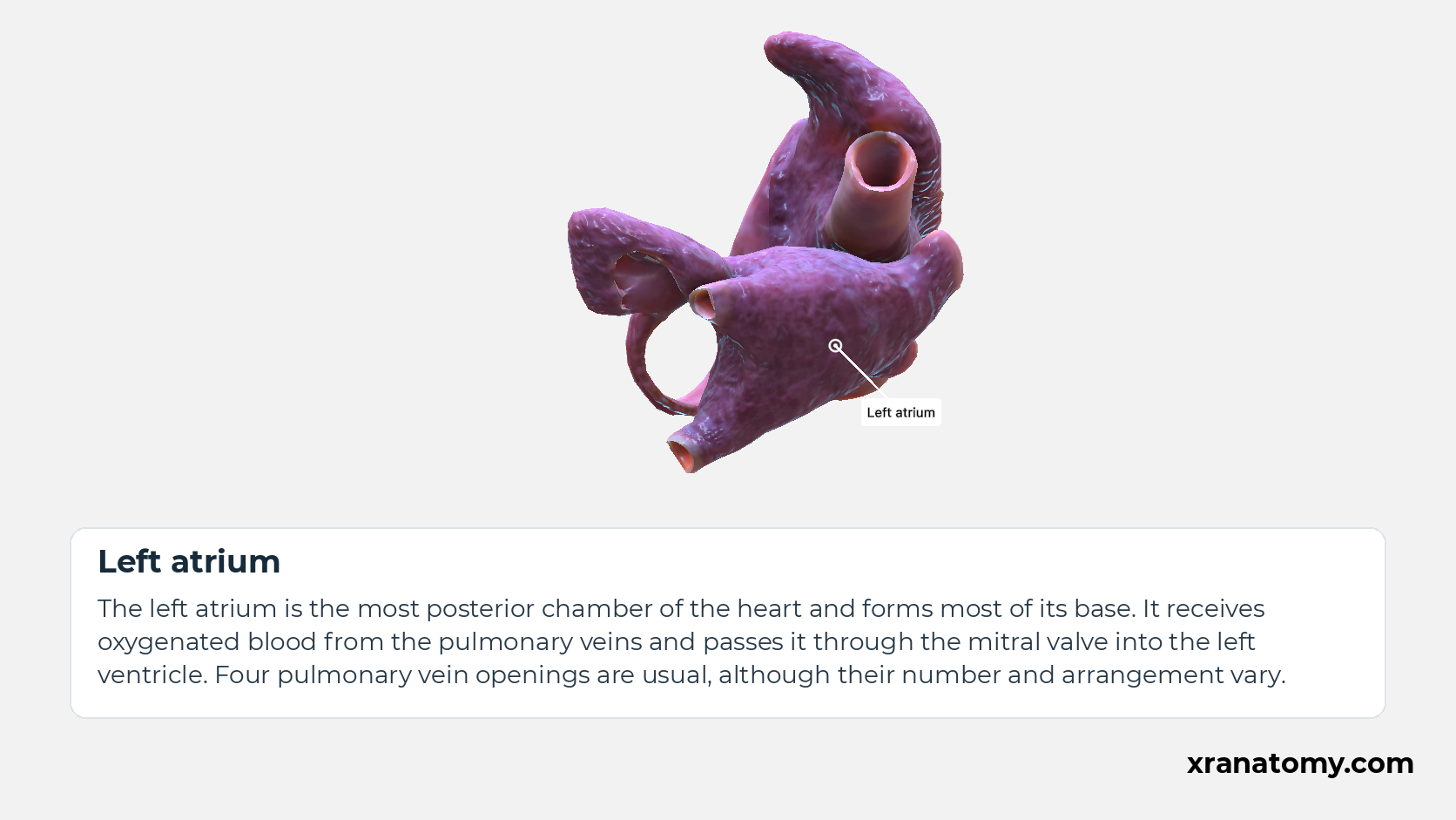
Left atrium
The left atrium is the most posterior chamber of the heart and forms most of its base. It receives oxygenated blood from the pulmonary veins and passes it through the mitral valve into the left ventricle. Four pulmonary vein openings are usual, although their number and arrangement vary.Show less
Study Pulmonary venous component and smooth body in this fixed image. Use the thumbnails to move between the 2D views; each view highlights one structure. The image does not rotate.
Illustration by XR Anatomy.
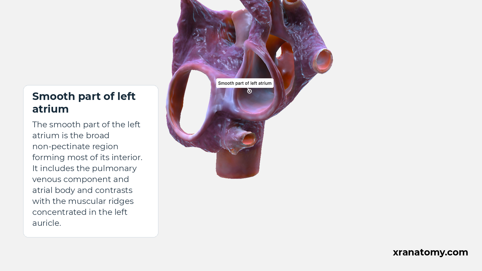
Smooth part of left atrium
The smooth part of the left atrium is the broad non-pectinate region forming most of its interior. It includes the pulmonary venous component and atrial body and contrasts with the muscular ridges concentrated in the left auricle.
Illustration by XR Anatomy.
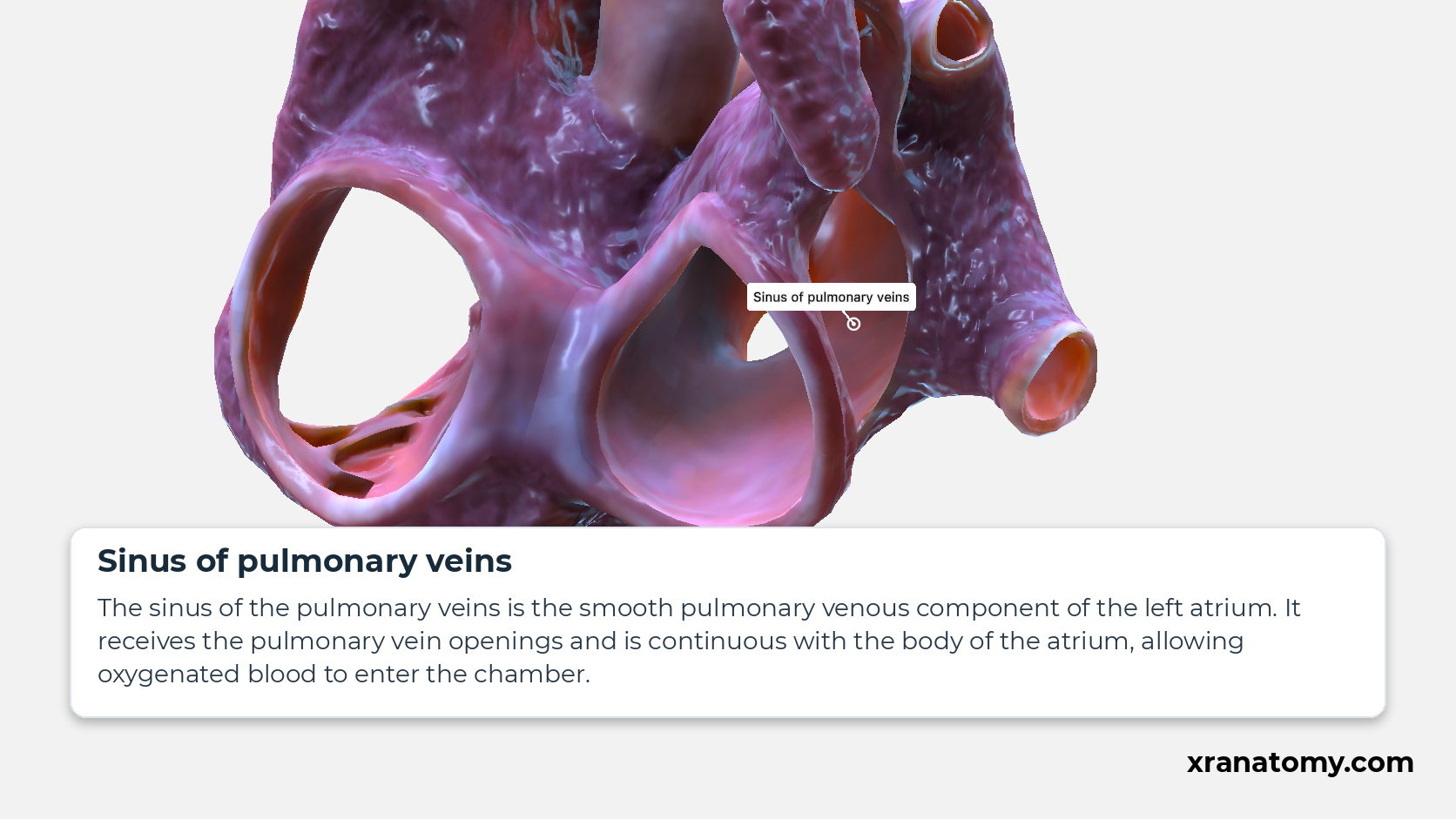
Sinus of pulmonary veins
The sinus of the pulmonary veins is the smooth pulmonary venous component of the left atrium. It receives the pulmonary vein openings and is continuous with the body of the atrium, allowing oxygenated blood to enter the chamber.
Illustration by XR Anatomy.
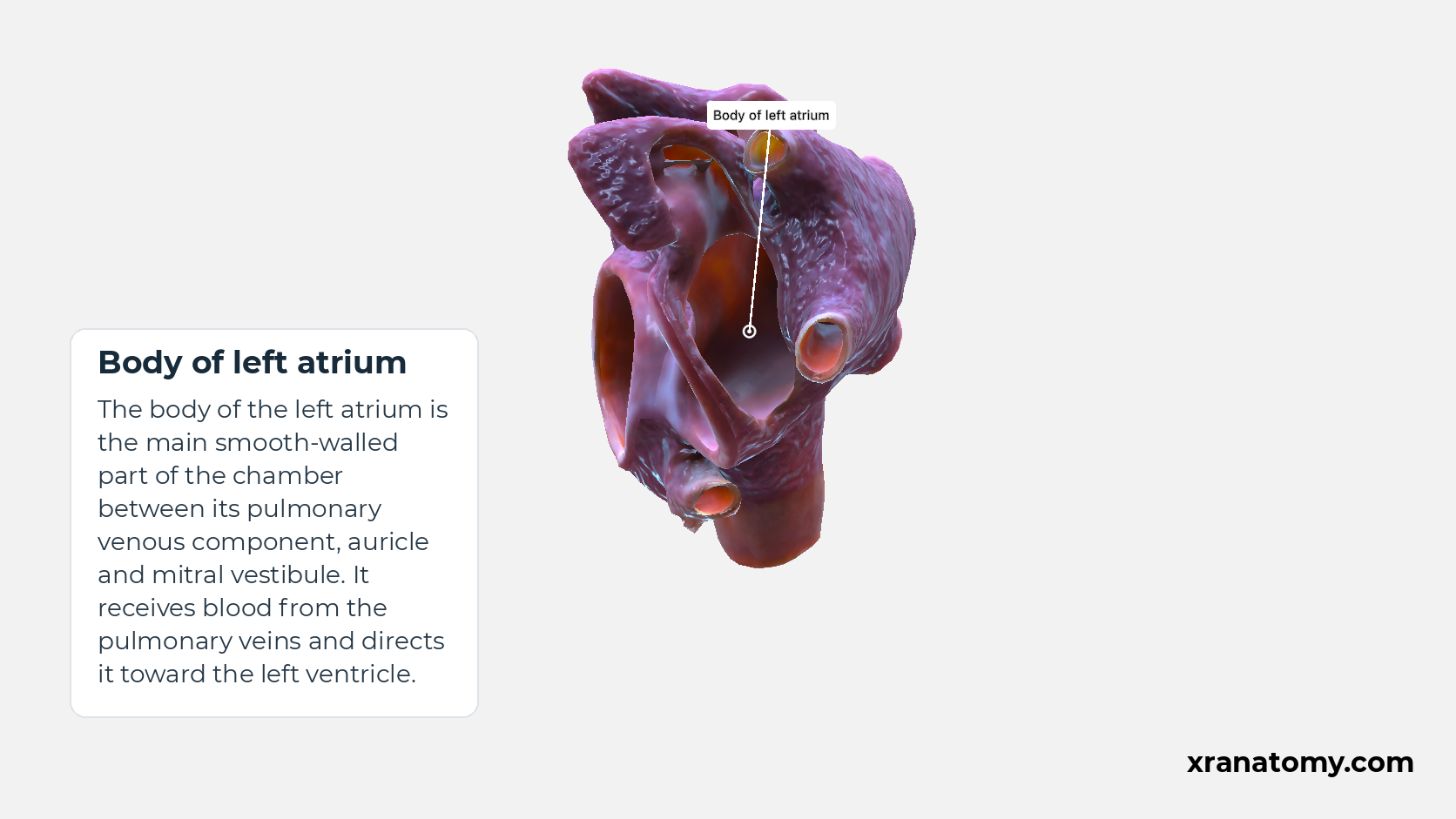
Body of left atrium
The body of the left atrium is the main smooth-walled part of the chamber between its pulmonary venous component, auricle and mitral vestibule. It receives blood from the pulmonary veins and directs it toward the left ventricle.
Illustration by XR Anatomy.
Position and venous return
The left atrium lies farther back than the other cardiac chambers and forms most of the base of the heart. Usually four pulmonary veins open into it, although their number and arrangement can vary. From the atrial cavity, blood passes through the mitral opening into the left ventricle. This route connects the incoming veins with the ventricular inlet.
In depth · 1 min read Smooth body and pulmonary venous component
The pulmonary venous component, also called the sinus of pulmonary veins, receives the pulmonary-vein openings and is continuous with the atrial body. The left atrium has no equivalent of the terminal crest and terminal groove of the right atrium. It does, however, have a left lateral ridge between the appendage opening and the left pulmonary veins. This is a different landmark, rather than a left-sided terminal crest.
The auricle and the region around the valve
Left auricle
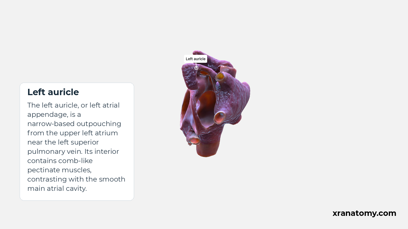
The left auricle, or left atrial appendage, is a narrow-based outpouching from the upper left atrium near the left superior pulmonary vein. Its interior contains comb-like pectinate muscles, contrasting with the smooth main atrial cavity.
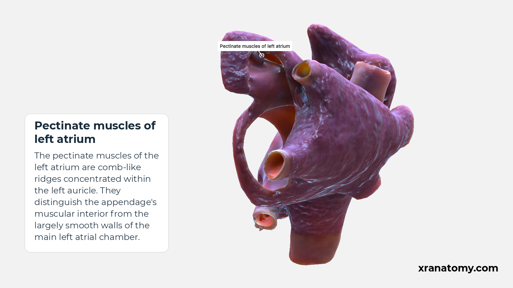
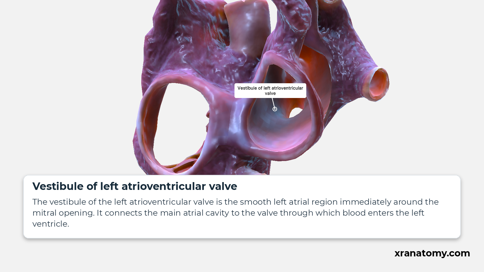
Study Auricle, pectinate muscles, and mitral vestibule in this fixed image. Use the thumbnails to move between the 2D views; each view highlights one structure. The image does not rotate.
Illustration by XR Anatomy.
Pectinate muscles of left atrium
The pectinate muscles of the left atrium are comb-like ridges concentrated within the left auricle. They distinguish the appendage's muscular interior from the largely smooth walls of the main left atrial chamber.
Illustration by XR Anatomy.
Vestibule of left atrioventricular valve
The vestibule of the left atrioventricular valve is the smooth left atrial region immediately around the mitral opening. It connects the main atrial cavity to the valve through which blood enters the left ventricle.
Illustration by XR Anatomy.
Most of the inside of the left atrium is smooth, but its auricle has a different appearance. The left auricle is a muscular pouch joined to the chamber by a narrow neck. Pectinate muscles form ridges within the pouch. Near the outlet, the smooth vestibule surrounds the mitral opening and leads towards the ventricle.
In depth · 2 sections · 1 min read2 more sections on this part2 sections open — reading In depth- Left auricle and pectinate muscles
- Mitral vestibule
Left auricle and pectinate muscles
The left auricle, or left atrial appendage, projects from the upper part of the left atrium, near and anterior to the left superior pulmonary vein. Its attachment is narrower than that of the broad-based right auricle. Pectinate muscles form raised muscular bands within the pouch.
Mitral vestibule
The vestibule of the left atrioventricular valve surrounds the mitral opening and continues towards the ventricular inlet. The chordal apparatus supports the leaflets of the mitral valve.
