Anatomy
Sacrum
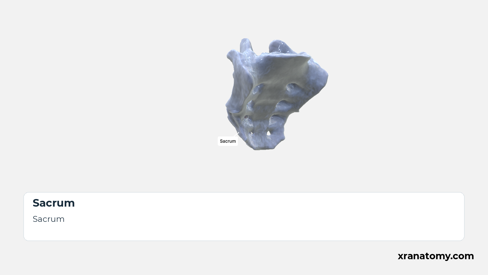
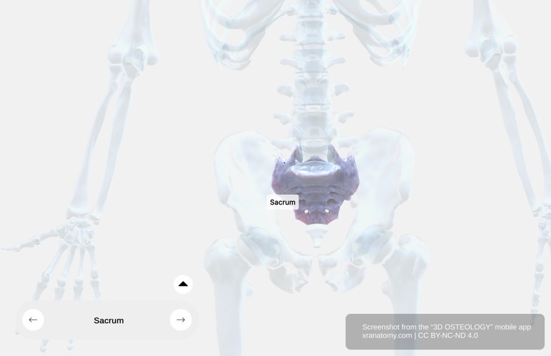
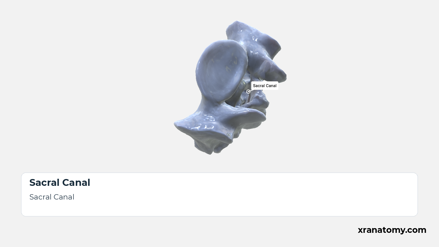
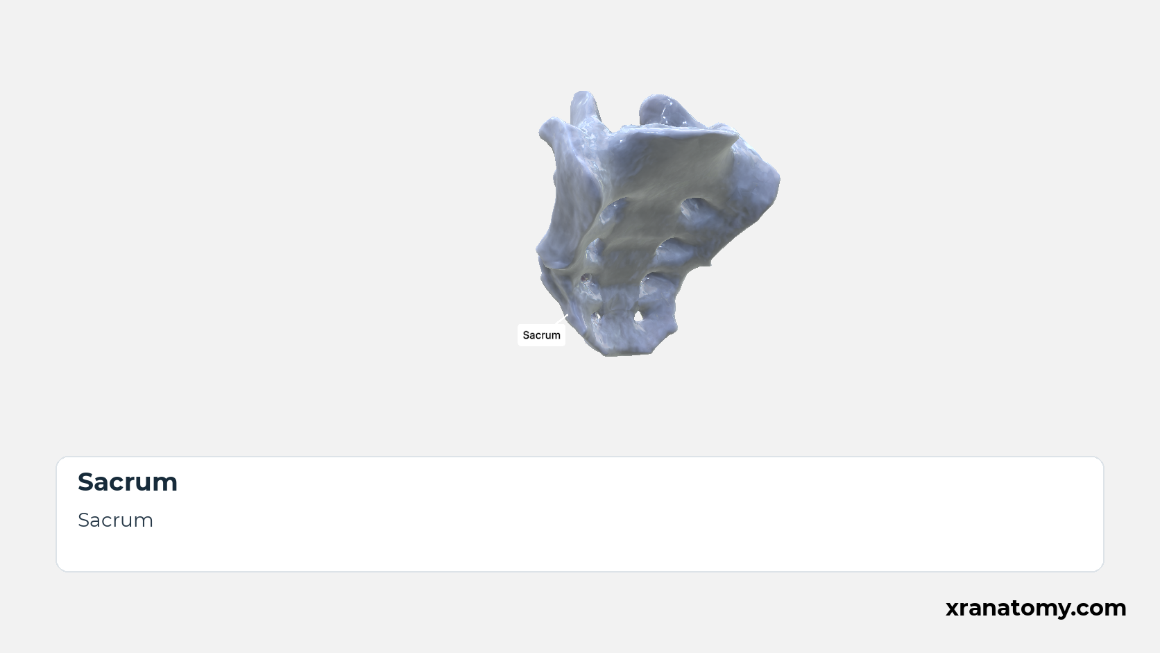
The sacrum (os sacrum) is the large triangular bone that closes the back of the pelvis, wedged between the two hip bones. It is usually formed by the fusion of five sacral vertebrae, so its broad base faces upwards towards the spine and its narrow apex downwards towards the coccyx; pelvic, dorsal, and lateral surfaces surround the sacral canal at its core.
General Structure
Base of Sacrum
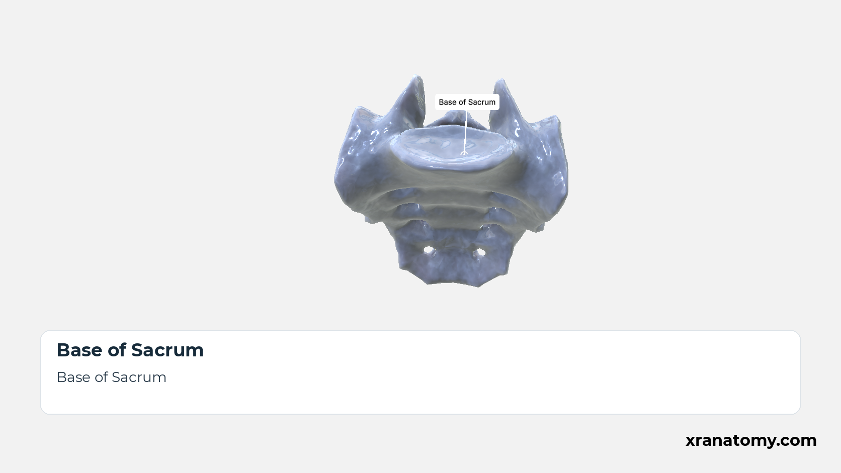
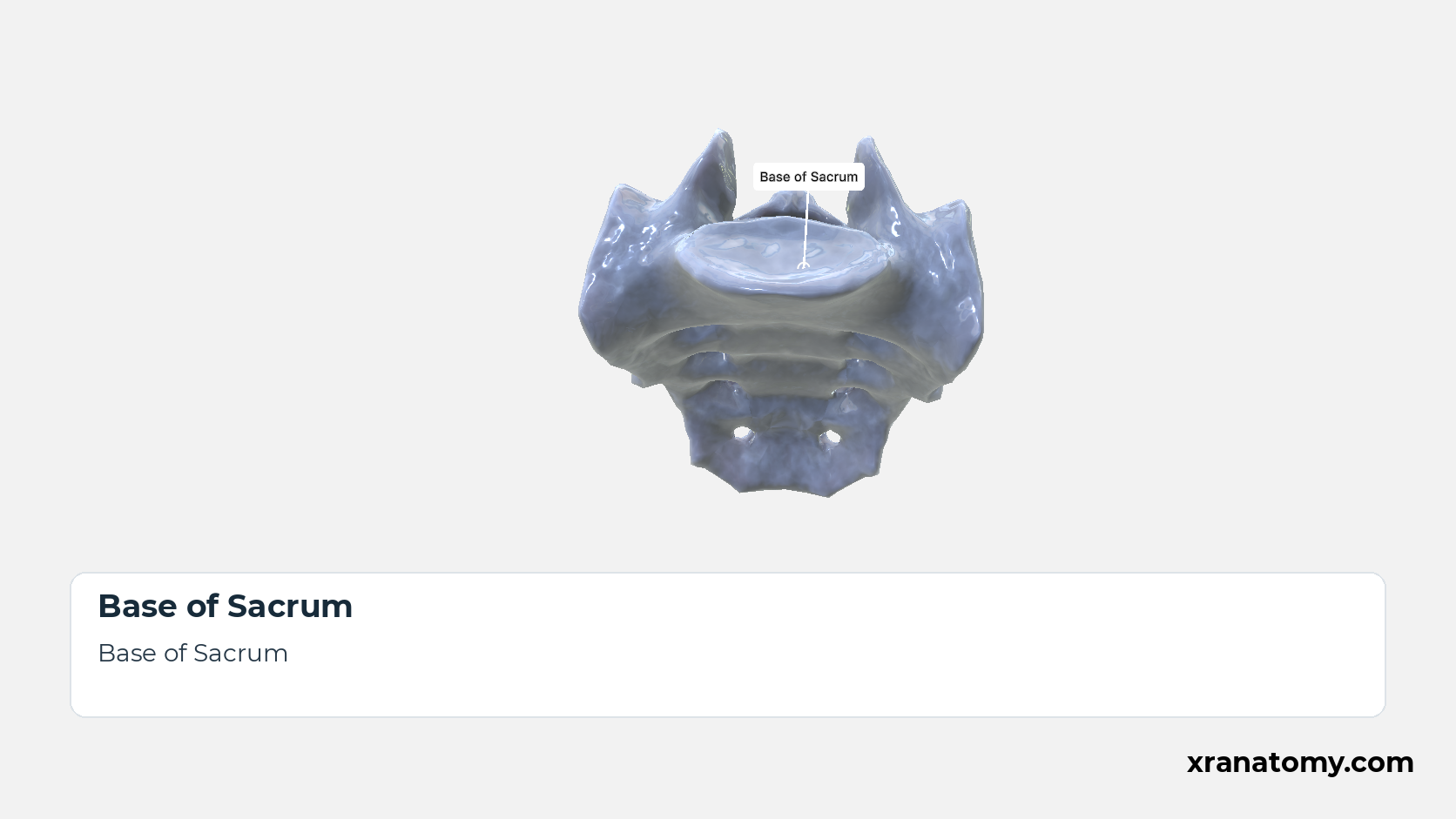
The sacral base is the broad superior end of the bone, on which the fifth lumbar vertebra rests. The upper surface of the S1 body forms its centre, the alae extend it to either side, and the sacral canal opens behind the body between the superior articular processes; the Superior View shows these from above. [2] [3] [1]Show less
Study Sacrum; General Structure in this fixed image. Use the thumbnails to move between the 2D views; each view highlights one structure. The image does not rotate.
Illustration by XR Anatomy.
Apex of Sacrum
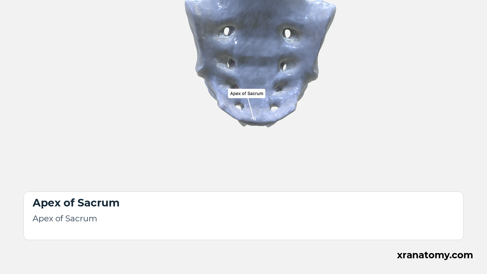
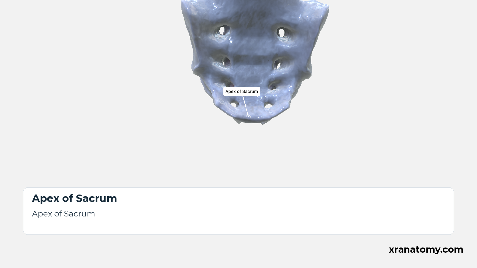
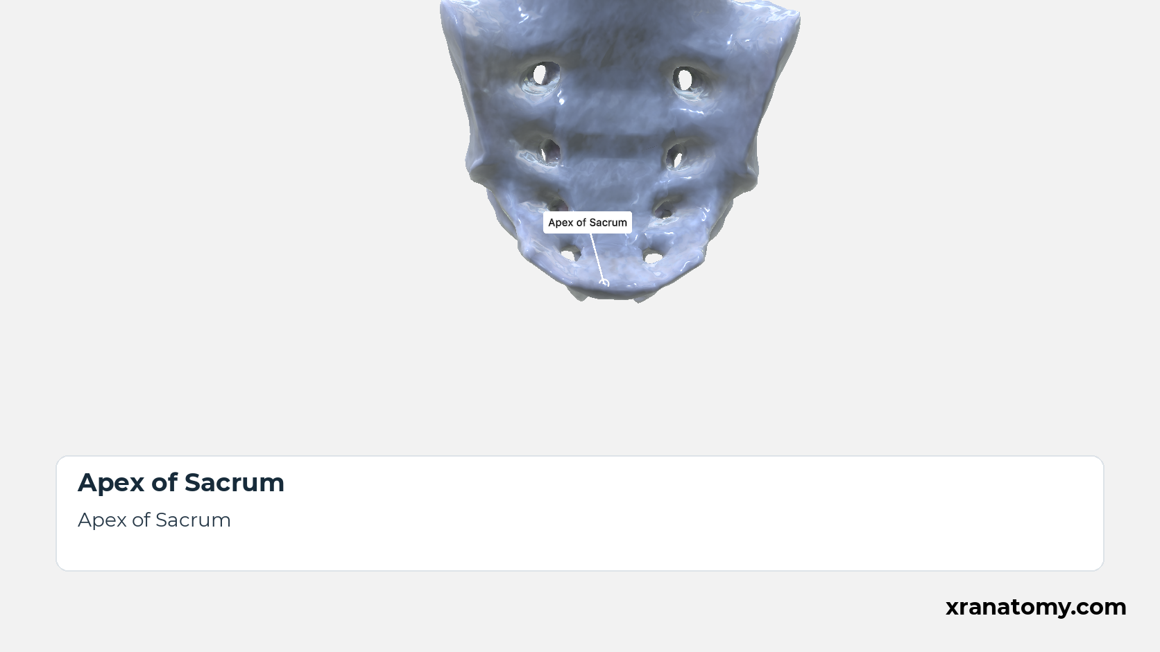
The sacral apex is the narrow inferior end, the lower surface of the S5 body. Its small oval facet meets the base of the coccyx at the sacrococcygeal joint. [2] [3] [1]
Illustration by XR Anatomy.
Pelvic Surface of Sacrum
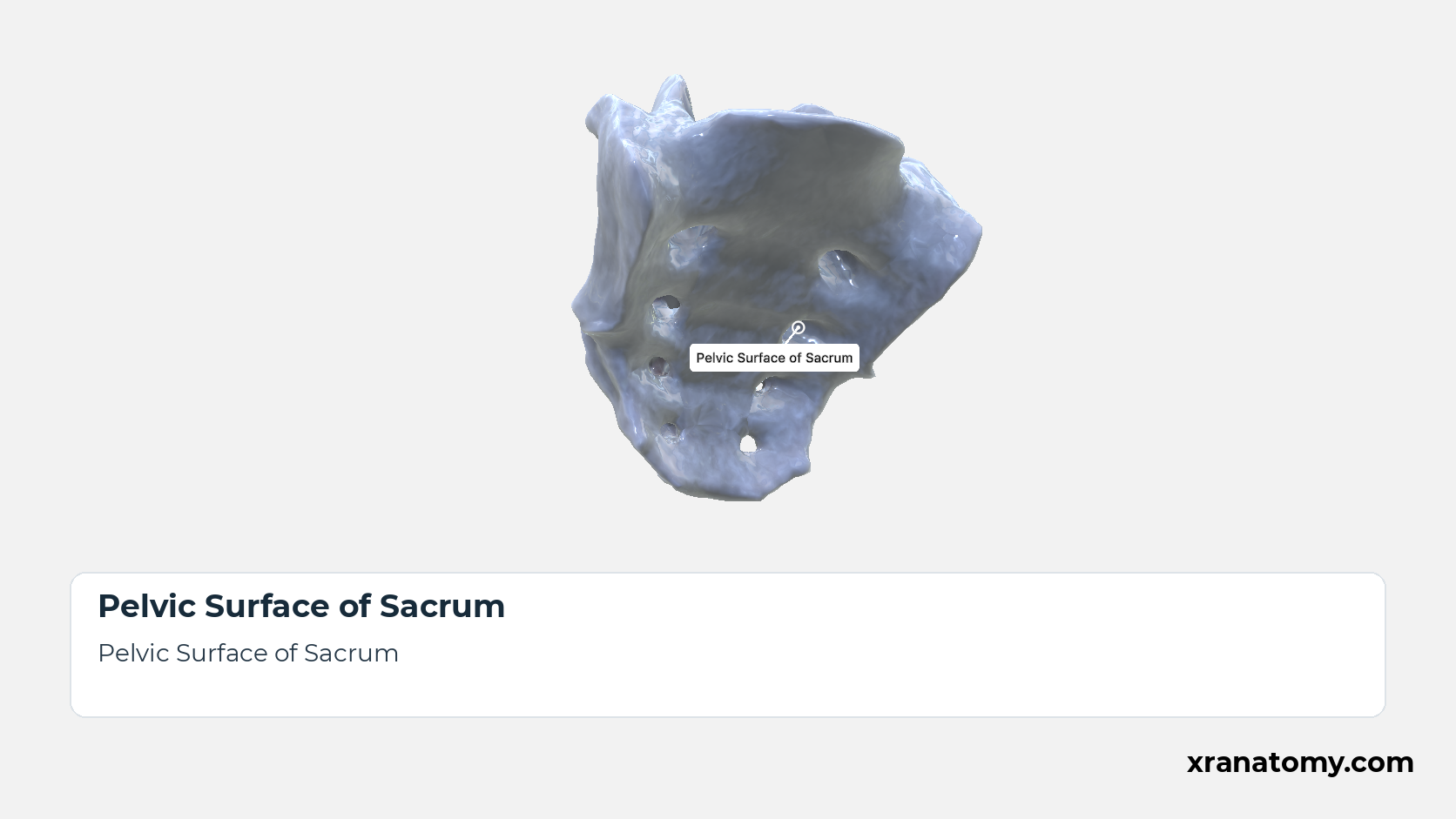
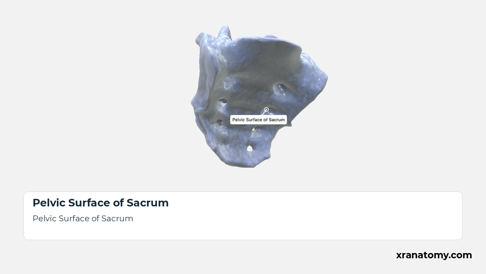
The pelvic surface faces forwards and downwards into the pelvic cavity and is smooth and concave in both directions. Its markings are the four transverse ridges and, at the lateral ends of the ridges, the four pairs of anterior sacral foramina, the pelvic openings of the intervertebral foramina that lead out of the sacral canal. [2] [3] [4]Show less
Illustration by XR Anatomy.
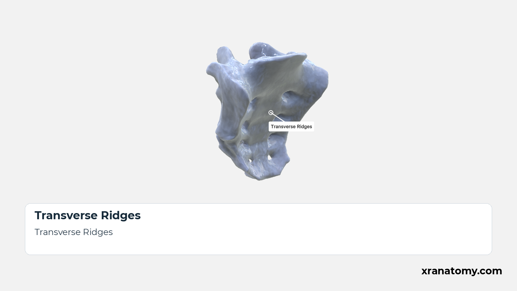
Transverse Ridges
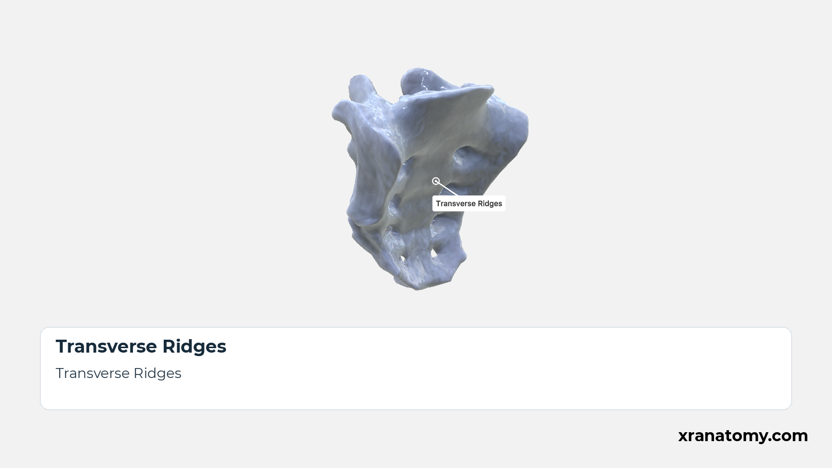
The four transverse ridges cross the pelvic surface at the lines of fusion between the bodies of the five sacral vertebrae, each ridge marking the site of a former intervertebral disc. [2] [3] [1]
Illustration by XR Anatomy.
Sacrum
The sacrum (os sacrum) is the large triangular bone that closes the back of the pelvis, wedged between the two hip bones. It is usually formed by the fusion of five sacral vertebrae, so its broad base faces upwards towards the spine and its narrow apex downwards towards the coccyx; pelvic, dorsal, and lateral surfaces surround the sacral canal at its core.Show less
Illustration by XR Anatomy.
Sacral Canal
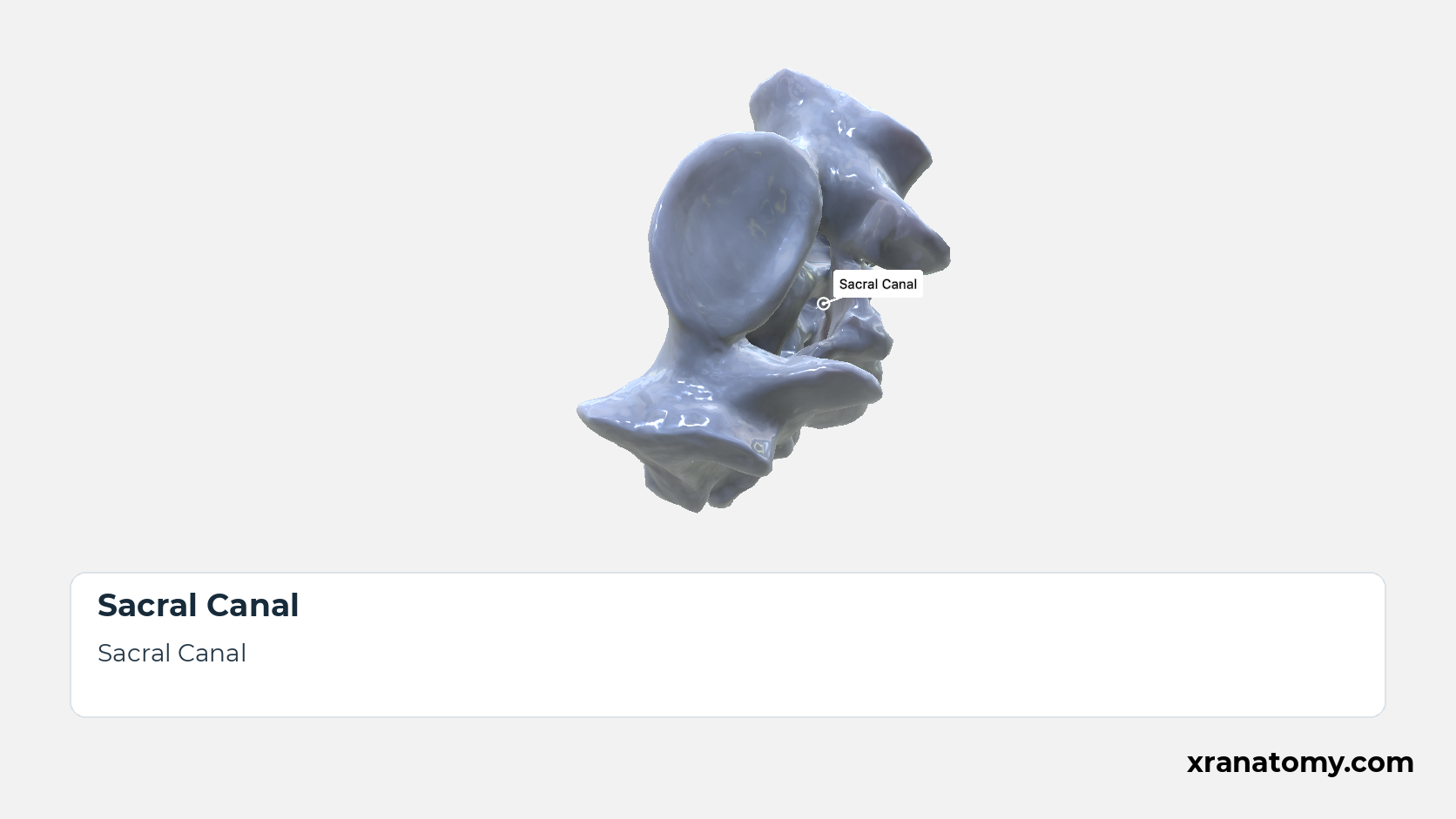
The sacral canal is the continuation of the vertebral canal through the sacrum. Triangular in section at the base, it narrows downwards and opens on the dorsal surface at the sacral hiatus. It lodges the sacral and coccygeal nerve roots: the upper four pairs of sacral nerves leave through the intervertebral foramina towards the anterior and posterior sacral foramina, while the fifth sacral and the coccygeal nerves emerge through the hiatus. [2] [3] [4]Show less
Illustration by XR Anatomy.
 ↺ drag to rotate · scroll to zoom
Sacrum; General Structure
Rotate the sacrum to connect its fused segments, broad base, tapered apex, and anterior and posterior surfaces.
Drag to rotate the sacrum and use the labels to compare its major surfaces and ends.
Sourced from 3D Osteology.
The fused bone keeps the plan of the five vertebrae it is made from: their bodies in front, their arches behind, and the canal between. This section covers that composition, the two ends of the fused column, the base above and the apex below, and the basic character of its two main surfaces.
Composition
The sacrum is usually made of five sacral vertebrae that gradually fuse into a single adult bone; the transverse ridges of the pelvic surface are the visible record of that fusion. The fused column has two ends, the broad base above and the narrow apex below, described next.
In depth · 2 sections · 1 min read2 more sections on this part2 sections open — reading In depth- Base of Sacrum
- Apex of Sacrum
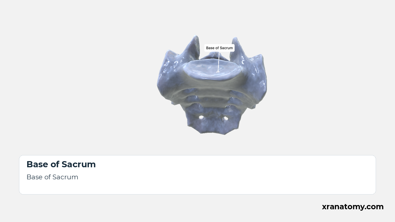
Base of Sacrum
The sacral base is the broad superior end of the bone, on which the fifth lumbar vertebra rests. The upper surface of the S1 body forms its centre, the alae extend it to either side, and the sacral canal opens behind the body between the superior articular processes; the Superior View shows these from above.
Apex of Sacrum
The sacral apex is the narrow inferior end, the lower surface of the S5 body. Its small oval facet meets the base of the coccyx at the sacrococcygeal joint.
Surface Configuration
The two main surfaces have contrasting contours and landmarks. The pelvic surface, facing the pelvic cavity, is smooth and concave; the dorsal surface, buried under the muscles and ligaments of the back, is rough and convex, raised into sacral crests and pierced by the posterior foramina. Both are described in detail under Surfaces and Features.
Practice · Active recall · 3 questions Recall: General Structure
In depth · 1 question Recall: Base of Sacrum
The broad superior end of the sacrum is the .
base
The sacral base is the broad superior end. It contains the upper surface of the S1 body, the superior opening of the sacral canal, the paired alae, and the superior articular processes.
Review this sectionHow well did you know it? · press 1–4
Nice work — revisit in a few days.
The former junctions between the sacral vertebral bodies are marked by .
The sacrum is usually made of five sacral vertebrae that fuse into a single bone during late adolescence and early adult life. The transverse ridges of the pelvic surface are the visible record of that fusion.
Review this sectionHow well did you know it? · press 1–4
The sacral surface that is concave and smooth is the surface.
pelvic
The pelvic surface is concave and smooth. The dorsal surface is convex and rough, with sacral crests and posterior foramina.
Review this sectionHow well did you know it? · press 1–4
Nice work — revisit in a few days.
Surfaces and Features
The pelvic surface is in front, the dorsal surface is behind, and the sacral canal lies between them. Each intervertebral foramen leaves the canal and splits to open on both surfaces, so the anterior and posterior sacral foramina, through which the sacral nerves leave, are the two ends of one passage.
Pelvic Surface of Sacrum
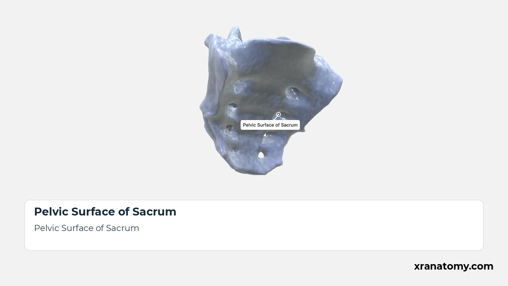
Pelvic Surface of Sacrum
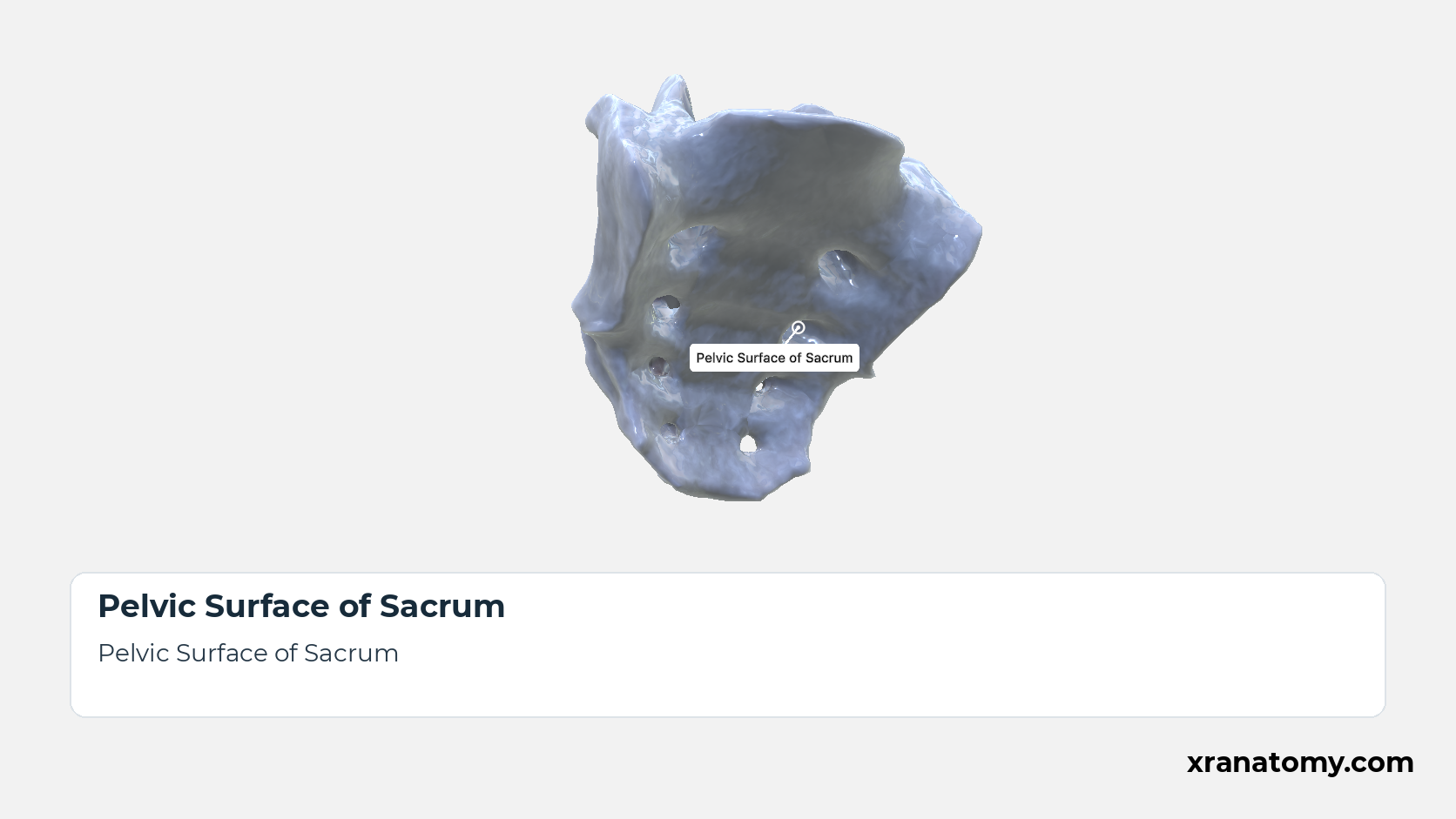
The pelvic surface faces forwards and downwards into the pelvic cavity and is smooth and concave in both directions. Its markings are the four transverse ridges and, at the lateral ends of the ridges, the four pairs of anterior sacral foramina, the pelvic openings of the intervertebral foramina that lead out of the sacral canal. [2] [3] [4]Show less
Study Pelvic Surface of the Sacrum in this fixed image. Use the thumbnails to move between the 2D views; each view highlights one structure. The image does not rotate.
Illustration by XR Anatomy.
Transverse Ridges
The four transverse ridges cross the pelvic surface at the lines of fusion between the bodies of the five sacral vertebrae, each ridge marking the site of a former intervertebral disc. [2] [3] [1]
Illustration by XR Anatomy.
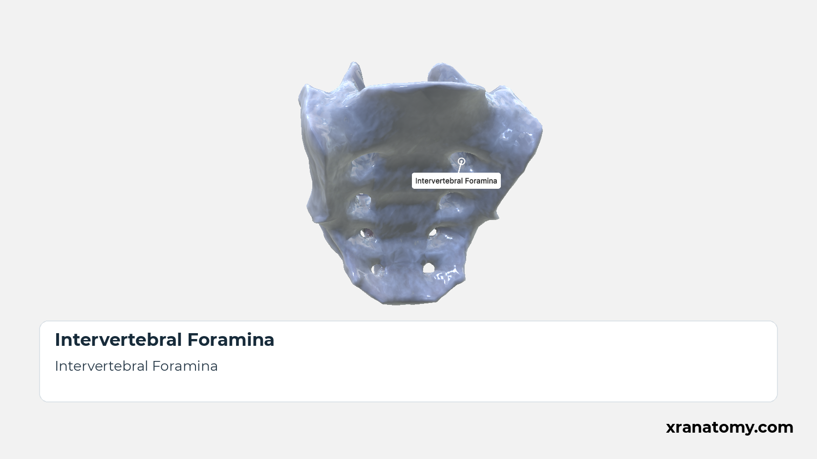
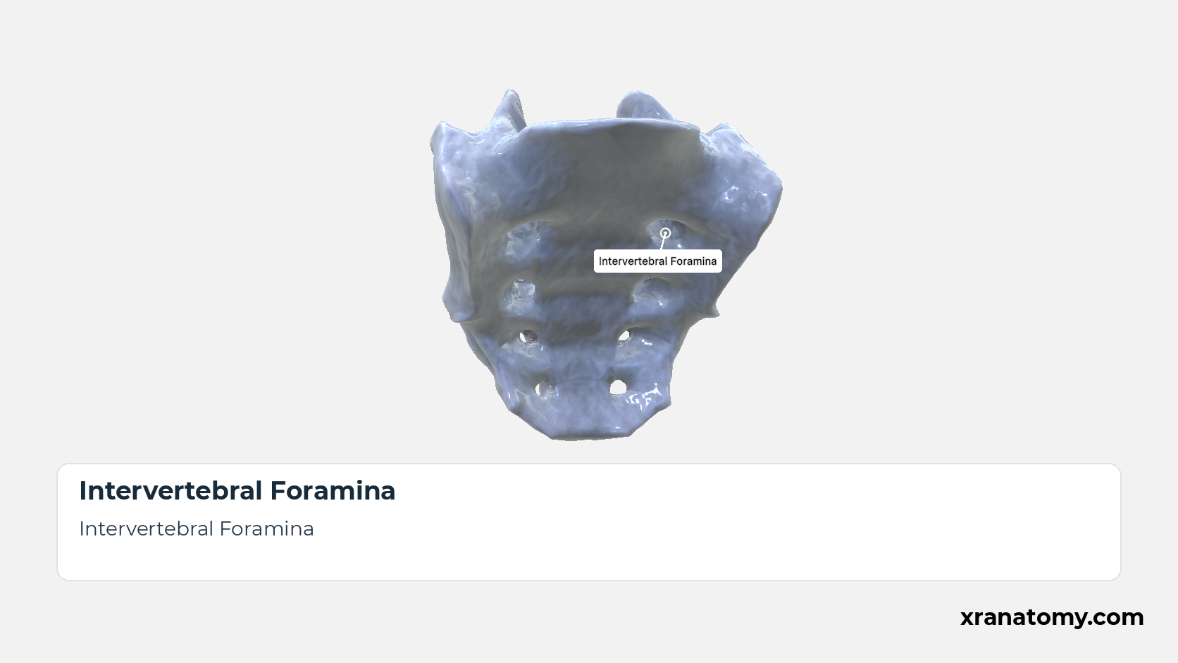
Intervertebral Foramina
Within the fused bone, each sacral intervertebral foramen leads laterally out of the sacral canal and divides, opening forwards as an anterior and backwards as a posterior sacral foramen. [2] [3] [1]
Illustration by XR Anatomy.
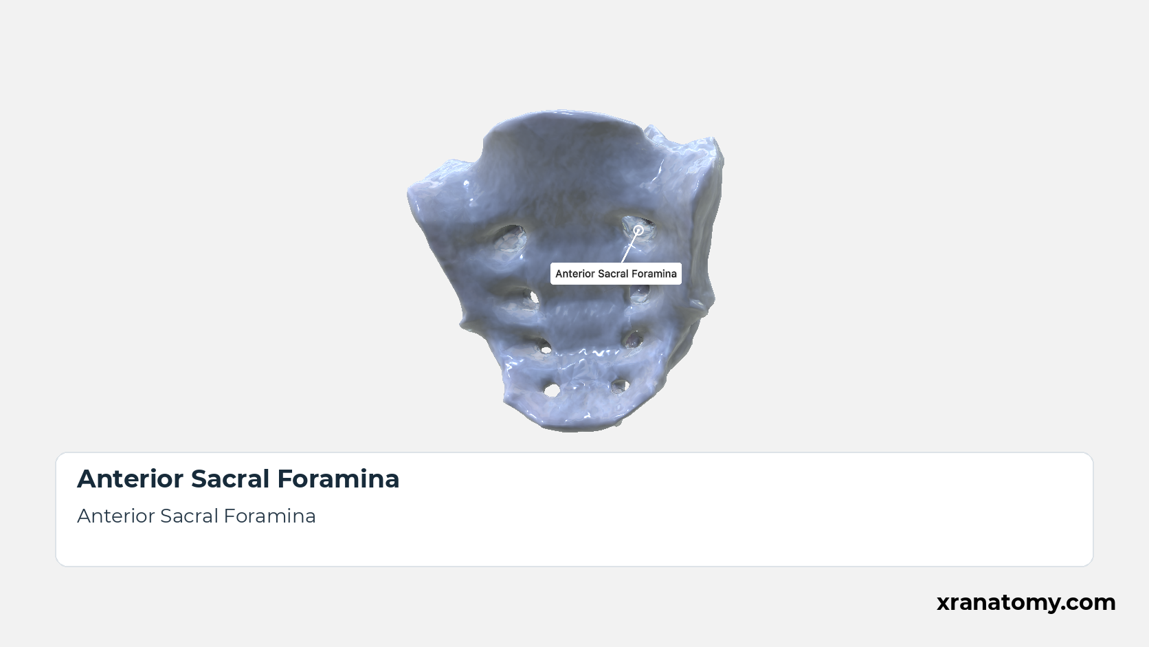
Anterior Sacral Foramina
The four pairs of anterior sacral foramina are the pelvic openings of that passage. Each lies at the lateral end of a transverse ridge on the pelvic surface, reaches the sacral canal through an intervertebral foramen, and transmits the anterior ramus of a sacral spinal nerve; they are larger than the posterior foramina. [2] [3] [1]Show less
Illustration by XR Anatomy.
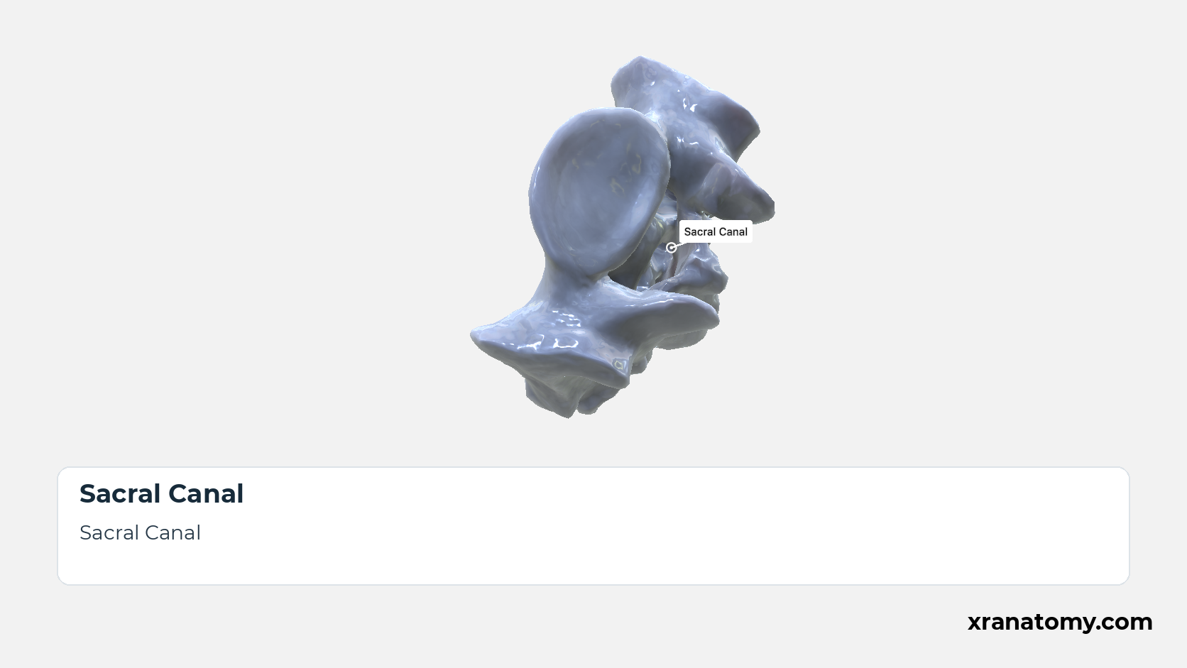
Sacral Canal
The sacral canal is the continuation of the vertebral canal through the sacrum. Triangular in section at the base, it narrows downwards and opens on the dorsal surface at the sacral hiatus. It lodges the sacral and coccygeal nerve roots: the upper four pairs of sacral nerves leave through the intervertebral foramina towards the anterior and posterior sacral foramina, while the fifth sacral and the coccygeal nerves emerge through the hiatus. [2] [3] [4]Show less
Illustration by XR Anatomy.
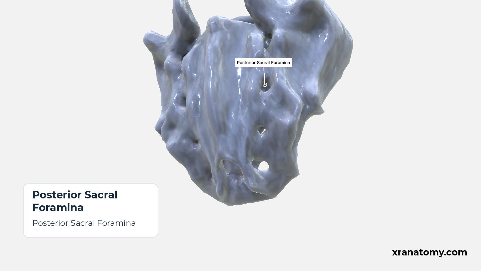
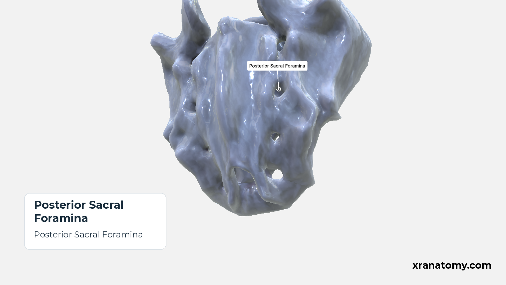
Posterior Sacral Foramina
The four pairs of posterior sacral foramina open onto the dorsal surface between the intermediate and lateral crests. They are smaller than the anterior foramina and transmit the posterior rami of the sacral spinal nerves. [2] [3] [1]
Illustration by XR Anatomy.
The pelvic surface faces forwards and downwards into the pelvic cavity and is smooth and concave in both directions. Its markings are the four transverse ridges and, at the lateral ends of the ridges, the four pairs of anterior sacral foramina, the pelvic openings of the intervertebral foramina that lead out of the sacral canal.
In depth · 3 sections · 1 min read3 more sections on this part3 sections open — reading In depth- Transverse Ridges
- Intervertebral Foramina
- Anterior Sacral Foramina
Transverse Ridges
The four transverse ridges cross the pelvic surface at the lines of fusion between the bodies of the five sacral vertebrae, each ridge marking the site of a former intervertebral disc.
Intervertebral Foramina
Within the fused bone, each sacral intervertebral foramen leads laterally out of the sacral canal and divides, opening forwards as an anterior and backwards as a posterior sacral foramen.
Anterior Sacral Foramina
The four pairs of anterior sacral foramina are the pelvic openings of that passage. Each lies at the lateral end of a transverse ridge on the pelvic surface, reaches the sacral canal through an intervertebral foramen, and transmits the anterior ramus of a sacral spinal nerve; they are larger than the posterior foramina.
Dorsal Surface of Sacrum
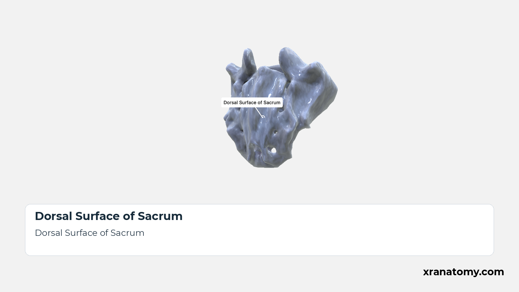
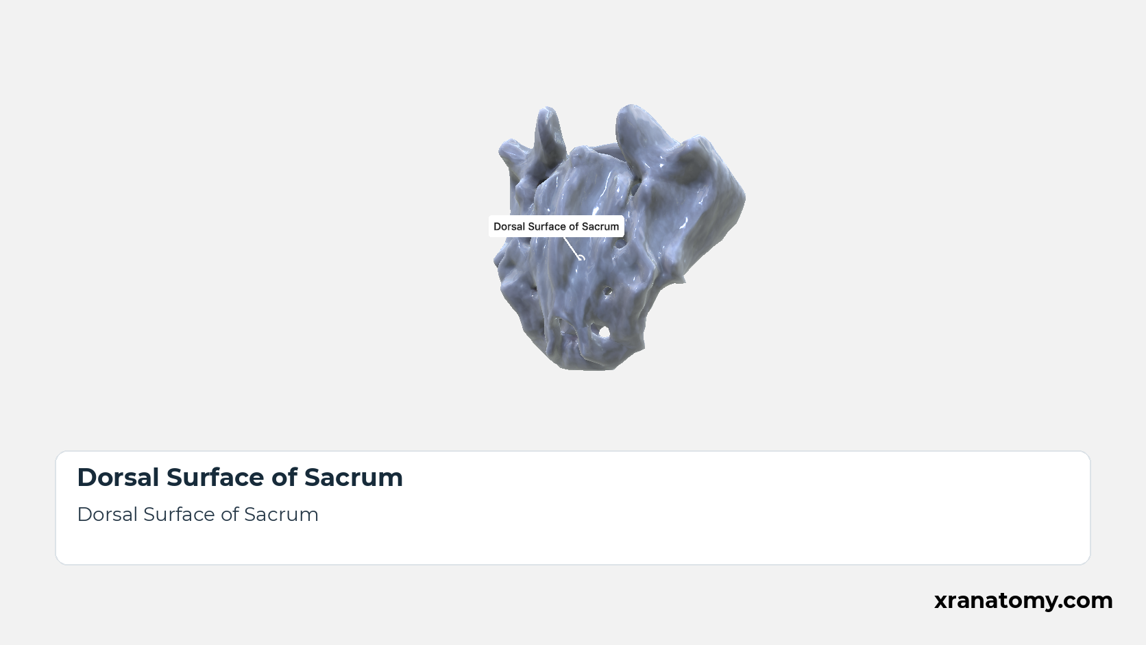
Dorsal Surface of Sacrum
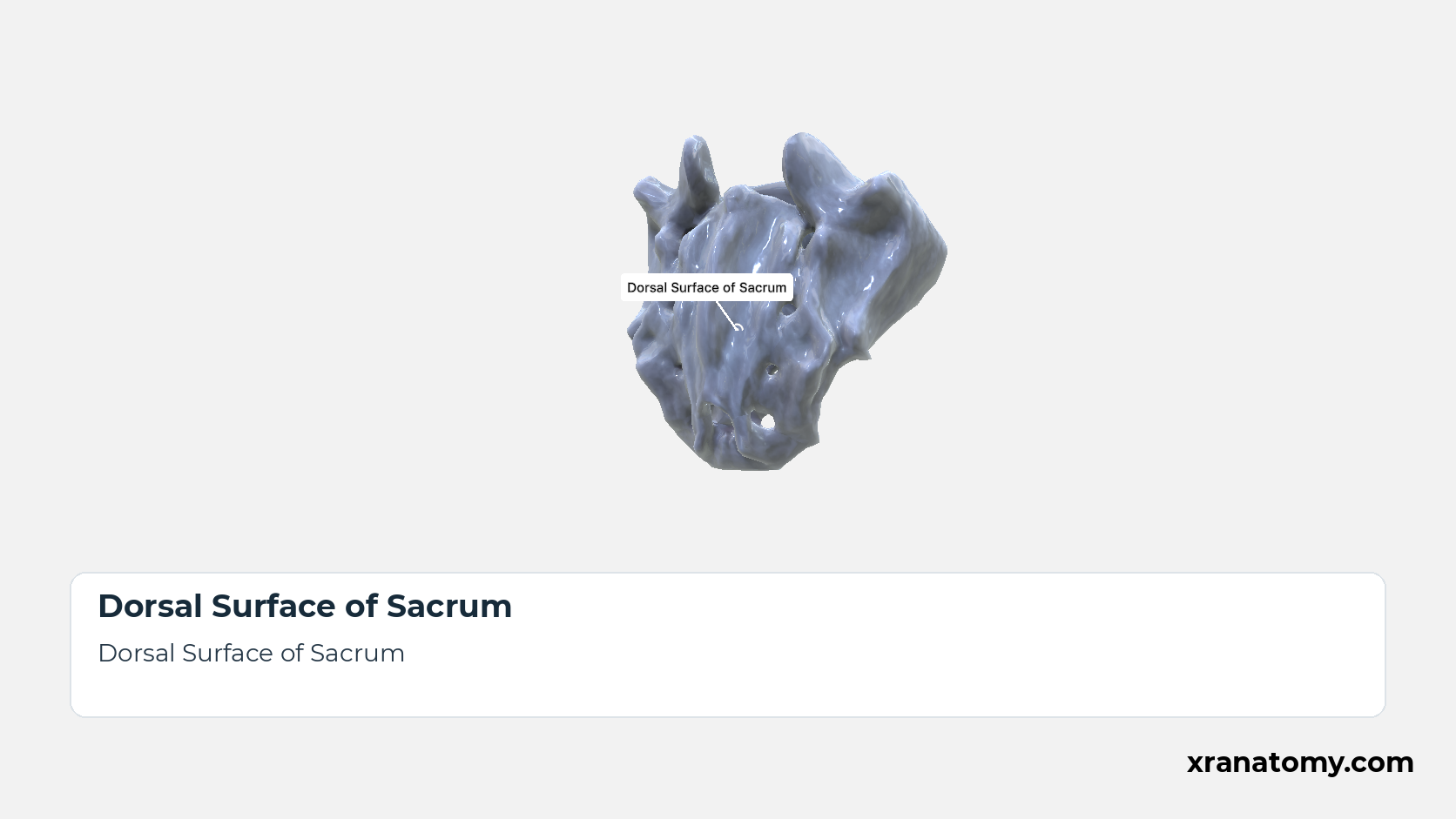
The dorsal surface is convex, narrower than the pelvic surface, and rough, because the back muscles and the sacroiliac ligaments attach to it. It is built from the fused posterior elements of the five vertebrae, which form five longitudinal crests: the median crest in the midline, an intermediate crest on each side, and the lateral sacral crests at the edges, with the four pairs of posterior sacral foramina between the intermediate and lateral crests. Between the median and intermediate crests the surface is made of the fused laminae; the lowest laminae fail to meet, so the canal opens below at the sacral hiatus, where the intermediate crests end as the sacral horns flanking the hiatus. [2] [3] [4]Show less
Study Posterior Surface of the Sacrum in this fixed image. Use the thumbnails to move between the 2D views; each view highlights one structure. The image does not rotate.
Illustration by XR Anatomy.
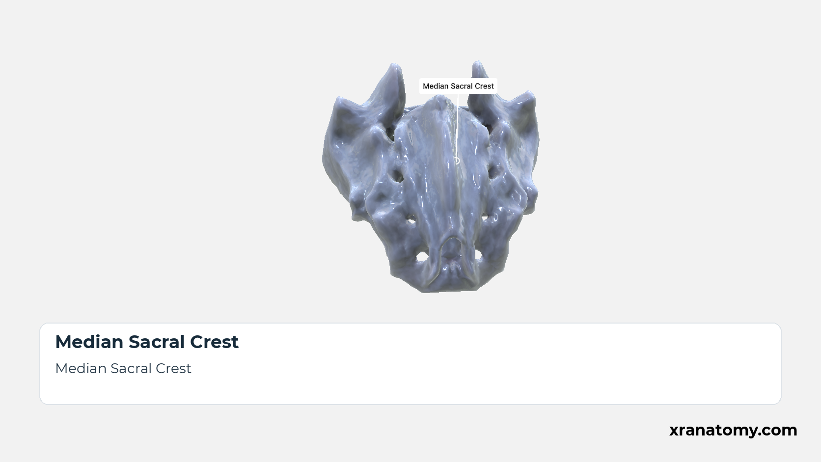
Median Sacral Crest
The median sacral crest runs down the posterior midline as a row of three or four tubercles, the fused rudimentary spinous processes of the upper sacral vertebrae. [2] [3] [1]
Illustration by XR Anatomy.
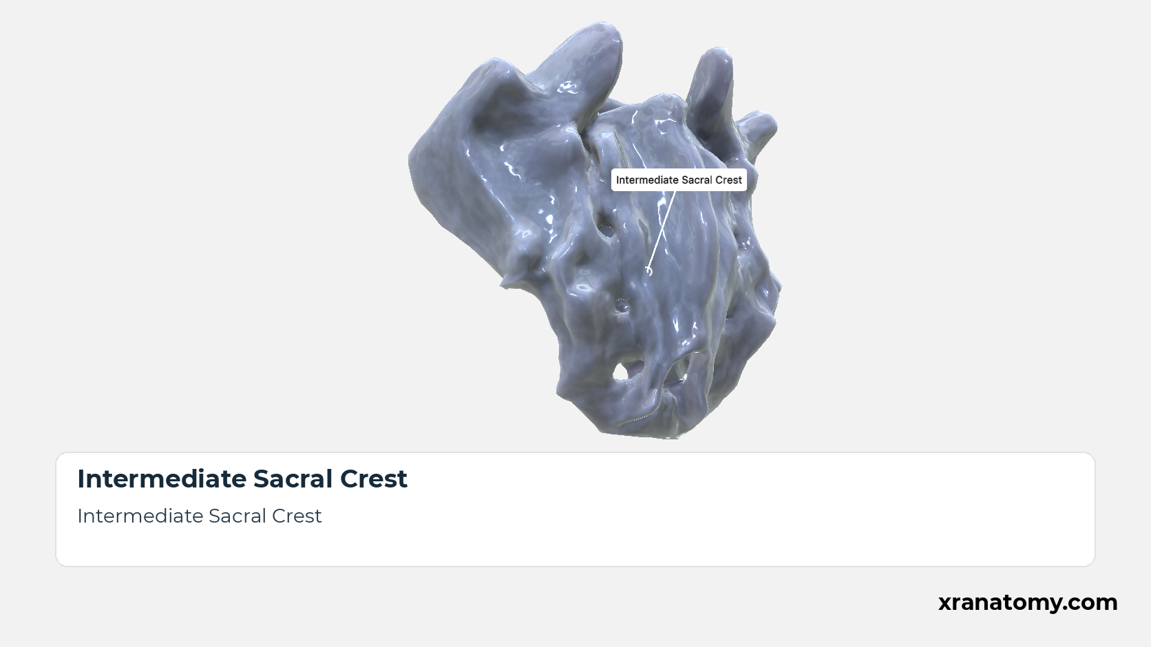
Intermediate Sacral Crest
An intermediate sacral crest runs down each side just medial to the row of posterior sacral foramina. It is the row of fused articular processes, and its lowest tubercle projects downwards as the sacral horn. [2] [3] [1]
Illustration by XR Anatomy.
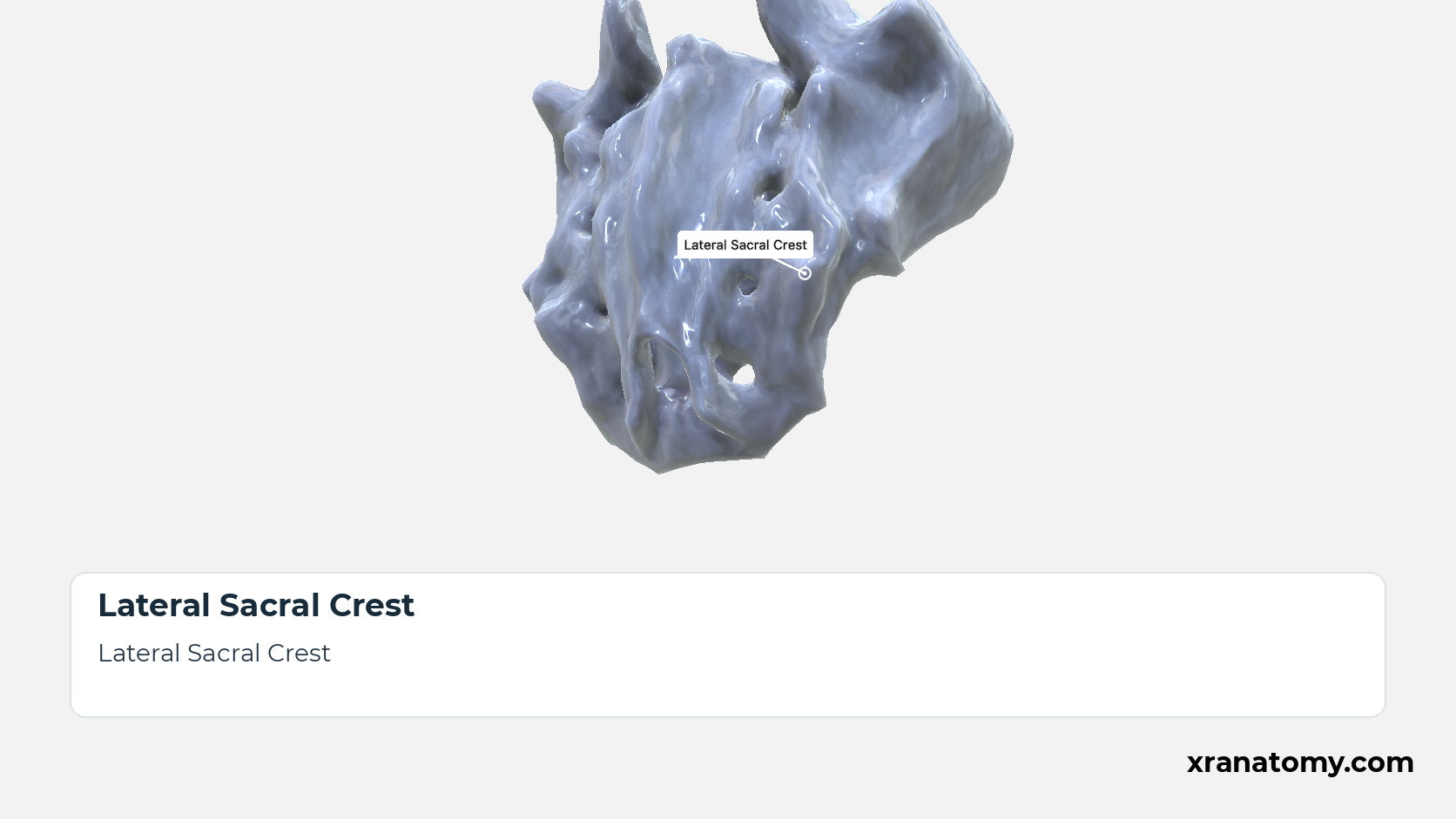
Lateral Sacral Crest
The lateral sacral crest runs down each side lateral to the posterior sacral foramina. Its tubercles are the fused transverse processes of the sacral vertebrae, and the posterior sacroiliac ligaments attach to them. [2] [3] [1]
Illustration by XR Anatomy.
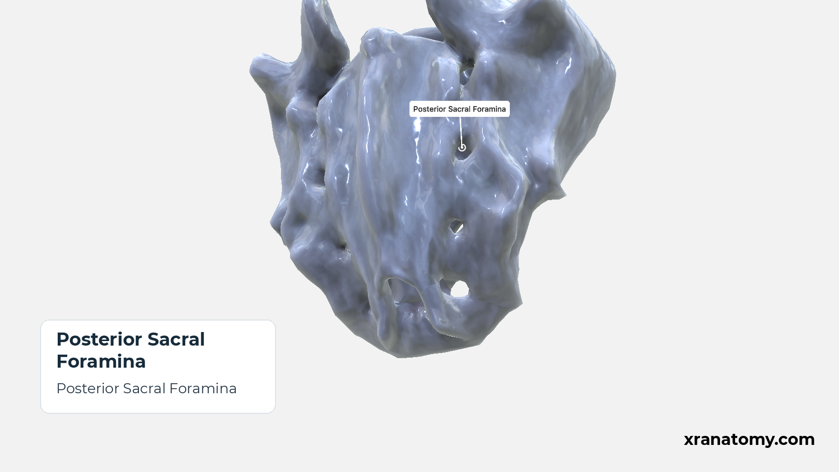
Posterior Sacral Foramina
The four pairs of posterior sacral foramina open onto the dorsal surface between the intermediate and lateral crests. They are smaller than the anterior foramina and transmit the posterior rami of the sacral spinal nerves. [2] [3] [1]
Illustration by XR Anatomy.
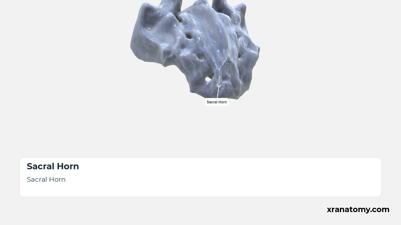
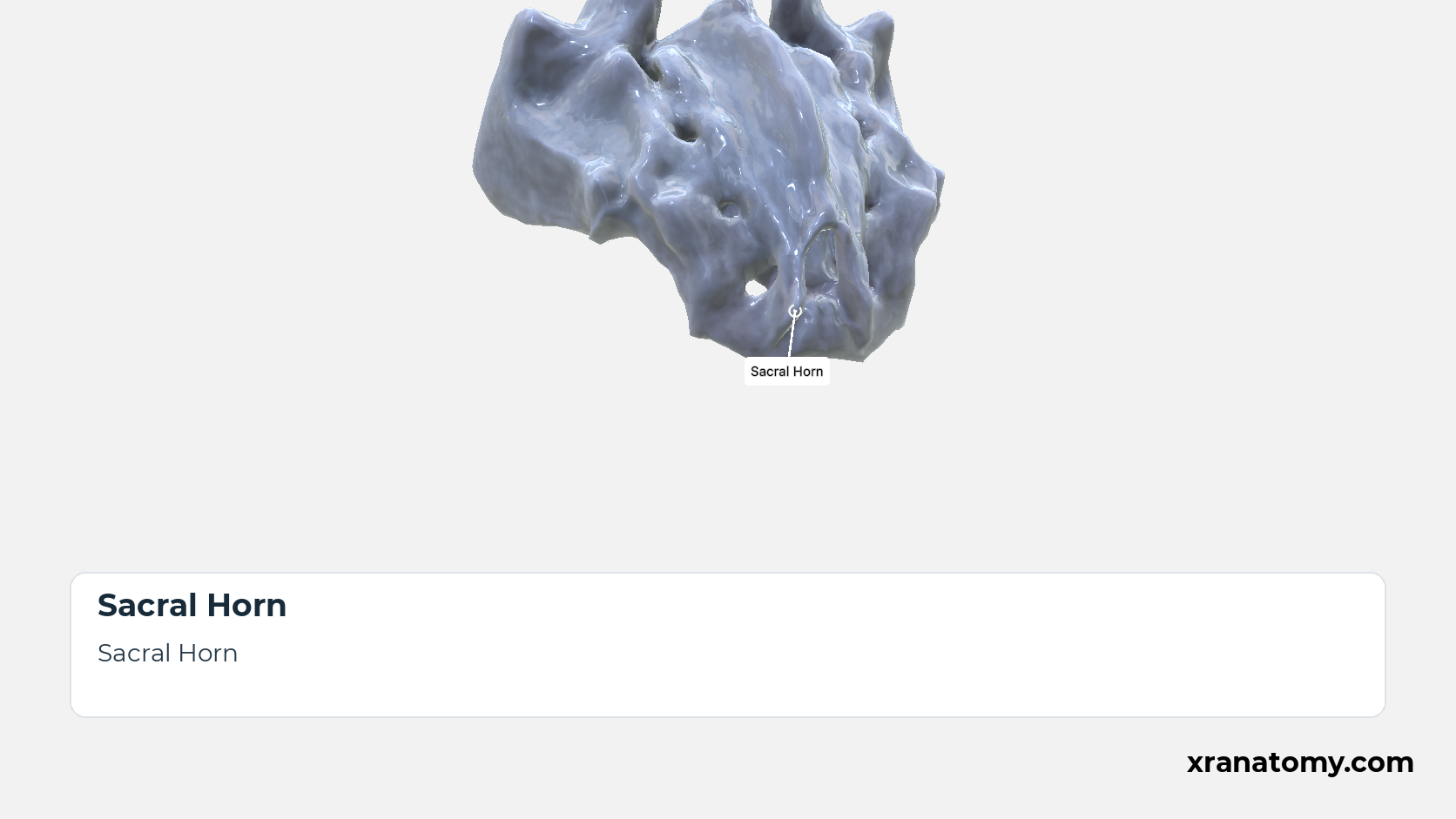
Sacral Horn
The paired sacral horns are the downward-projecting lower ends of the intermediate sacral crests, the inferior articular processes of S5. They flank the sacral hiatus and are joined by ligaments to the coccygeal horns below. [2] [3] [1]
Illustration by XR Anatomy.
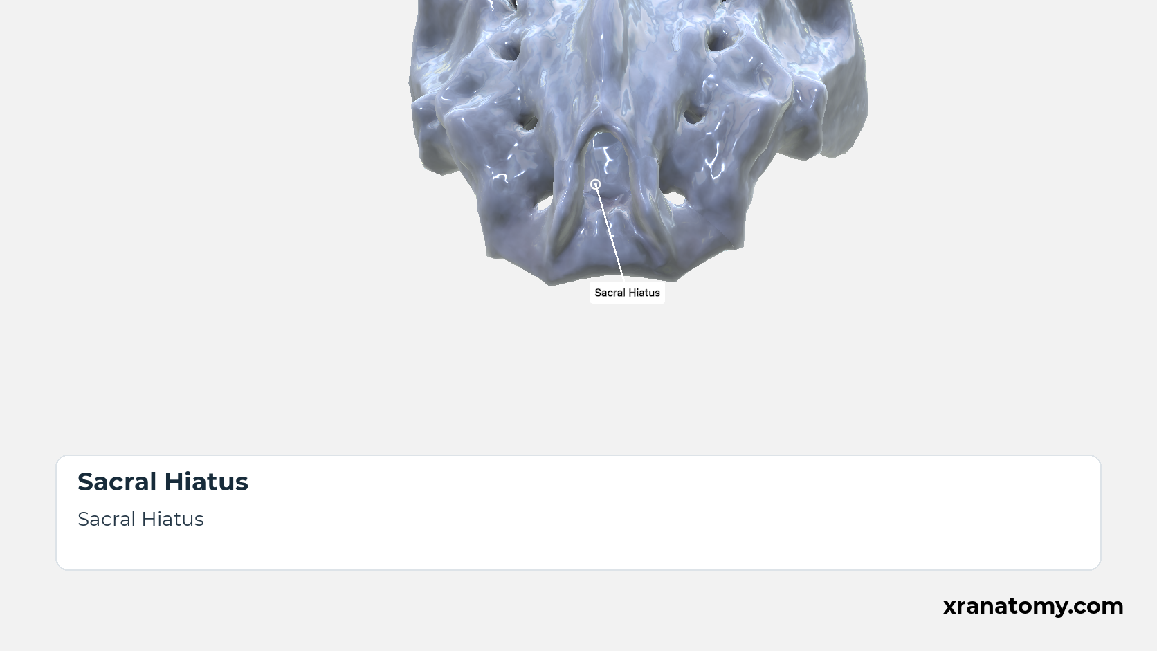
Sacral Hiatus
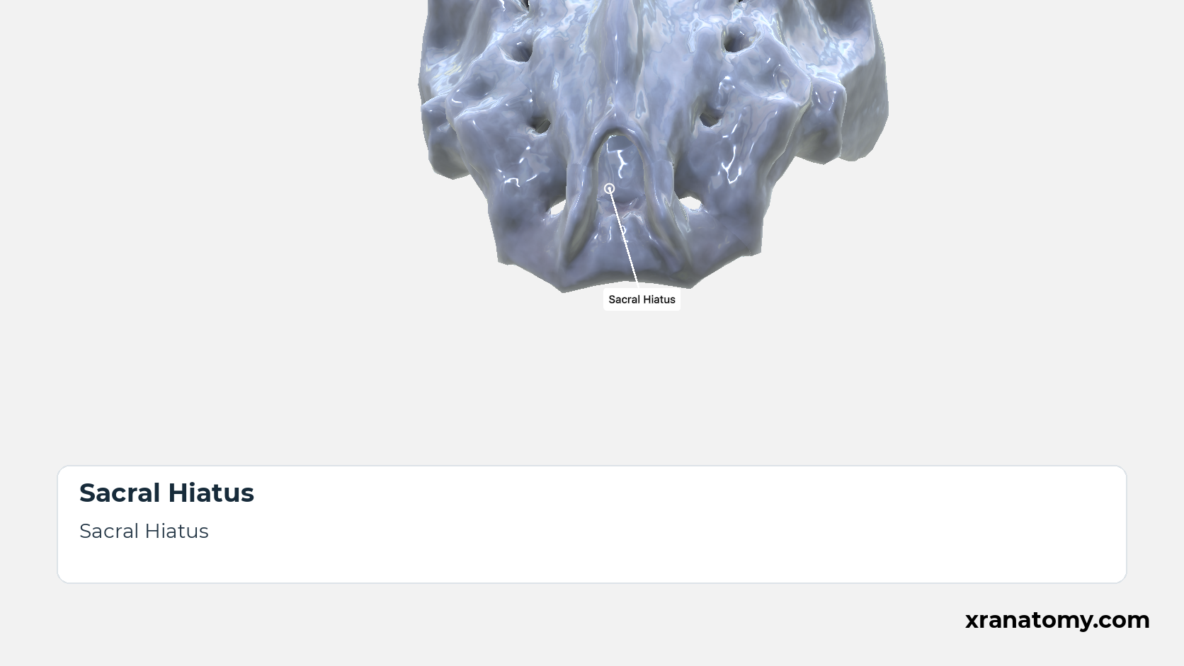
The sacral hiatus is the gap at the lower end of the posterior wall of the sacral canal. It arises because the laminae of S5, and sometimes the laminae of S4 as well, fail to meet in the midline; the sacral horns flank it on either side. [2] [3] [4] [1]Show less
Illustration by XR Anatomy.
Sacral Canal
The sacral canal is the continuation of the vertebral canal through the sacrum. Triangular in section at the base, it narrows downwards and opens on the dorsal surface at the sacral hiatus. It lodges the sacral and coccygeal nerve roots: the upper four pairs of sacral nerves leave through the intervertebral foramina towards the anterior and posterior sacral foramina, while the fifth sacral and the coccygeal nerves emerge through the hiatus. [2] [3] [4]Show less
Illustration by XR Anatomy.
The dorsal surface is convex, narrower than the pelvic surface, and rough. Back muscles and sacroiliac ligaments attach to this surface. It is built from the fused posterior elements of the five vertebrae, which form five longitudinal crests: the median crest in the midline, an intermediate crest on each side, and the lateral sacral crests at the edges, with the four pairs of posterior sacral foramina between the intermediate and lateral crests. Between the median and intermediate crests the surface is made of the fused laminae; the lowest laminae fail to meet, so the canal opens below at the sacral hiatus, where the intermediate crests end as the sacral horns flanking the hiatus.
In depth · 5 sections · 1 min read5 more sections on this part5 sections open — reading In depth- Median Sacral Crest
- Intermediate Sacral Crest
- Lateral Sacral Crest
- Posterior Sacral Foramina
- Sacral Horn
Median Sacral Crest
The median sacral crest runs down the posterior midline as a row of three or four tubercles, the fused rudimentary spinous processes of the upper sacral vertebrae.
Intermediate Sacral Crest
An intermediate sacral crest runs down each side just medial to the row of posterior sacral foramina. It is the row of fused articular processes, and its lowest tubercle projects downwards as the sacral horn.
Lateral Sacral Crest
The lateral sacral crest runs down each side lateral to the posterior sacral foramina. Its tubercles are the fused transverse processes of the sacral vertebrae, and posterior sacroiliac and sacrotuberous ligaments attach to them.
Posterior Sacral Foramina
The four pairs of posterior sacral foramina open onto the dorsal surface between the intermediate and lateral crests. They are smaller than the anterior foramina and transmit the posterior rami of the sacral spinal nerves.
Sacral Horn
The paired sacral horns are the downward-projecting lower ends of the intermediate sacral crests, the inferior articular processes of S5. They flank the sacral hiatus and are joined by ligaments to the coccygeal horns below.
Sacral Canal
Sacral Canal
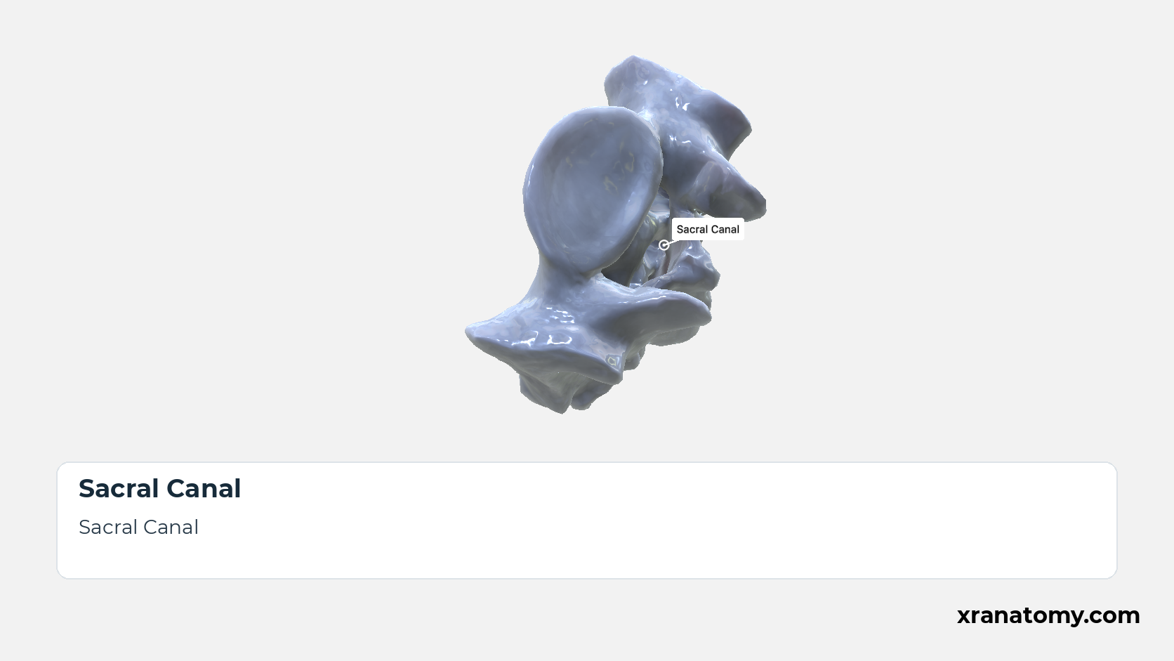
The sacral canal is the continuation of the vertebral canal through the sacrum. Triangular in section at the base, it narrows downwards and opens on the dorsal surface at the sacral hiatus. It lodges the sacral and coccygeal nerve roots: the upper four pairs of sacral nerves leave through the intervertebral foramina towards the anterior and posterior sacral foramina, while the fifth sacral and the coccygeal nerves emerge through the hiatus. [2] [3] [4]Show less
Study Sacral Canal in this fixed image. Use the thumbnails to move between the 2D views; each view highlights one structure. The image does not rotate.
Illustration by XR Anatomy.
Sacral Hiatus
The sacral hiatus is the gap at the lower end of the posterior wall of the sacral canal. It arises because the laminae of S5, and sometimes the laminae of S4 as well, fail to meet in the midline; the sacral horns flank it on either side. [2] [3] [4] [1]Show less
Illustration by XR Anatomy.
Sacrum
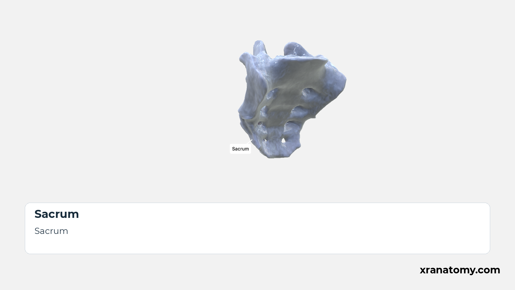
The sacrum (os sacrum) is the large triangular bone that closes the back of the pelvis, wedged between the two hip bones. It is usually formed by the fusion of five sacral vertebrae, so its broad base faces upwards towards the spine and its narrow apex downwards towards the coccyx; pelvic, dorsal, and lateral surfaces surround the sacral canal at its core.Show less
Illustration by XR Anatomy.
Intervertebral Foramina
Within the fused bone, each sacral intervertebral foramen leads laterally out of the sacral canal and divides, opening forwards as an anterior and backwards as a posterior sacral foramen. [2] [3] [1]
Illustration by XR Anatomy.
Posterior Sacral Foramina
The four pairs of posterior sacral foramina open onto the dorsal surface between the intermediate and lateral crests. They are smaller than the anterior foramina and transmit the posterior rami of the sacral spinal nerves. [2] [3] [1]
Illustration by XR Anatomy.
Sacral Horn
The paired sacral horns are the downward-projecting lower ends of the intermediate sacral crests, the inferior articular processes of S5. They flank the sacral hiatus and are joined by ligaments to the coccygeal horns below. [2] [3] [1]
Illustration by XR Anatomy.
The sacral canal is the continuation of the vertebral canal through the sacrum. Triangular in section at the base, it narrows downwards and opens on the dorsal surface at the sacral hiatus. It lodges the sacral and coccygeal nerve roots: the upper four pairs of sacral nerves leave through the intervertebral foramina towards the anterior and posterior sacral foramina, while the fifth sacral and the coccygeal nerves emerge through the hiatus.
In depth · 1 min read Sacral Hiatus
The sacral hiatus is the gap at the lower end of the posterior wall of the sacral canal. It arises because the laminae of S5, and sometimes the laminae of S4 as well, fail to meet in the midline; the sacral horns flank it on either side.
Practice · Active recall · 4 questions Recall: Surfaces and Features
Why this helps
Testing yourself before revealing the answer (active recall) strengthens memory far more than re-reading — so each prompt stays hidden until you choose to reveal it.
Which surface is concave and crossed by four transverse ridges?
pelvic surface
The pelvic surface is concave and crossed by four transverse ridges. Four pairs of anterior sacral foramina open onto it; intervertebral foramina connect these openings with the sacral canal.
Review this sectionHow well did you know it? · press 1–4
In depth · 1 question Recall: Transverse Ridges, Intervertebral Foramina, Anterior Sacral Foramina
The pelvic surface is crossed by transverse ridges.
four
The pelvic surface is concave and crossed by four transverse ridges. Four pairs of anterior sacral foramina open onto it; intervertebral foramina connect these openings with the sacral canal.
Review this sectionHow well did you know it? · press 1–4
Nice work — revisit in a few days.
The dorsal surface bears the median, intermediate, and sacral crests.
lateral
The dorsal surface is convex and bears the median, intermediate, and lateral sacral crests, with four pairs of posterior sacral foramina. Inferiorly, the intermediate crests end as the sacral horns flanking the sacral hiatus, where the lowest laminae fail to meet.
Review this sectionHow well did you know it? · press 1–4
The sacral canal opens posteriorly at the .
The sacral canal continues the vertebral canal through most of the sacrum. It is triangular superiorly, narrows inferiorly, and opens posteriorly at the sacral hiatus. Intervertebral foramina connect it laterally to the anterior and posterior sacral foramina.
Review this sectionHow well did you know it? · press 1–4
Nice work — revisit in a few days.
Anatomical Views
Two more views finish the description of the sacrum. From above, you see the base, the broad upper end of the bone, with the parts that meet the last lumbar vertebra. From the side, you see the lateral part, which is broad above and narrows below. It has the joint surface for the hip bone and, behind this surface, a rough area where the sacroiliac ligaments attach.
Superior View
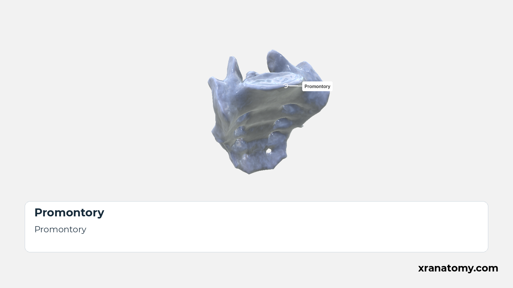
Promontory
The promontory is the projecting anterior edge of the upper surface of the S1 vertebral body, at the front of the sacral base. It forms the midpoint of the posterior boundary of the pelvic inlet. [2] [3] [1]
Study Superior View of the Sacrum in this fixed image. Use the thumbnails to move between the 2D views; each view highlights one structure. The image does not rotate.
Illustration by XR Anatomy.
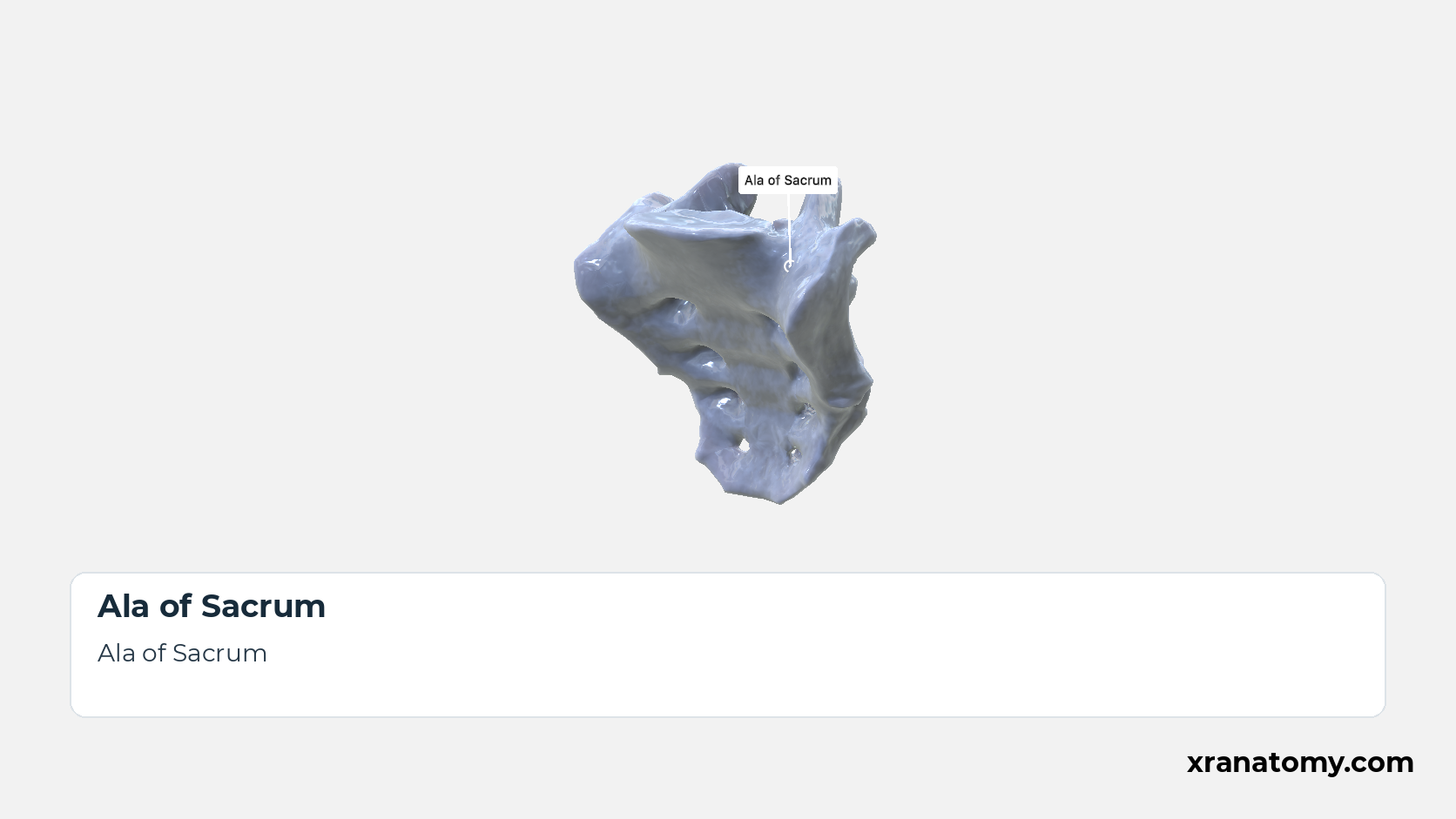
Ala of Sacrum
Each ala is the wing-like expansion lateral to the body of S1 and the upper end of the lateral part. The alae complete the broad base and continue downwards as the sacrum's lateral parts. [2] [3] [1]
Illustration by XR Anatomy.
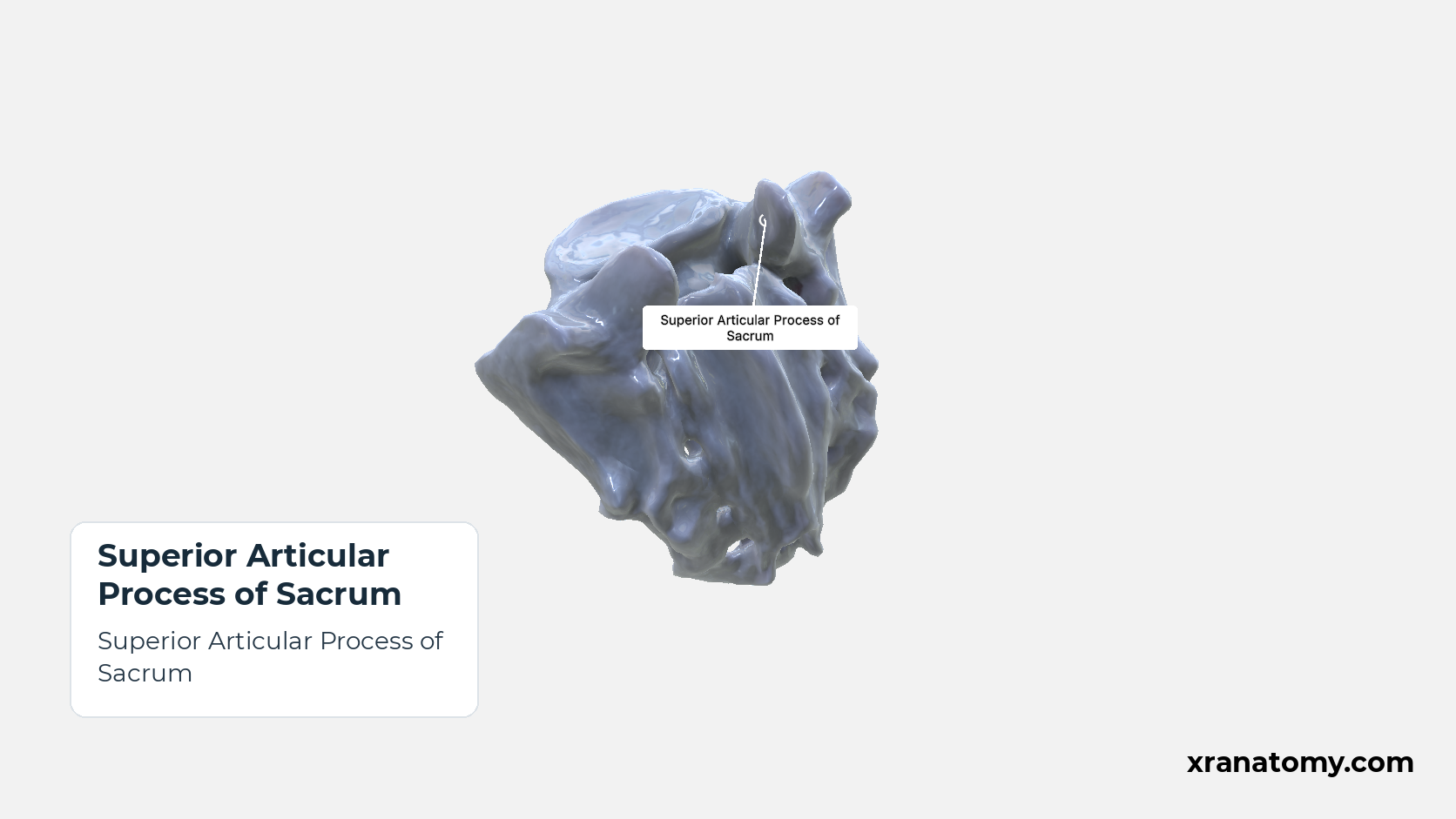
Superior Articular Process of Sacrum
The paired superior articular processes rise from the back of the base, one on each side of the canal opening. Their facets are concave and face posteromedially to receive the inferior articular processes of the fifth lumbar vertebra. [2] [3] [1]Show less
Illustration by XR Anatomy.
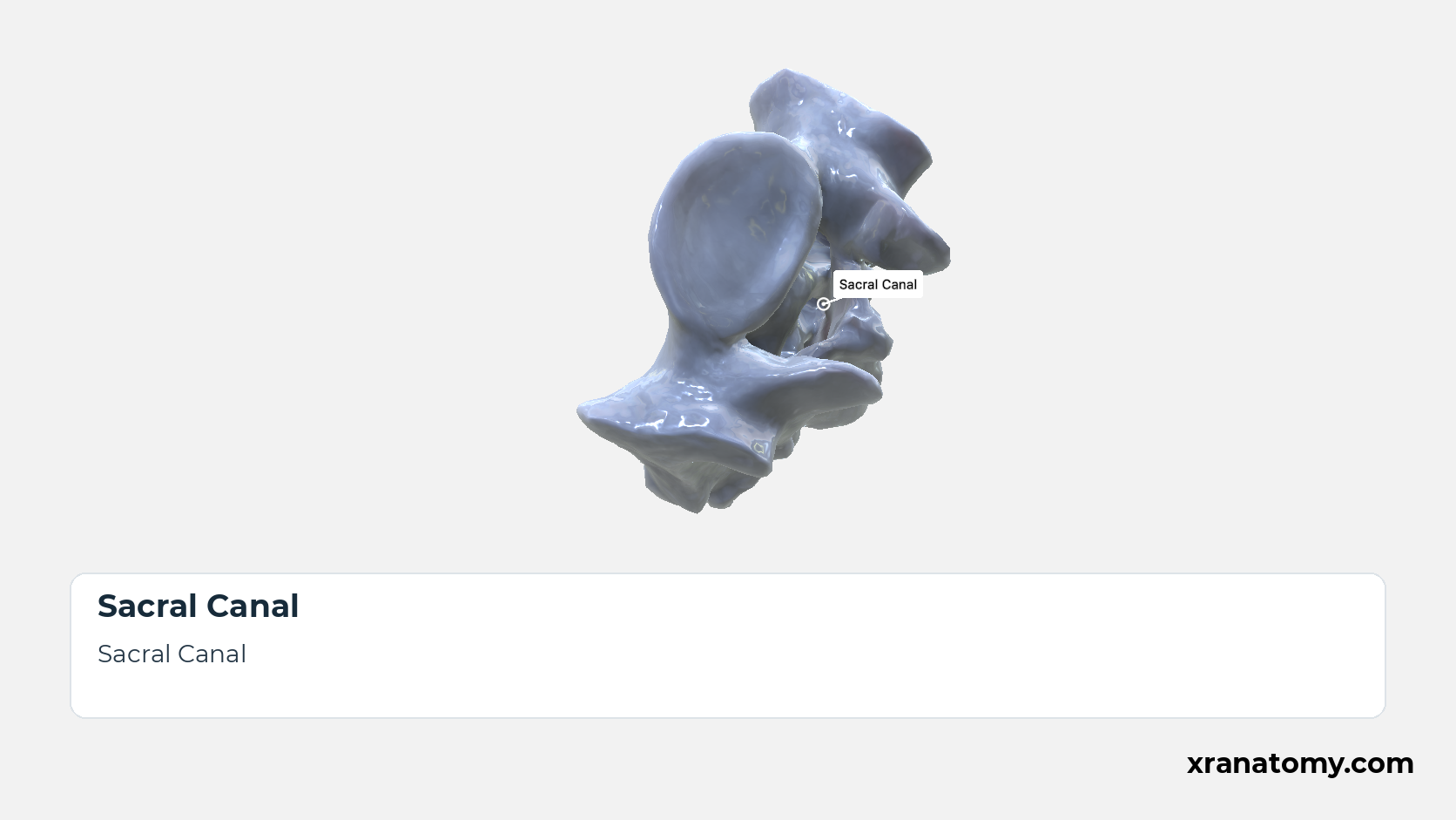
Sacral Canal
The sacral canal is the continuation of the vertebral canal through the sacrum. Triangular in section at the base, it narrows downwards and opens on the dorsal surface at the sacral hiatus. It lodges the sacral and coccygeal nerve roots: the upper four pairs of sacral nerves leave through the intervertebral foramina towards the anterior and posterior sacral foramina, while the fifth sacral and the coccygeal nerves emerge through the hiatus. [2] [3] [4]Show less
Illustration by XR Anatomy.
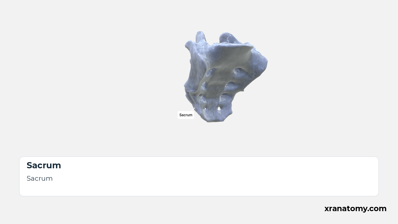
Sacrum
The sacrum (os sacrum) is the large triangular bone that closes the back of the pelvis, wedged between the two hip bones. It is usually formed by the fusion of five sacral vertebrae, so its broad base faces upwards towards the spine and its narrow apex downwards towards the coccyx; pelvic, dorsal, and lateral surfaces surround the sacral canal at its core.Show less
Illustration by XR Anatomy.
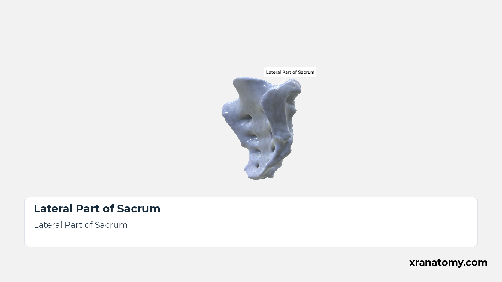
Lateral Part of Sacrum
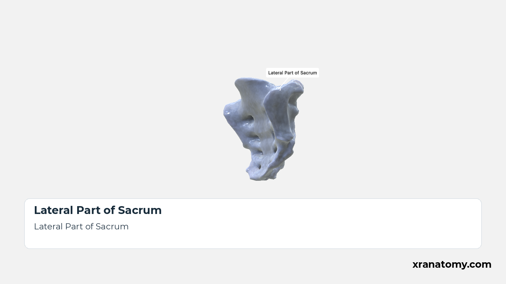
The lateral part is the mass of bone on each side of the fused bodies, formed by the fused transverse processes and costal elements of the sacral vertebrae. Broad above, where it carries the auricular surface and the sacral tuberosity, it tapers to a thin edge below. [2] [3] [1]Show less
Illustration by XR Anatomy.
Base of Sacrum
The sacral base is the broad superior end of the bone, on which the fifth lumbar vertebra rests. The upper surface of the S1 body forms its centre, the alae extend it to either side, and the sacral canal opens behind the body between the superior articular processes; the Superior View shows these from above. [2] [3] [1]Show less
Illustration by XR Anatomy.
Pelvic Surface of Sacrum
The pelvic surface faces forwards and downwards into the pelvic cavity and is smooth and concave in both directions. Its markings are the four transverse ridges and, at the lateral ends of the ridges, the four pairs of anterior sacral foramina, the pelvic openings of the intervertebral foramina that lead out of the sacral canal. [2] [3] [4]Show less
Illustration by XR Anatomy.
Dorsal Surface of Sacrum
The dorsal surface is convex, narrower than the pelvic surface, and rough, because the back muscles and the sacroiliac ligaments attach to it. It is built from the fused posterior elements of the five vertebrae, which form five longitudinal crests: the median crest in the midline, an intermediate crest on each side, and the lateral sacral crests at the edges, with the four pairs of posterior sacral foramina between the intermediate and lateral crests. Between the median and intermediate crests the surface is made of the fused laminae; the lowest laminae fail to meet, so the canal opens below at the sacral hiatus, where the intermediate crests end as the sacral horns flanking the hiatus. [2] [3] [4]Show less
Illustration by XR Anatomy.
Apex of Sacrum
The sacral apex is the narrow inferior end, the lower surface of the S5 body. Its small oval facet meets the base of the coccyx at the sacrococcygeal joint. [2] [3] [1]
Illustration by XR Anatomy.
Seen from above, the base presents the upper surface of the S1 body, whose projecting front edge is the promontory; the alae spread out on either side of it, the triangular opening of the sacral canal lies behind it, and the two superior articular processes stand one on each side of that opening. The body surface and the articular processes are what meet the fifth lumbar vertebra and its disc.
In depth · 3 sections · 1 min read3 more sections on this part3 sections open — reading In depth- Promontory
- Ala of Sacrum
- Superior Articular Process of Sacrum
Promontory
The promontory is the projecting anterior edge of the upper surface of the S1 vertebral body, at the front of the sacral base. It forms the midpoint of the posterior boundary of the pelvic inlet.
Ala of Sacrum
Each ala is the wing-like expansion lateral to the body of S1 and the upper end of the lateral part. The alae complete the broad base and continue downwards as the sacrum's lateral parts.
Superior Articular Process of Sacrum
The paired superior articular processes rise from the back of the base, one on each side of the canal opening. Their facets are concave and face posteromedially to receive the inferior articular processes of the fifth lumbar vertebra.
Lateral View
Lateral Part of Sacrum
The lateral part is the mass of bone on each side of the fused bodies, formed by the fused transverse processes and costal elements of the sacral vertebrae. Broad above, where it carries the auricular surface and the sacral tuberosity, it tapers to a thin edge below. [2] [3] [1]Show less
Study Lateral View of the Sacrum in this fixed image. Use the thumbnails to move between the 2D views; each view highlights one structure. The image does not rotate.
Illustration by XR Anatomy.
Auricular Surface of Sacrum
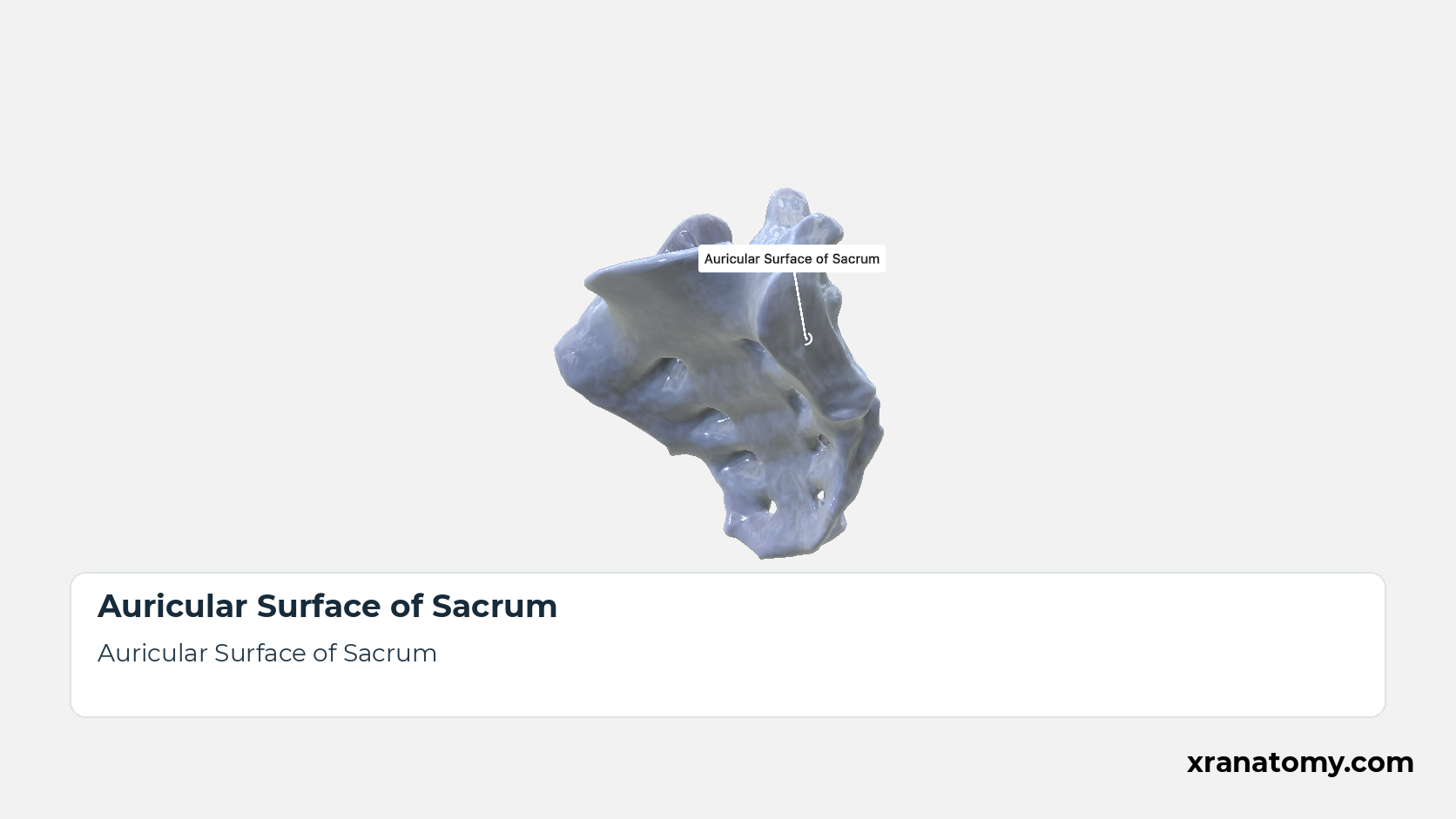
The auricular surface is the ear-shaped articular area on the upper part of the lateral surface of the sacrum, covered with cartilage in life, that meets the matching surface of the ilium at the sacroiliac joint. [2] [3] [1]
Illustration by XR Anatomy.
Sacral Tuberosity
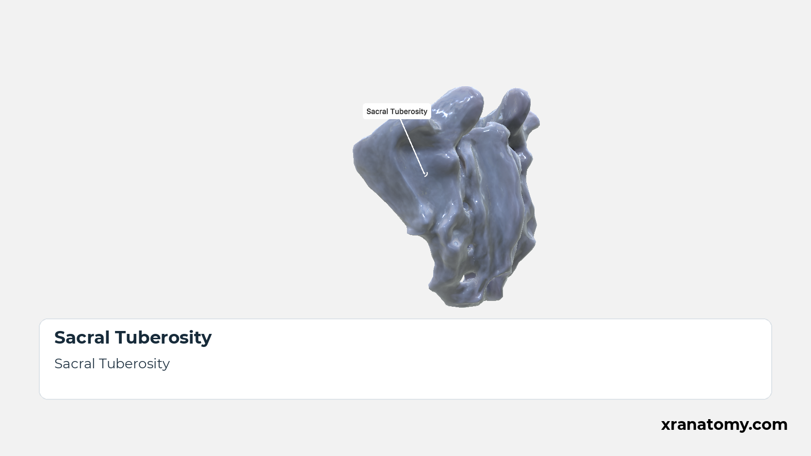
The sacral tuberosity is the rough, pitted area behind the auricular surface; the interosseous and posterior sacroiliac ligaments attach to it. [2] [3] [1]
Illustration by XR Anatomy.
Sacrum
The sacrum (os sacrum) is the large triangular bone that closes the back of the pelvis, wedged between the two hip bones. It is usually formed by the fusion of five sacral vertebrae, so its broad base faces upwards towards the spine and its narrow apex downwards towards the coccyx; pelvic, dorsal, and lateral surfaces surround the sacral canal at its core.Show less
Illustration by XR Anatomy.
Base of Sacrum
The sacral base is the broad superior end of the bone, on which the fifth lumbar vertebra rests. The upper surface of the S1 body forms its centre, the alae extend it to either side, and the sacral canal opens behind the body between the superior articular processes; the Superior View shows these from above. [2] [3] [1]Show less
Illustration by XR Anatomy.
Pelvic Surface of Sacrum
The pelvic surface faces forwards and downwards into the pelvic cavity and is smooth and concave in both directions. Its markings are the four transverse ridges and, at the lateral ends of the ridges, the four pairs of anterior sacral foramina, the pelvic openings of the intervertebral foramina that lead out of the sacral canal. [2] [3] [4]Show less
Illustration by XR Anatomy.
Dorsal Surface of Sacrum
The dorsal surface is convex, narrower than the pelvic surface, and rough, because the back muscles and the sacroiliac ligaments attach to it. It is built from the fused posterior elements of the five vertebrae, which form five longitudinal crests: the median crest in the midline, an intermediate crest on each side, and the lateral sacral crests at the edges, with the four pairs of posterior sacral foramina between the intermediate and lateral crests. Between the median and intermediate crests the surface is made of the fused laminae; the lowest laminae fail to meet, so the canal opens below at the sacral hiatus, where the intermediate crests end as the sacral horns flanking the hiatus. [2] [3] [4]Show less
Illustration by XR Anatomy.
Apex of Sacrum
The sacral apex is the narrow inferior end, the lower surface of the S5 body. Its small oval facet meets the base of the coccyx at the sacrococcygeal joint. [2] [3] [1]
Illustration by XR Anatomy.
From the side, the lateral part is broad above and narrows below. Its upper part shows the two markings of the sacroiliac joint: in front, the ear-shaped auricular surface that articulates with the hip bone, and behind it the rough sacral tuberosity for the ligaments that hold the joint together.
In depth · 3 sections · 1 min read3 more sections on this part3 sections open — reading In depth- Lateral Part of Sacrum
- Auricular Surface of Sacrum
- Sacral Tuberosity
Lateral Part of Sacrum
The lateral part is the mass of bone on each side of the fused bodies, formed by the fused transverse processes and costal elements of the sacral vertebrae. Broad above, where it carries the auricular surface and the sacral tuberosity, it tapers to a thin edge below.
Auricular Surface of Sacrum
The auricular surface is the ear-shaped articular area on the upper part of the lateral surface of the sacrum, covered with cartilage in life, that meets the matching surface of the ilium at the sacroiliac joint.
Sacral Tuberosity
The sacral tuberosity is the rough, pitted area behind the auricular surface; the interosseous and posterior sacroiliac ligaments attach to it.
Practice · Active recall · 3 questions Recall: Anatomical Views
In depth · 1 question Recall: Promontory
The projecting anterior margin of the S1 body is the .
promontory
The base contains the upper surface of the S1 body, whose projecting anterior margin is the promontory. The alae spread laterally, and the superior opening of the sacral canal lies behind the S1 body.
Review this sectionHow well did you know it? · press 1–4
Nice work — revisit in a few days.
Behind the S1 body lies the superior opening of the .
The base contains the upper surface of the S1 body, whose projecting anterior margin is the promontory. The alae spread laterally, and the superior opening of the sacral canal lies behind the S1 body.
Review this sectionHow well did you know it? · press 1–4
Nice work — revisit in a few days.
What rough area lies posterior to the auricular surface?
sacral tuberosity
Each lateral part bears an ear-shaped auricular surface. The rough sacral tuberosity lies posterior to it.
Review this sectionHow well did you know it? · press 1–4
Bibliography
Every claim on this page is sourced against the standard anatomical references.
- FIPAT. Terminologia Anatomica, 2nd edition, Part 2: Bones. https://cdn.dal.ca/content/dam/dalhousie/pdf/library/FIPAT/TA2/FIPAT-TA2-Part-2.pdf
- Gray H. Anatomy of the Human Body. 21st ed. Philadelphia: Lea & Febiger; 1924.
- Drake RL, Vogl AW, Mitchell AWM. Gray's Anatomy for Students. Elsevier.
- Betts JG, et al. Anatomy and Physiology 2e. OpenStax; Rice University.
