Anatomy
Heart wall and myocardium
The heart wall comprises three layers: from outside in, the epicardium, the myocardium and the endocardium, which lines the chambers. The myocardium is its thick cardiac-muscle layer.
The heart wall brings together three kinds of tissue: a covering on the outside, muscle in the middle and a lining on the inside. These layers have different roles. Their arrangement is shared by the chambers, but the amount of muscle varies according to the work each chamber performs.
Layers and chamber thickness
Three wall layers
Moving from the outside of the heart towards a chamber cavity, the three layers are the epicardium, myocardium and endocardium. The outer epicardium forms the visceral serous covering. On the inner surface, the endocardium lines the chambers and extends over the valves. The layers therefore occupy different positions and perform different functions, even when a gross anatomical model does not show their boundaries separately.
The muscle that drives blood forwards
Between the outer covering and inner lining lies the myocardium, the layer made of cardiac muscle. When this muscle contracts, it generates the force that moves blood through the heart. In the ventricles, that force propels blood onwards into the pulmonary and systemic circulations.
Why wall thickness differs between chambers
The thickness of the muscle reflects the demands placed on each chamber. The atrial walls are thinner than the ventricular walls. The difference also continues between the two ventricles: the left ventricular myocardium is thicker than the right. This is because the left ventricle works against the greater resistance of the systemic circulation, while the right pumps into the lower-resistance pulmonary circulation.
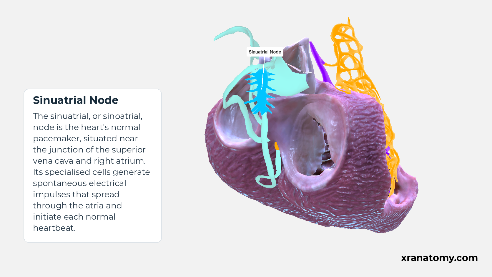
Sinuatrial Node
The sinuatrial, or sinoatrial, node is the heart's normal pacemaker, situated near the junction of the superior vena cava and right atrium. Its specialised cells generate spontaneous electrical impulses that spread through the atria and initiate each normal heartbeat.Show less
Study Vortex and fibre arrangement in this fixed image. Use the thumbnails to move between the 2D views; each view highlights one structure. The image does not rotate.
Illustration by XR Anatomy.
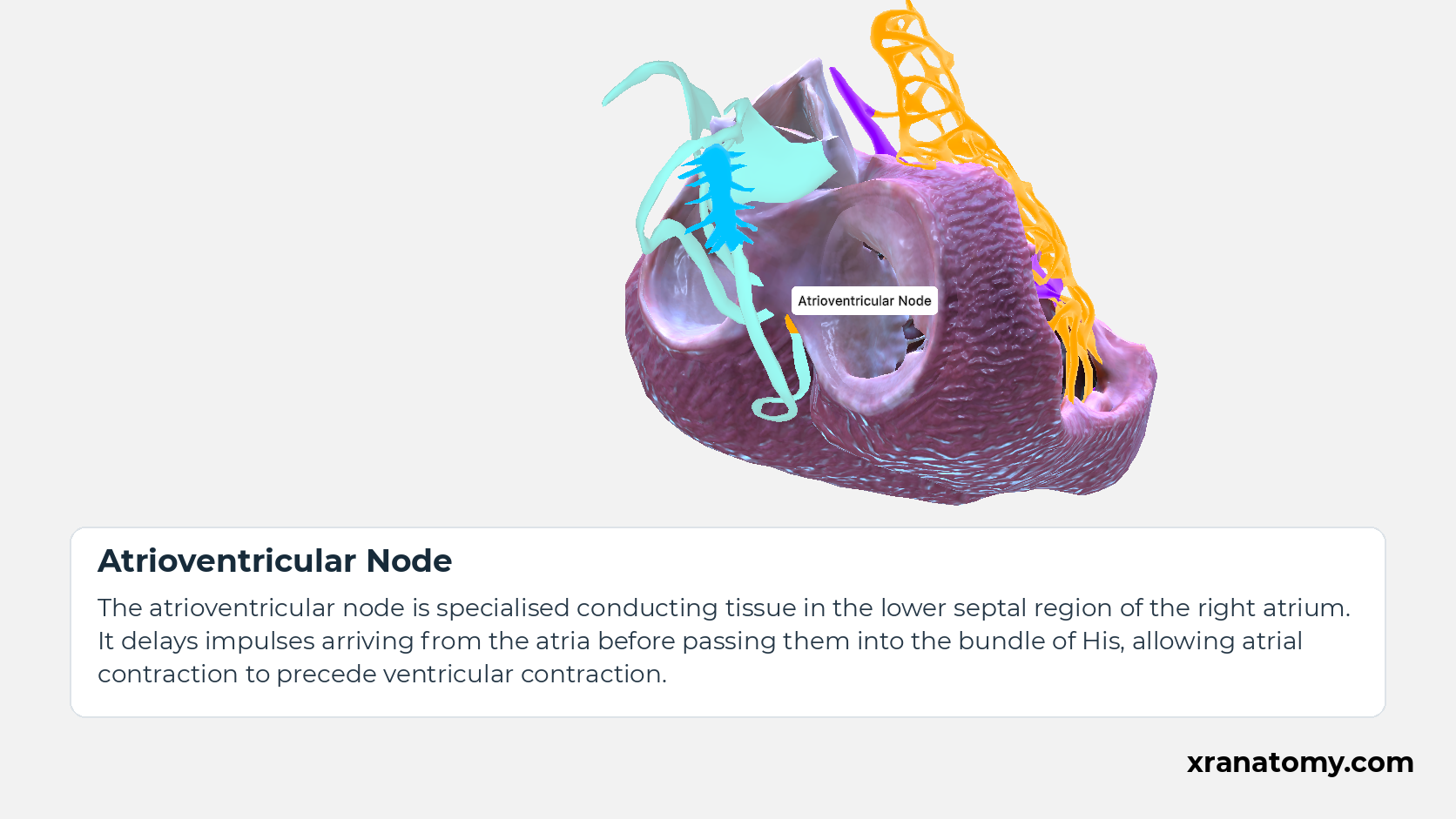
Atrioventricular Node
The atrioventricular node is specialised conducting tissue in the lower septal region of the right atrium. It delays impulses arriving from the atria before passing them into the bundle of His, allowing atrial contraction to precede ventricular contraction.Show less
Illustration by XR Anatomy.
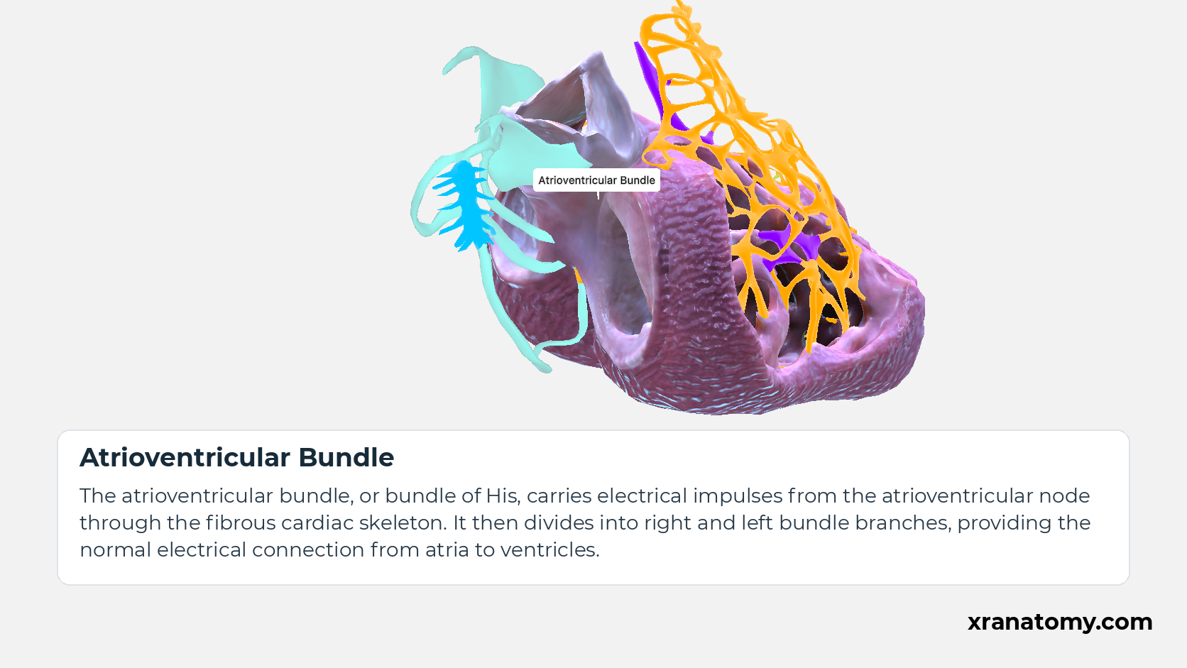
Atrioventricular Bundle
The atrioventricular bundle, or bundle of His, carries electrical impulses from the atrioventricular node through the fibrous cardiac skeleton. It then divides into right and left bundle branches, providing the normal electrical connection from atria to ventricles.Show less
Illustration by XR Anatomy.
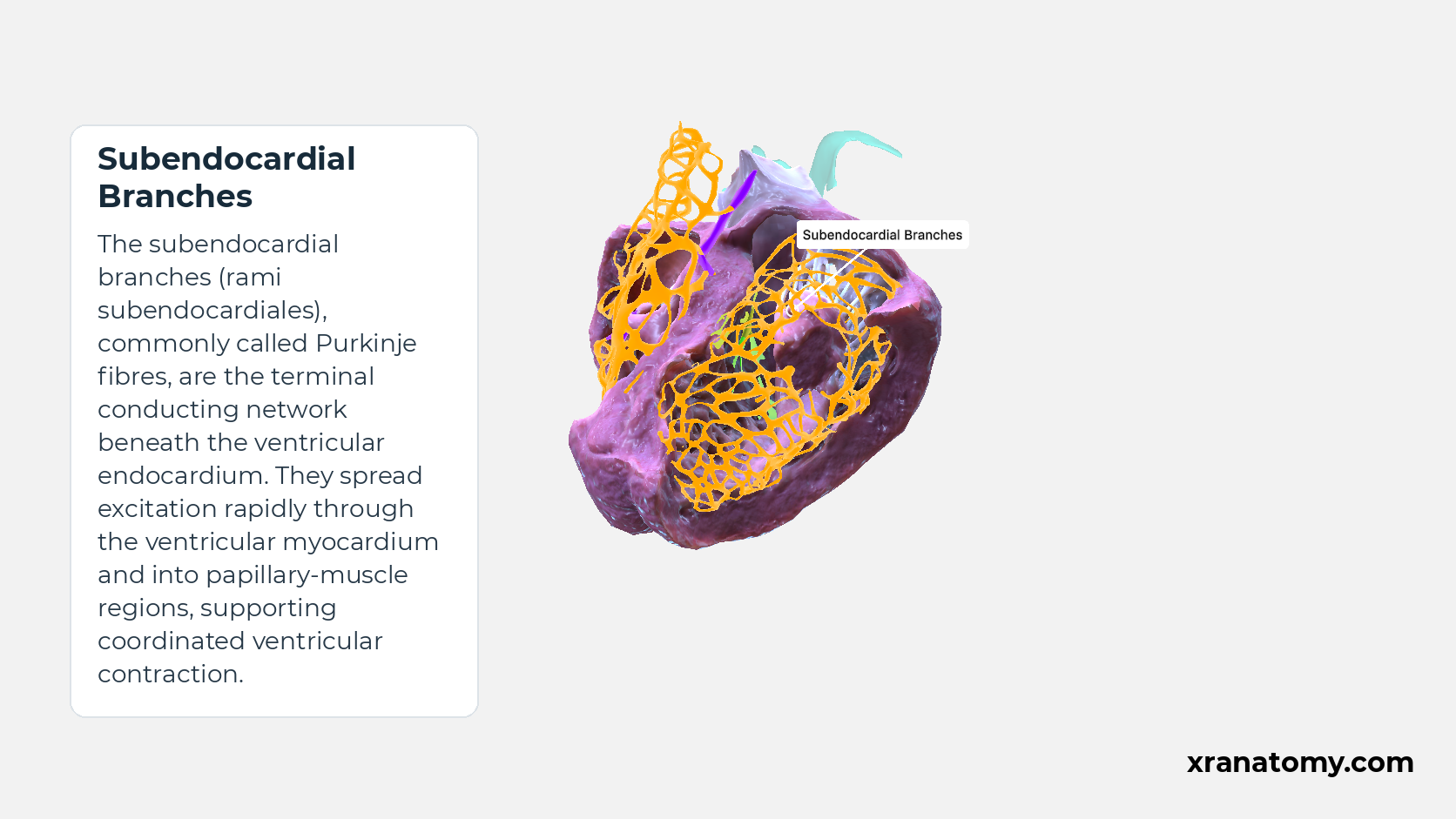
Subendocardial Branches
The subendocardial branches, commonly called Purkinje fibres, form the terminal conducting network beneath the ventricular endocardium. They distribute electrical impulses rapidly to the ventricular muscle and papillary-muscle regions, helping coordinate ventricular contraction.Show less
Illustration by XR Anatomy.
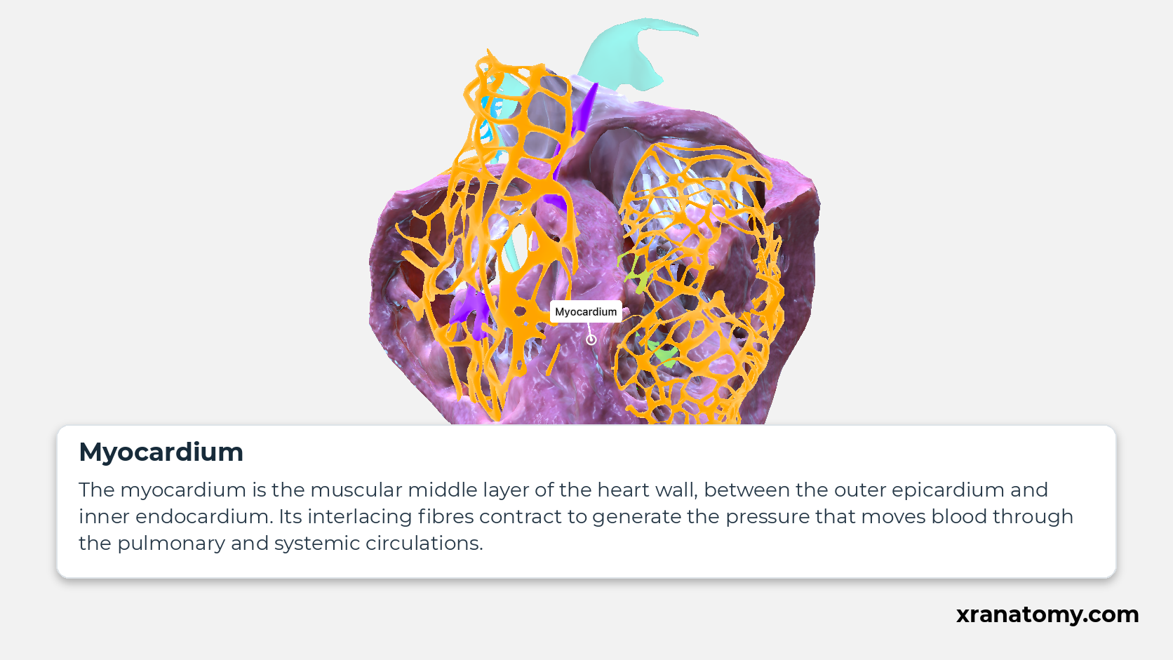
Myocardium
The myocardium is the muscular middle layer of the heart wall, between the outer epicardium and inner endocardium. Its interlacing fibres contract to generate the pressure that moves blood through the pulmonary and systemic circulations.
Illustration by XR Anatomy.
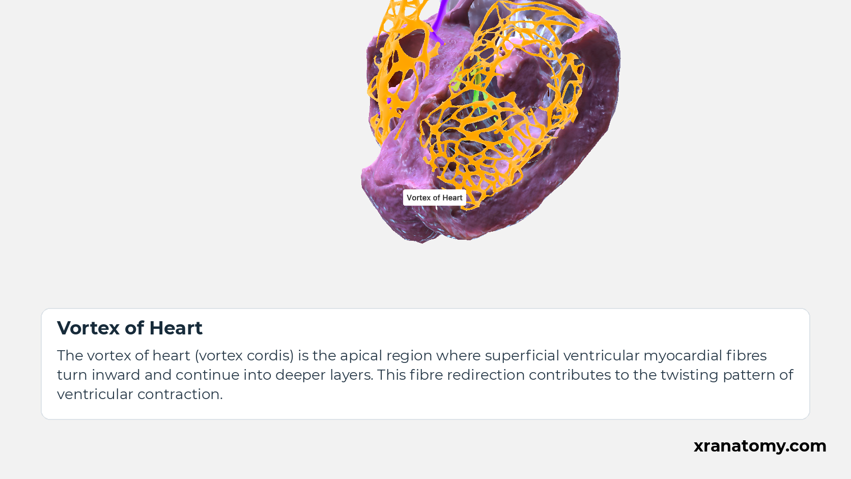
Vortex of Heart
The apical region where superficial ventricular myocardial fibres turn inward and continue into deeper layers, contributing to the twisting pattern of ventricular contraction.
Illustration by XR Anatomy.
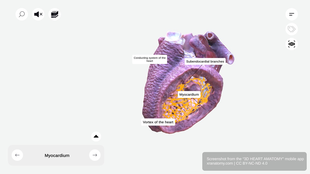 ↺ drag to rotate · scroll to zoom
Vortex and fibre arrangement
Vortex and fibre arrangement
Rotate the model to locate the ventricular myocardium and apical vortex.
Sourced from 3D Heart Anatomy.
In depth · 1 min read Vortex and fibre arrangement
Vortex of Heart
Ventricular muscle fibres form an interlacing, spiralling arrangement. At the vortex of heart (vortex cordis), superficial fibres turn inwards and continue into deeper layers. This redirection contributes to the twisting movement of ventricular contraction.
