Anatomy
Mandible
The mandible (mandibula) is the single movable lower-jaw bone and the largest and strongest bone of the face. Its curved body holds the lower teeth, and two upright branches (rami) rise from the posterior ends of the body.
Articulations
Mandible
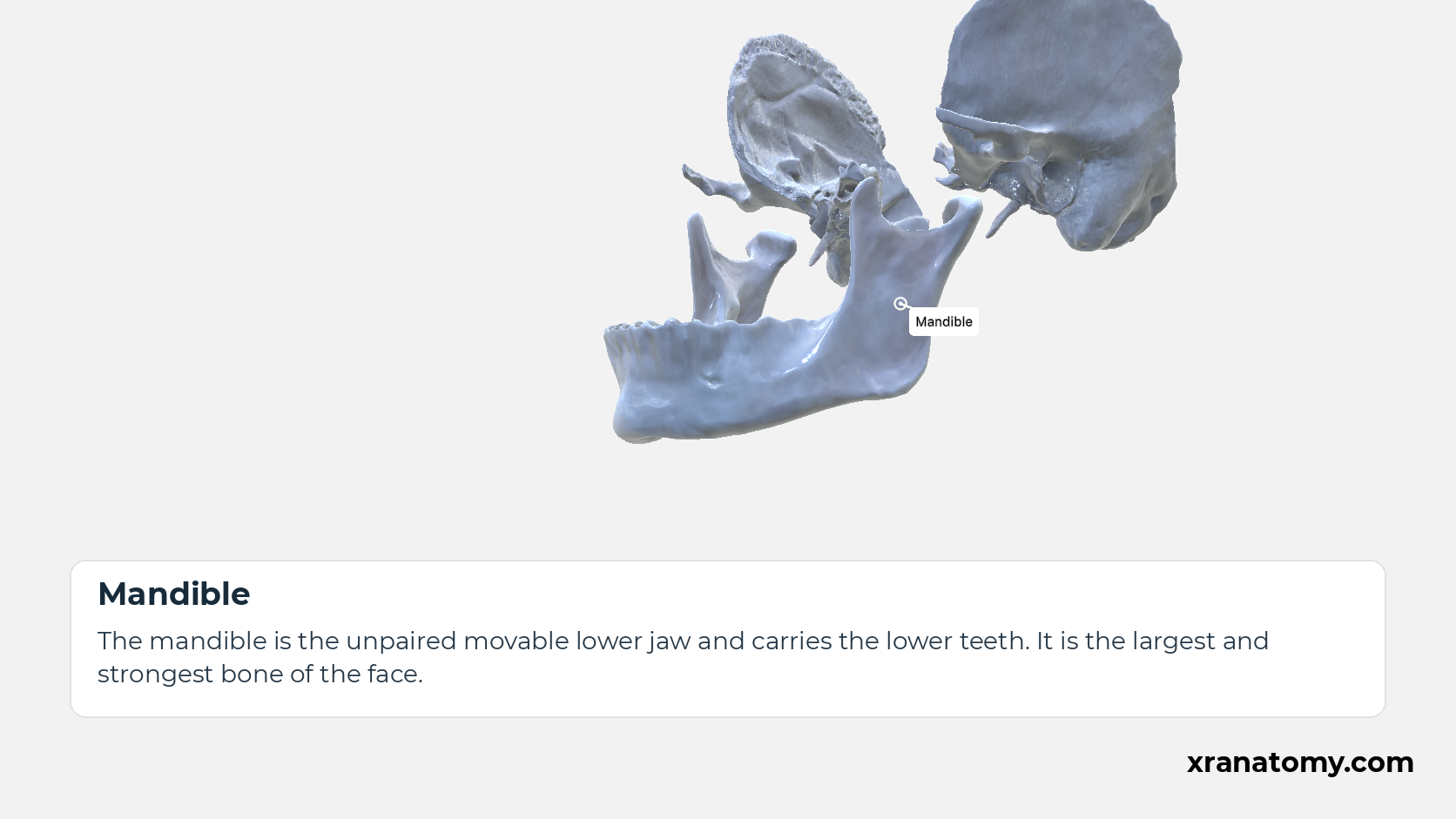
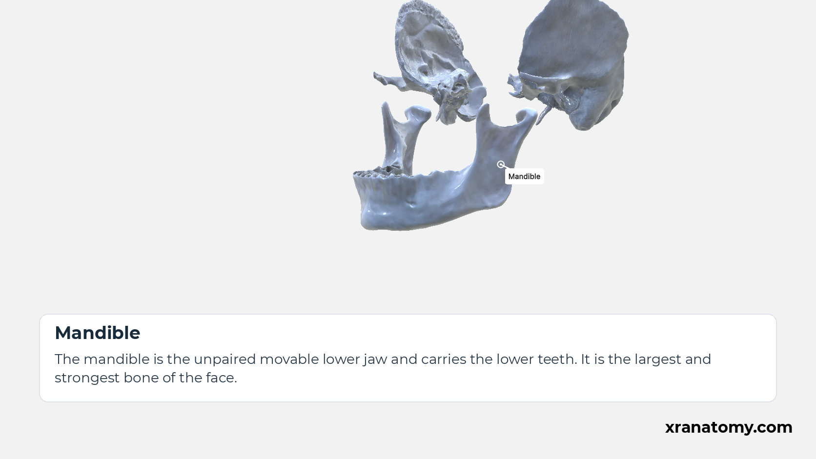
The mandible is the unpaired movable lower jaw and carries the lower teeth. It is the largest and strongest bone of the face.
Study Mandibular Articulations in this fixed image. Use the thumbnails to move between the 2D views; each view highlights one structure. The image does not rotate.
Illustration by XR Anatomy.
Temporal Bone
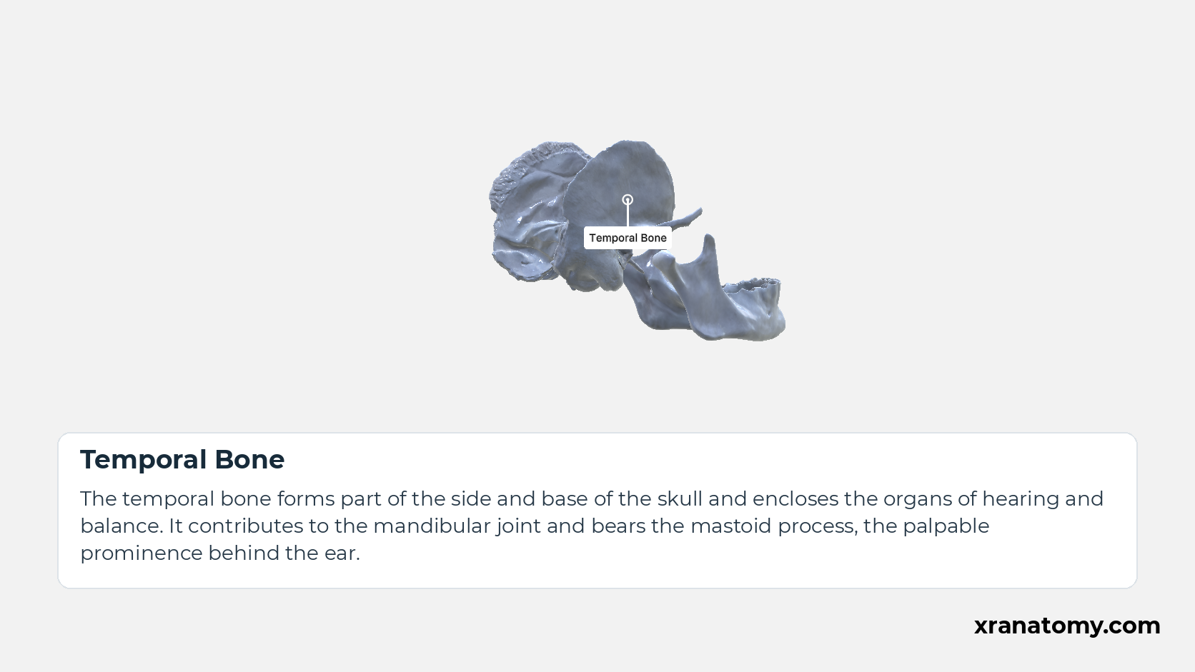
The temporal bone forms part of the side and base of the skull and encloses the organs of hearing and balance. It contributes to the mandibular joint and bears the mastoid process, the palpable prominence behind the ear.
Illustration by XR Anatomy.
The right and left mandibular condyles form temporomandibular joints with the temporal bones. Each condyle meets the underside of its joint's articular disc.
Practice · Active recall · 1 question Recall: Articulations
The mandibular condyles form temporomandibular joints with the bones.
temporal
The right and left mandibular condyles form temporomandibular joints with the temporal bones. Each condyle meets the underside of its joint's articular disc.
Review this sectionHow well did you know it? · press 1–4
Nice work — revisit in a few days.
Overview
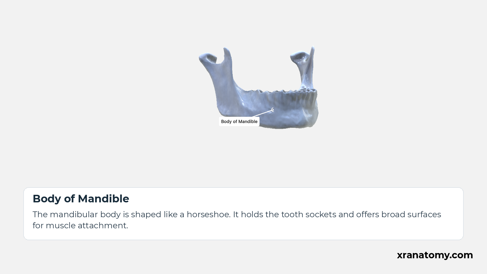
Body of Mandible
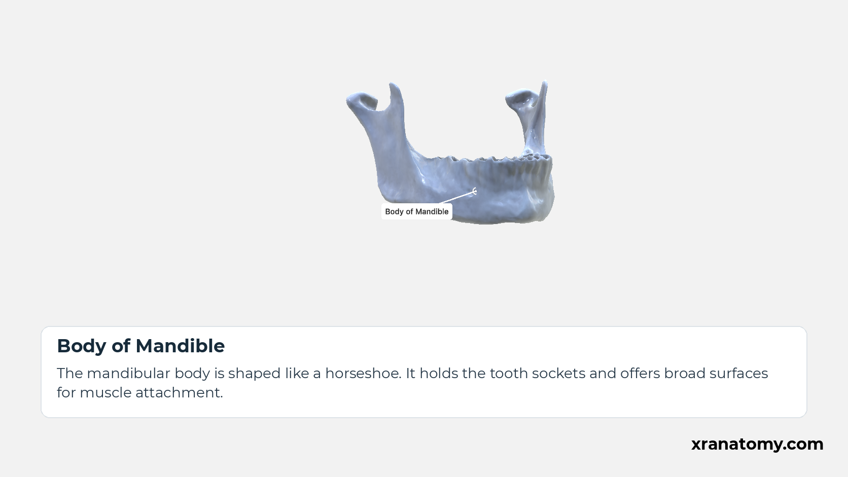
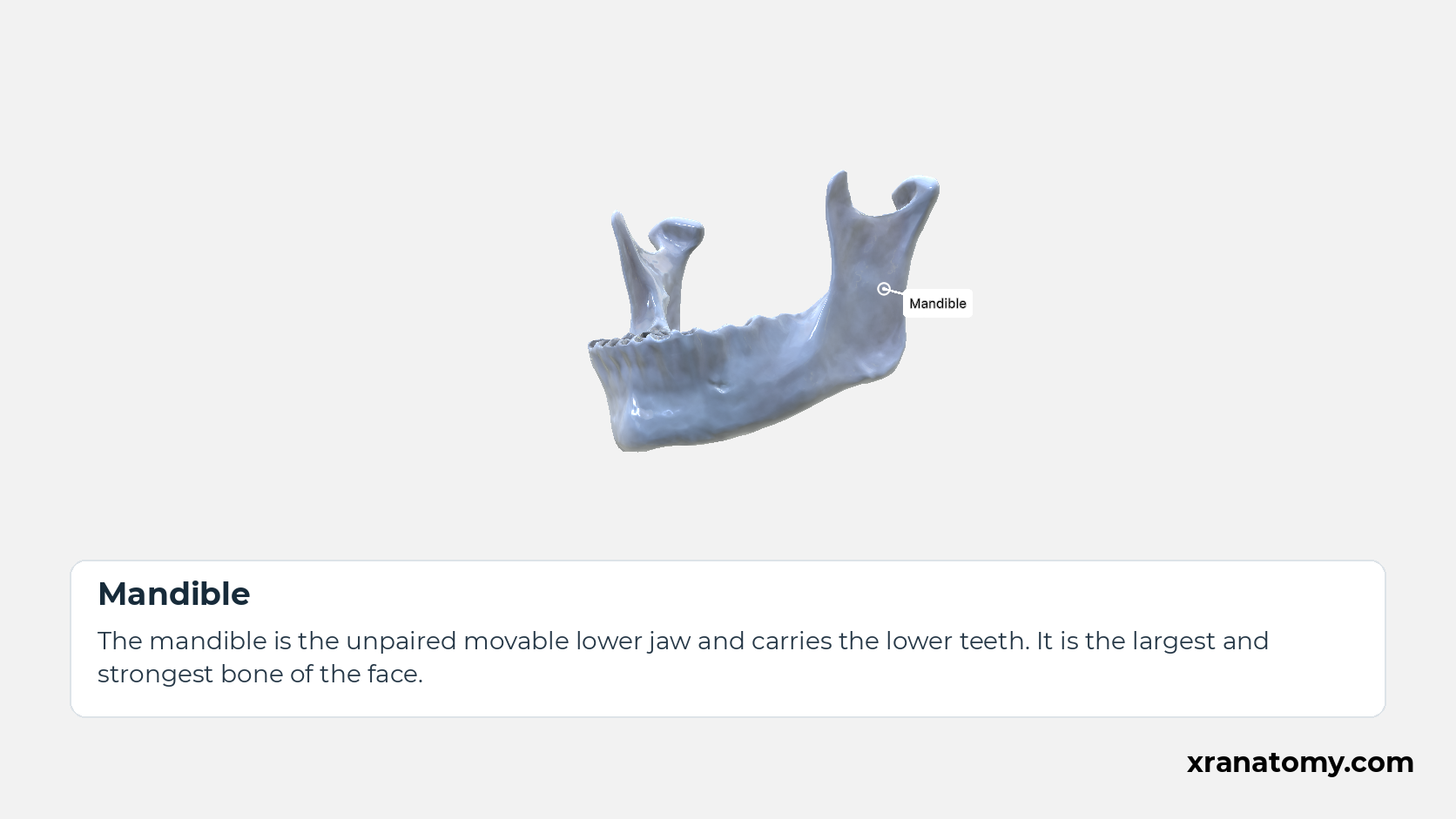
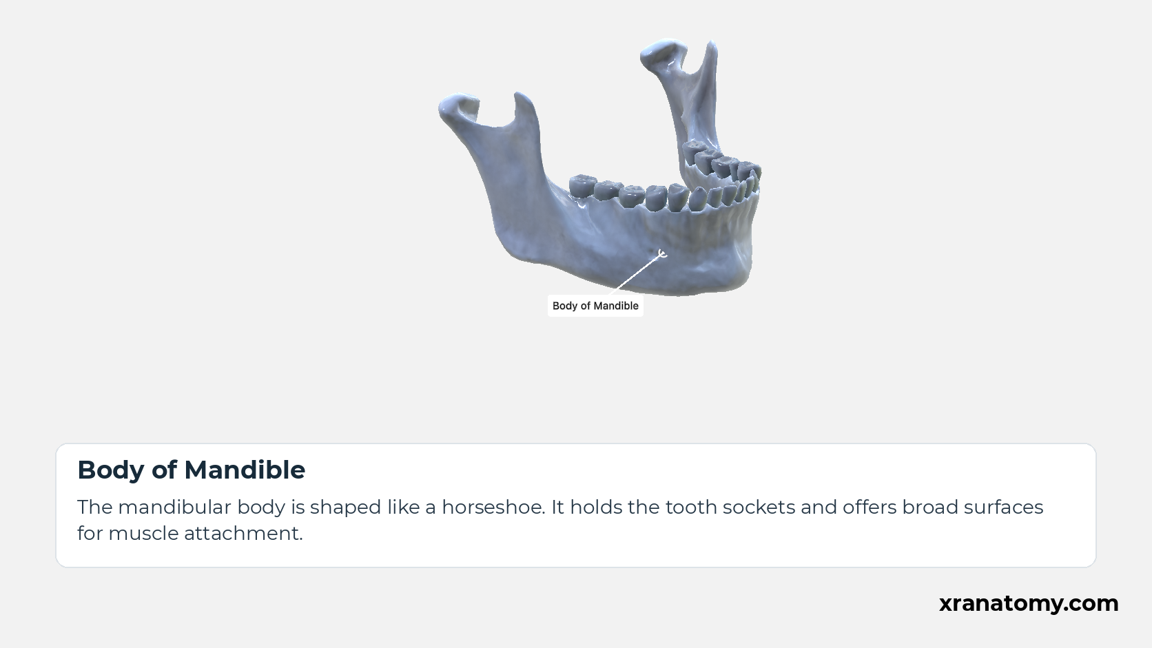
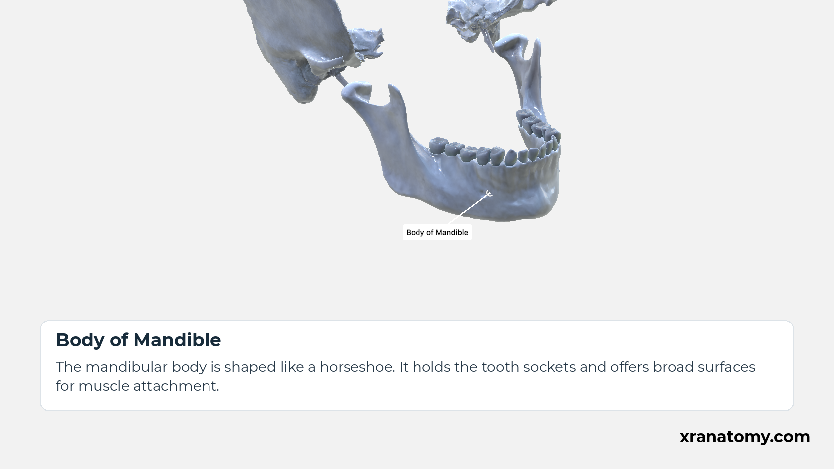
The mandibular body is shaped like a horseshoe. It holds the tooth sockets and offers broad surfaces for muscle attachment.
Study Main Parts of the Mandible in this fixed image. Use the thumbnails to move between the 2D views; each view highlights one structure. The image does not rotate.
Illustration by XR Anatomy.
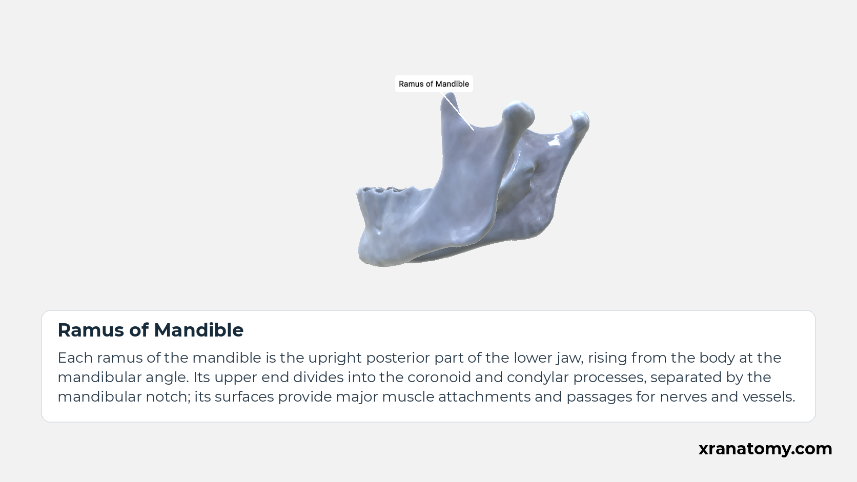
Ramus of Mandible
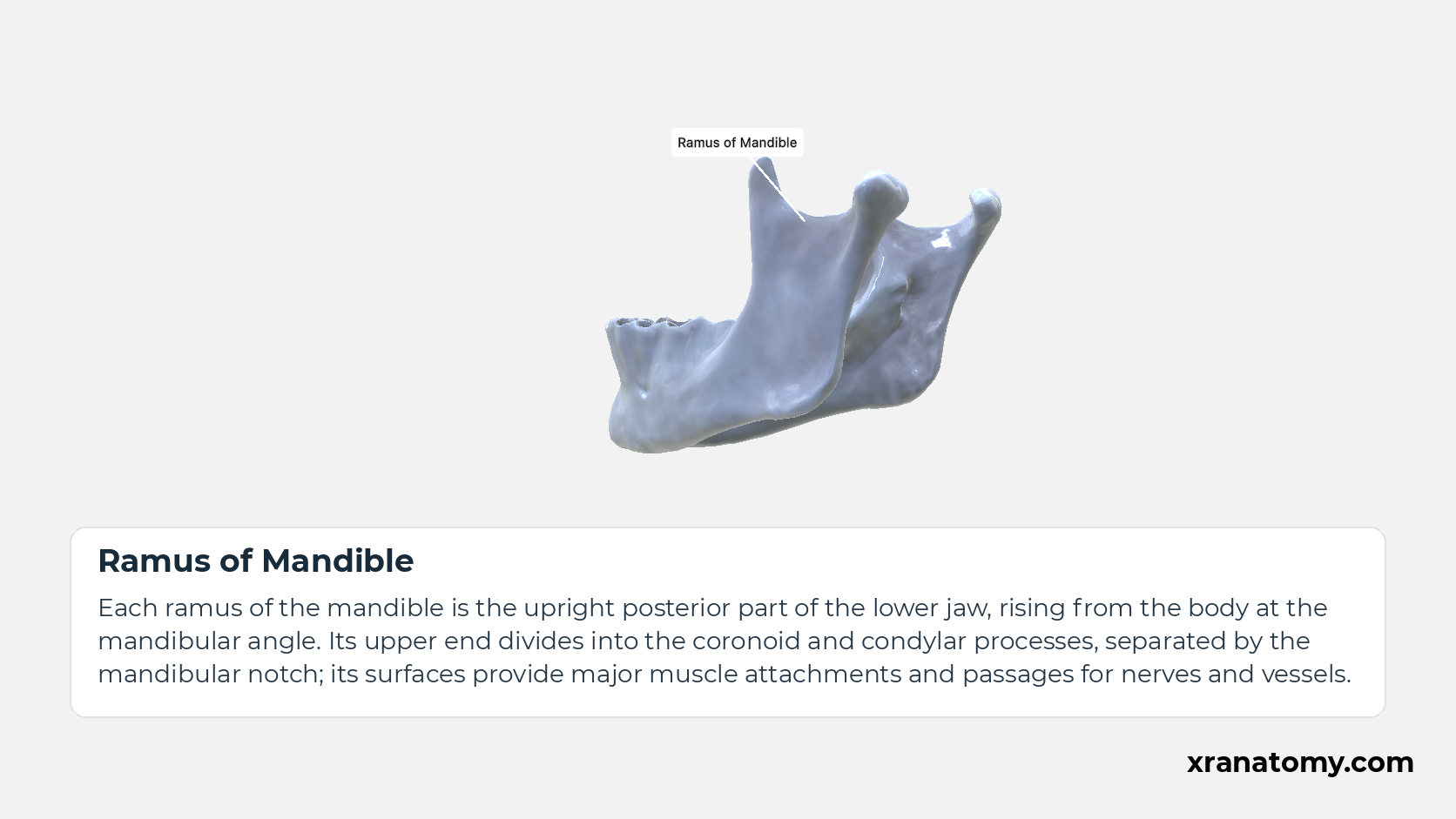
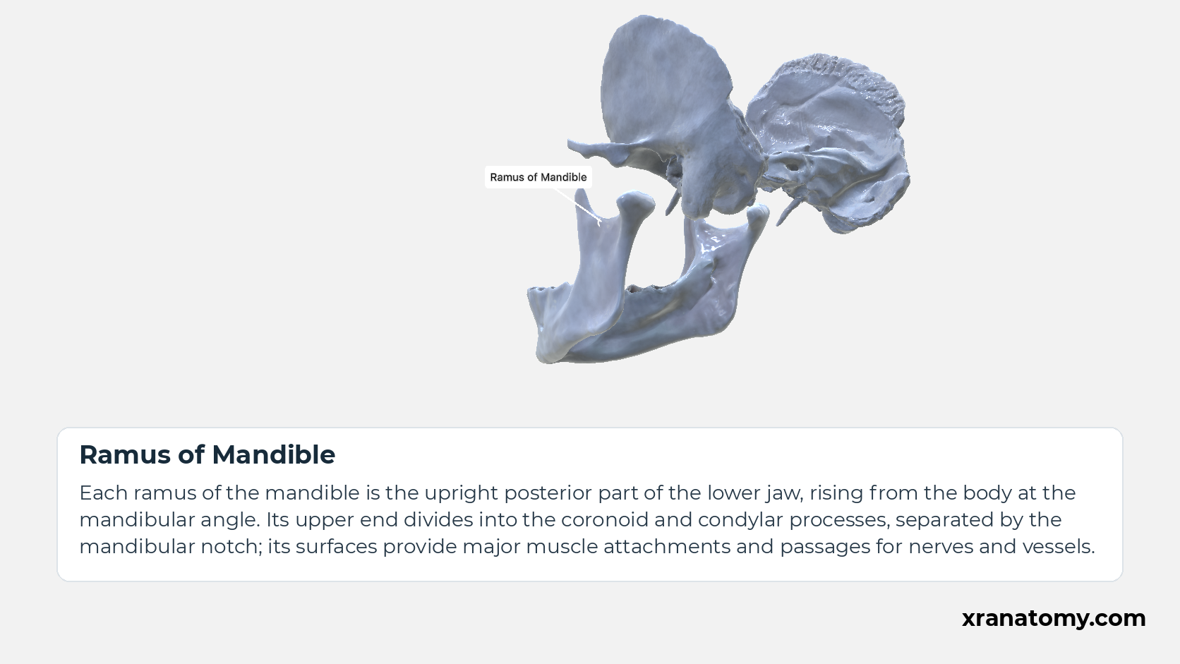
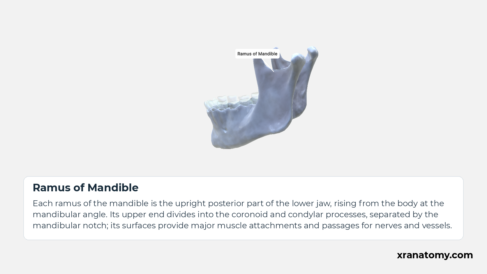
Each ramus of the mandible is the upright posterior part of the lower jaw, rising from the body at the mandibular angle. Its upper end divides into the coronoid and condylar processes, separated by the mandibular notch; its surfaces provide major muscle attachments and passages for nerves and vessels.Show less
Illustration by XR Anatomy.
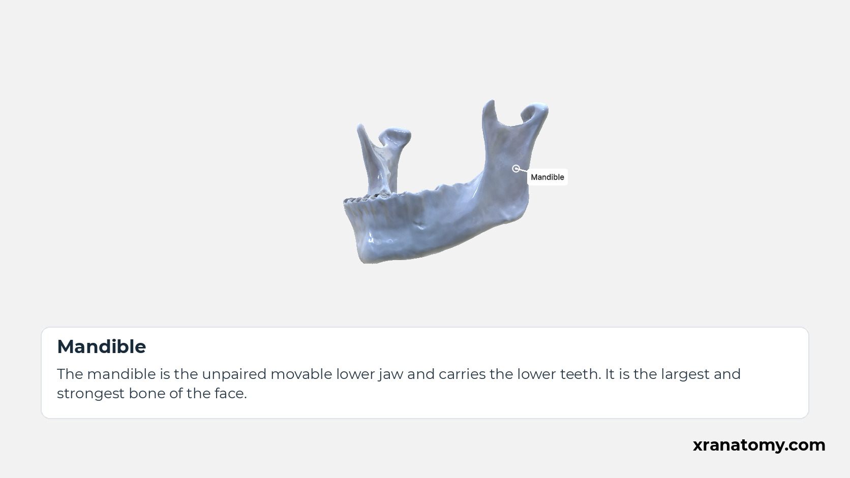
Mandible
The mandible is the unpaired movable lower jaw and carries the lower teeth. It is the largest and strongest bone of the face.
Illustration by XR Anatomy.
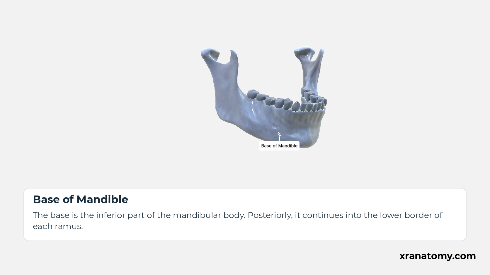
Base of Mandible
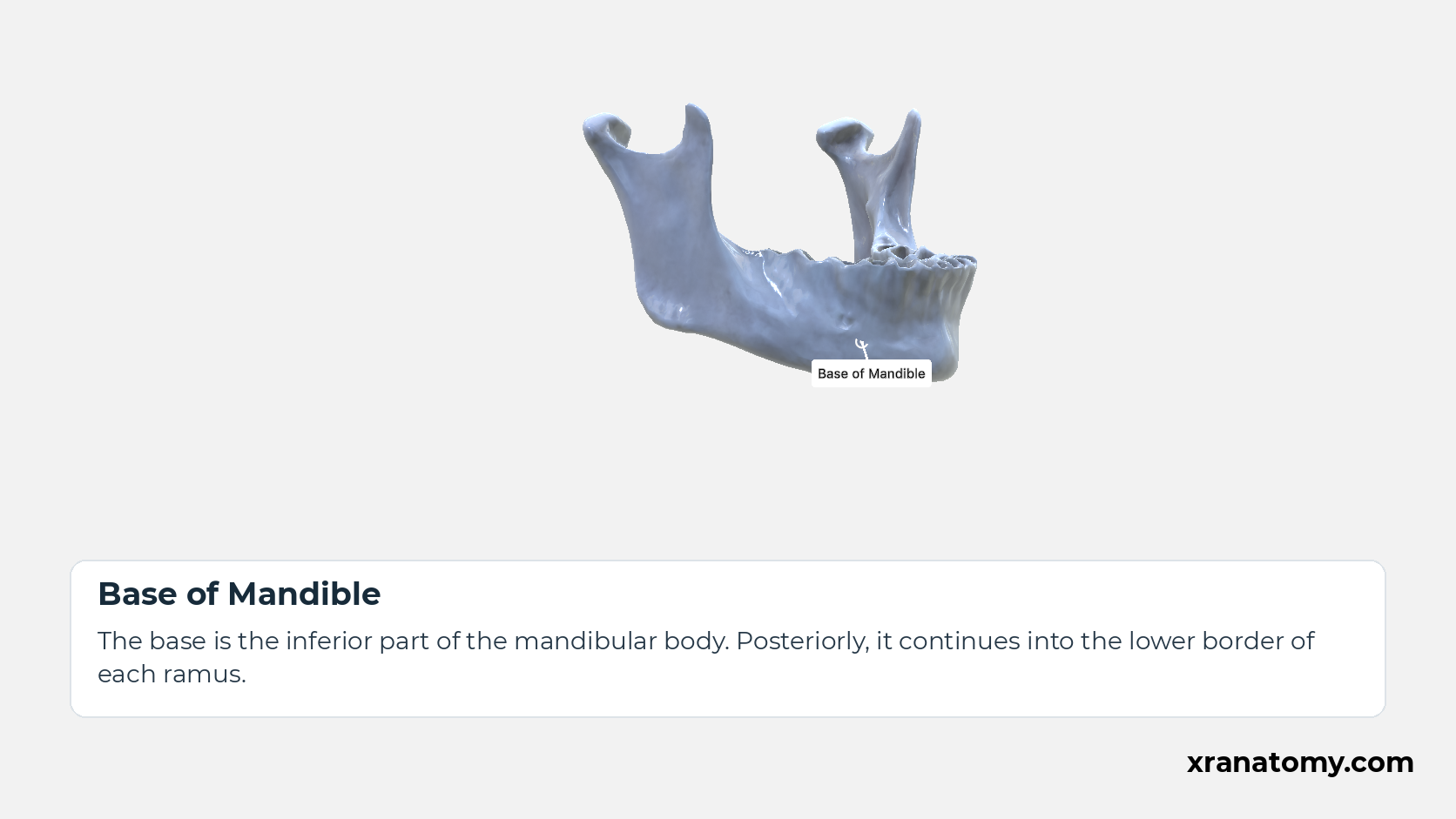
The base is the inferior part of the mandibular body. Posteriorly, it continues into the lower border of each ramus.
Illustration by XR Anatomy.
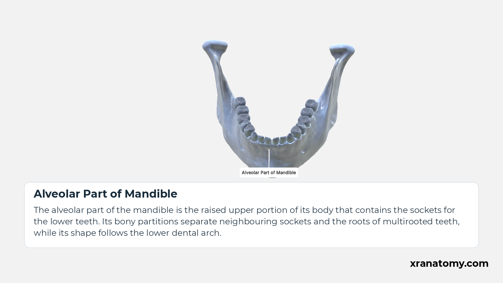
Alveolar Part of Mandible
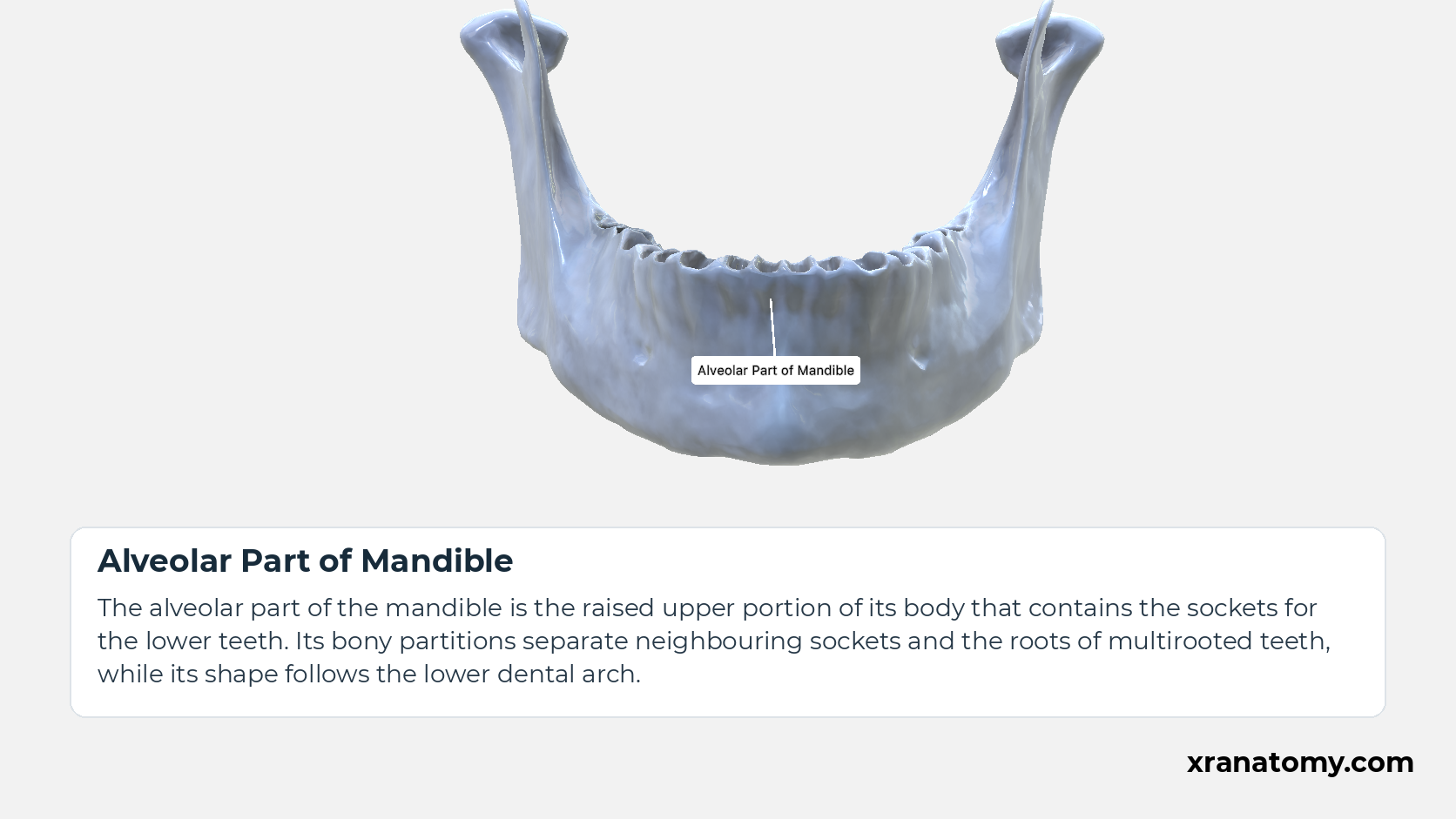
The alveolar part of the mandible is the raised upper portion of its body that contains the sockets for the lower teeth. Its bony partitions separate neighbouring sockets and the roots of multirooted teeth, while its shape follows the lower dental arch.Show less
Illustration by XR Anatomy.
Coronoid Process of Mandible
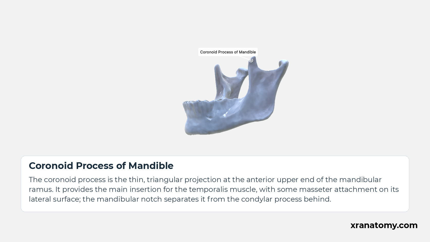
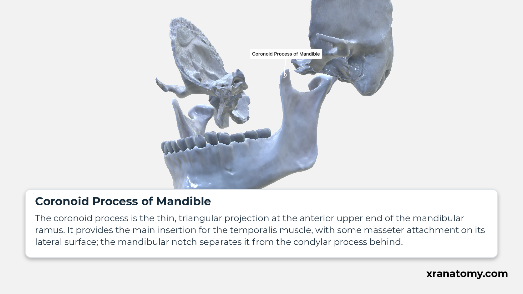
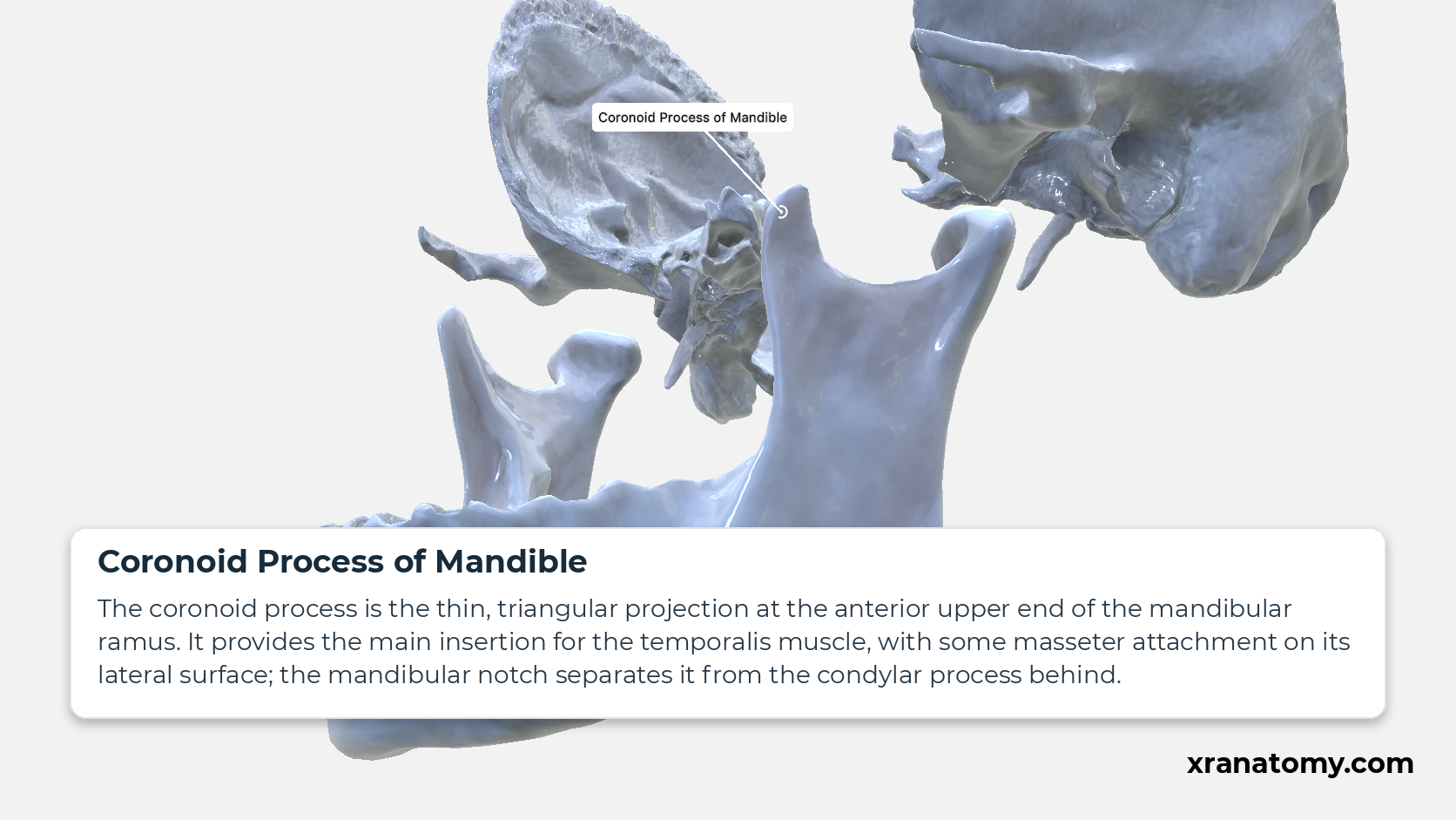
The coronoid process is the thin, triangular projection at the anterior upper end of the mandibular ramus. It provides the main insertion for the temporalis muscle, with some masseter attachment on its lateral surface; the mandibular notch separates it from the condylar process behind.Show less
Illustration by XR Anatomy.
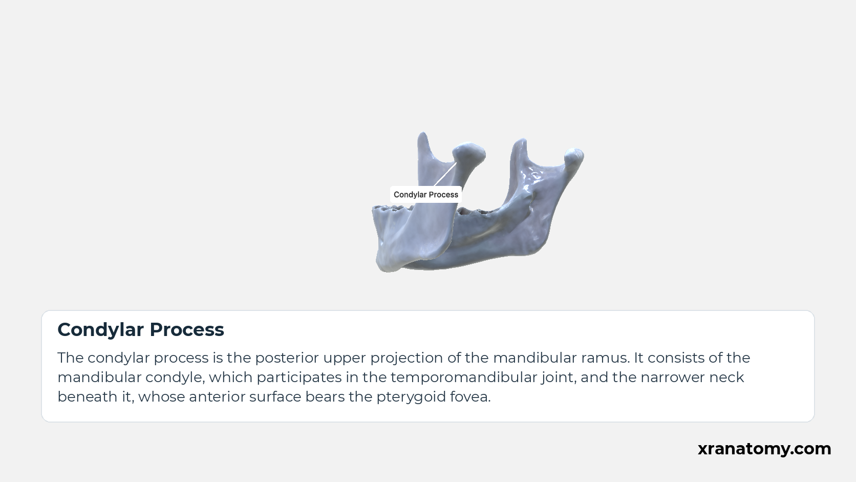
Condylar Process
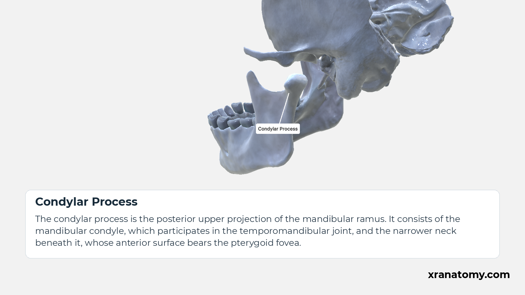
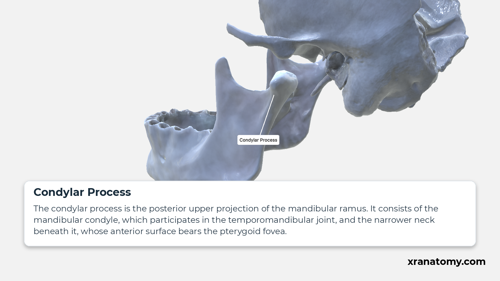
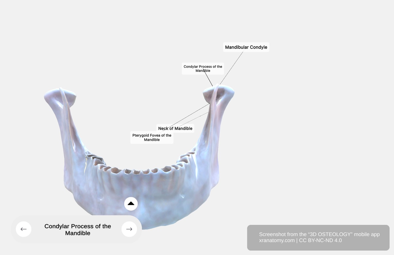
The condylar process is the posterior upper projection of the mandibular ramus. It consists of the mandibular condyle, which participates in the temporomandibular joint, and the narrower neck beneath it, whose anterior surface bears the pterygoid fovea.Show less
Illustration by XR Anatomy.
 ↺ drag to rotate · scroll to zoom
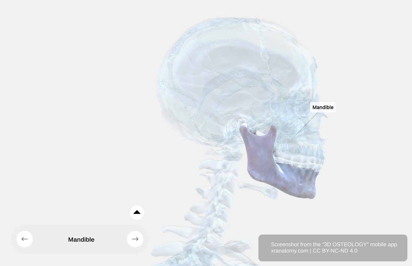
Main Parts of the Mandible
Main Parts of the Mandible
Drag or use the viewer controls to inspect the highlighted mandible in skull context.
Sourced from 3D Osteology.
| Branch | Location and role |
|---|---|
| Body of mandible | curved horizontal part; bears the lower teeth and chin landmarks |
| Ramus of mandible | paired ascending part; carries the coronoid and condylar processes |
Start with the body, its base and alveolar part; then follow the ramus to the coronoid and condylar processes.
Practice · Active recall · 1 question Recall: Overview
The body and paired rami form the mandible's main regions.
two
The body and paired rami form the mandible's two main regions.
Review this sectionHow well did you know it? · press 1–4
Nice work — revisit in a few days.
Body of Mandible
Body of Mandible
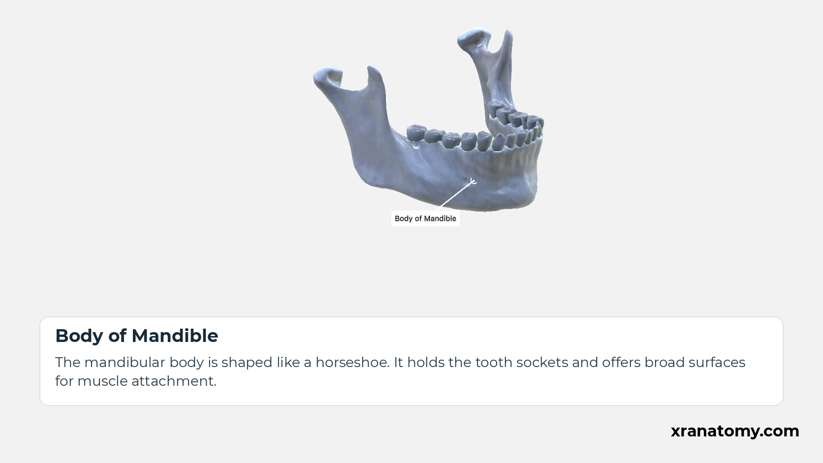
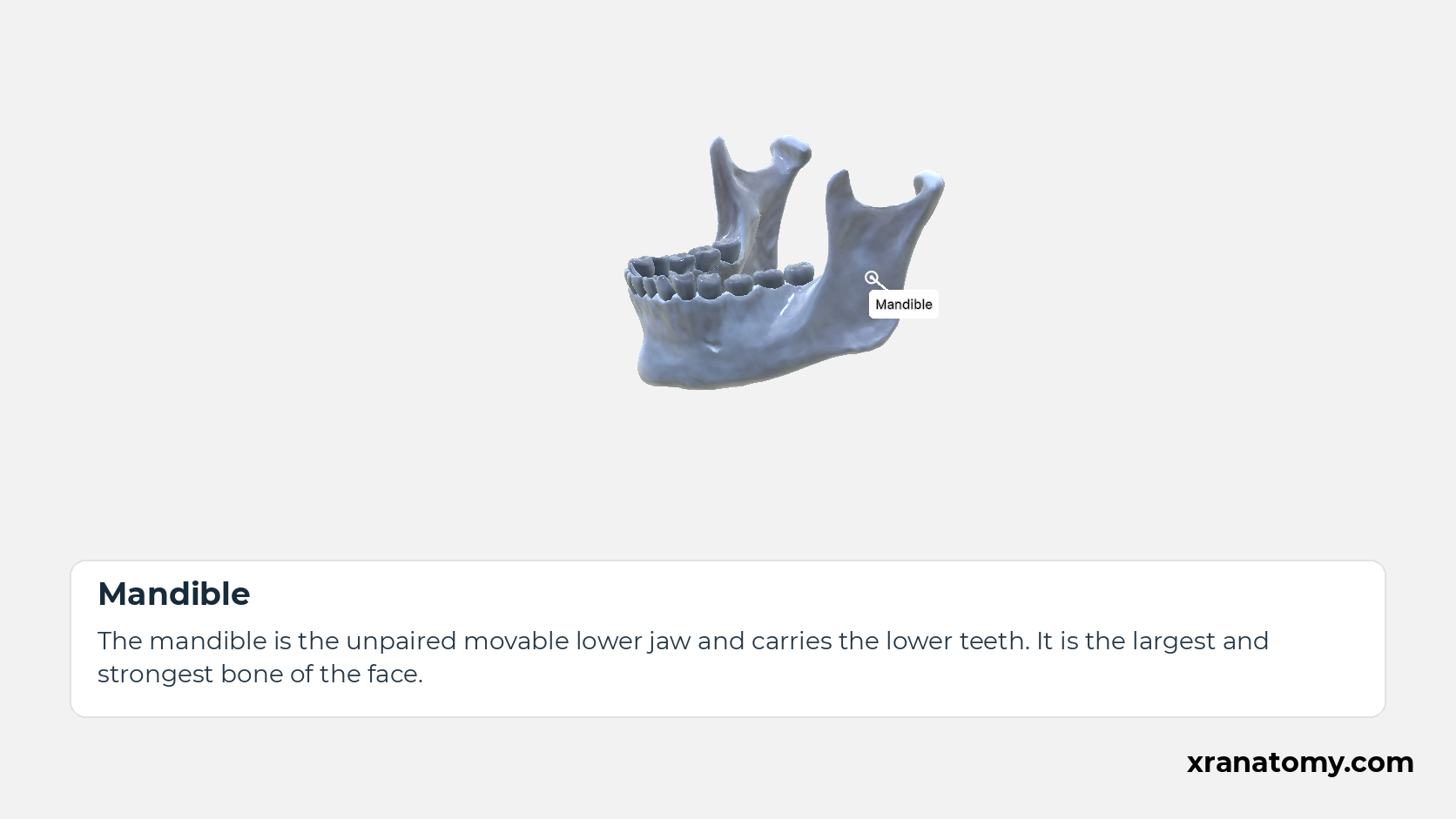
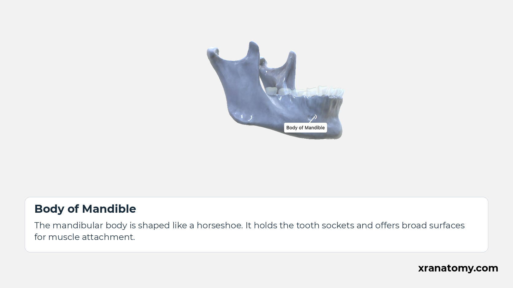
The mandibular body is shaped like a horseshoe. It holds the tooth sockets and offers broad surfaces for muscle attachment.
Study Body of Mandible in this fixed image. The image does not rotate.
Illustration by XR Anatomy.
Body of Mandible
The mandibular body is shaped like a horseshoe. It holds the tooth sockets and offers broad surfaces for muscle attachment.
Illustration by XR Anatomy.
Base of Mandible
The base is the inferior part of the mandibular body. Posteriorly, it continues into the lower border of each ramus.
Illustration by XR Anatomy.
Alveolar Part of Mandible
The alveolar part of the mandible is the raised upper portion of its body that contains the sockets for the lower teeth. Its bony partitions separate neighbouring sockets and the roots of multirooted teeth, while its shape follows the lower dental arch.Show less
Illustration by XR Anatomy.
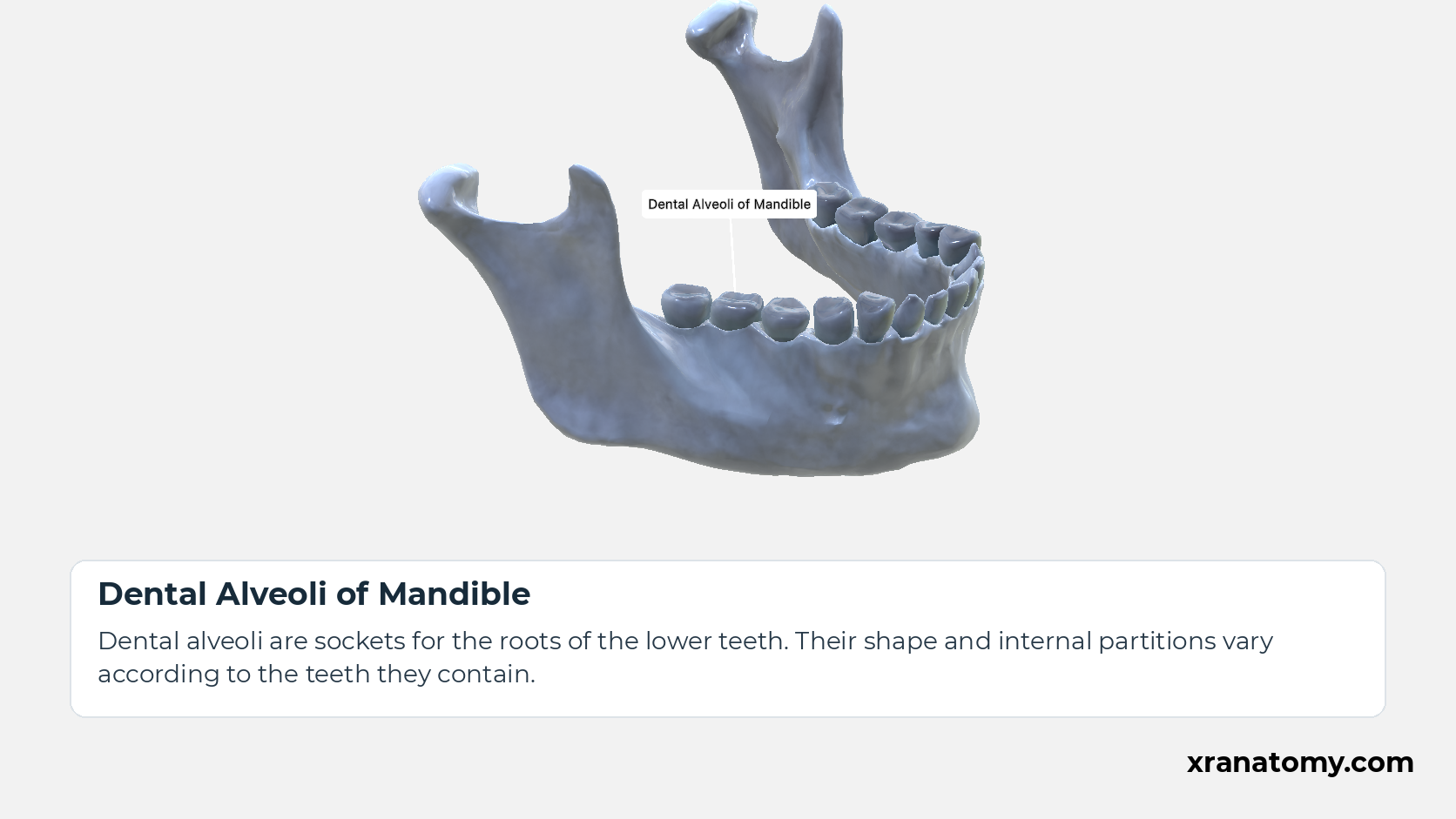
Dental Alveoli of Mandible
Dental alveoli are sockets for the roots of the lower teeth. Their shape and internal partitions vary according to the teeth they contain.
Illustration by XR Anatomy.
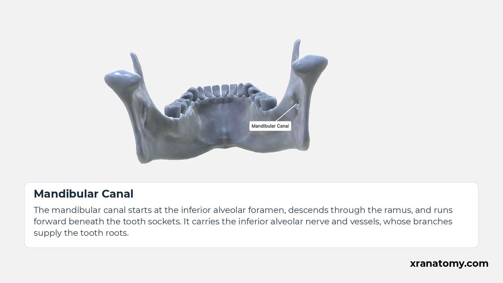
Mandibular Canal
The mandibular canal starts at the inferior alveolar foramen, descends through the ramus, and runs forward beneath the tooth sockets. It carries the inferior alveolar nerve and vessels, whose branches supply the tooth roots.
Illustration by XR Anatomy.
Mandible
The mandible is the unpaired movable lower jaw and carries the lower teeth. It is the largest and strongest bone of the face.
Illustration by XR Anatomy.
 ↺ drag to rotate · scroll to zoom
Body of the Mandible
Body of the Mandible
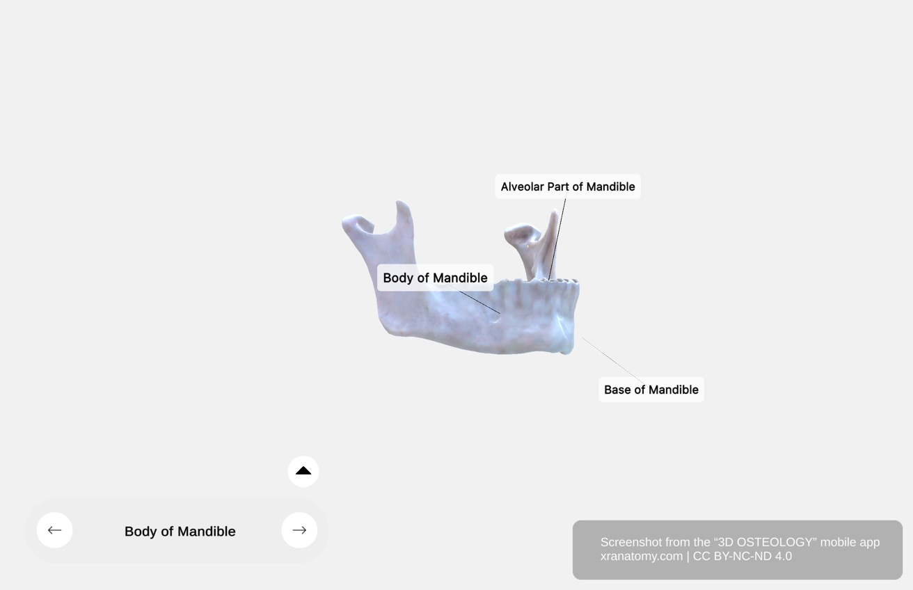
Drag or use the viewer controls to inspect the body of the mandible from different sides.
Sourced from 3D Osteology.
The mandibular body is shaped like a horseshoe. It holds the tooth sockets and offers broad surfaces for muscle attachment.
General Features of the Body of the Mandible
The body has a lower base and a raised alveolar part. Landmarks on its outer and inner surfaces relate to the chin, glands, muscles, teeth, and neurovascular pathways.
Subdivisions
The base is the lower part of the body, while the alveolar part is the raised upper part that bears the teeth.
Base of Mandible
Base of Mandible
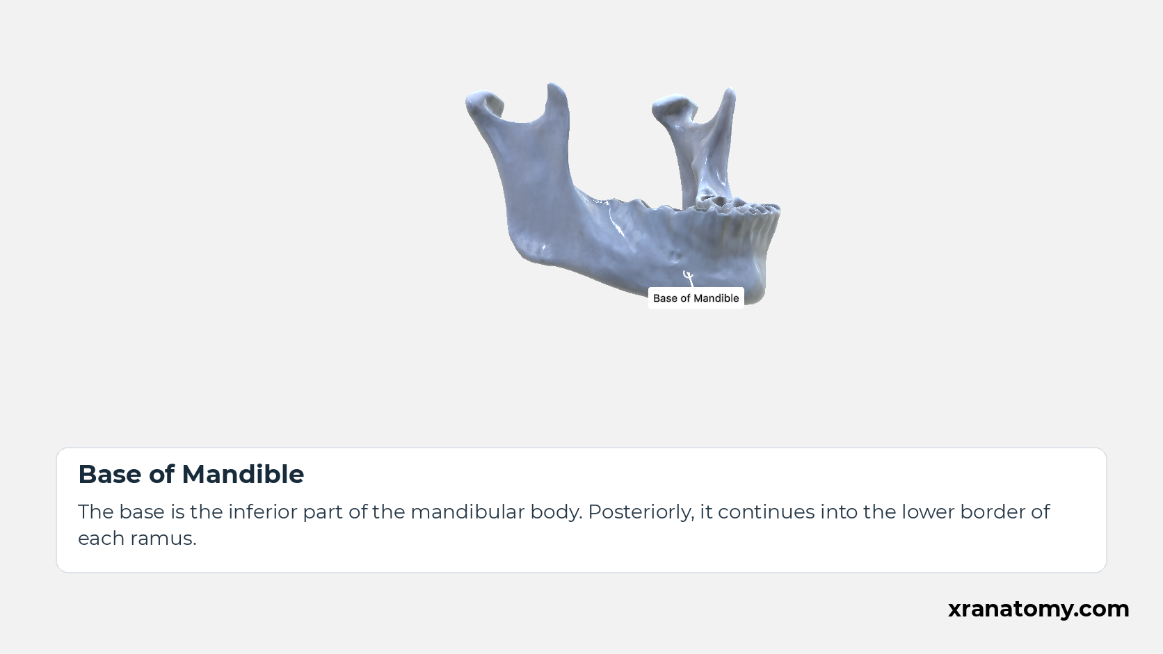
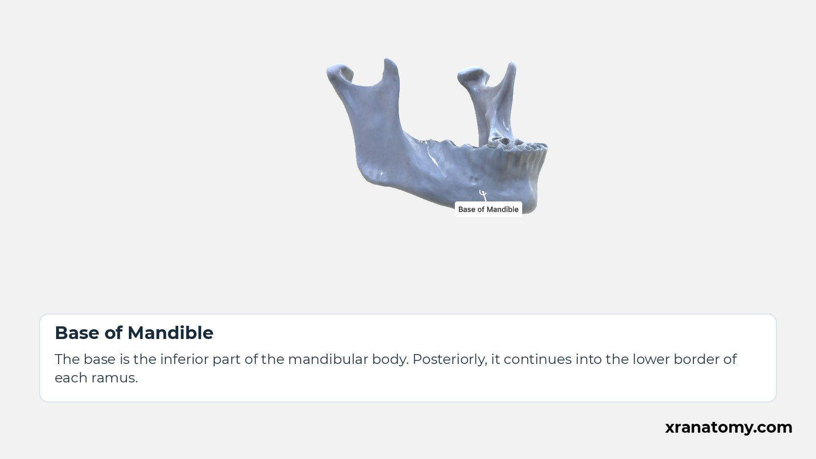
The base is the inferior part of the mandibular body. Posteriorly, it continues into the lower border of each ramus.
Study Base of Mandible in this fixed image. The image does not rotate.
Illustration by XR Anatomy.
Base of Mandible
The base is the inferior part of the mandibular body. Posteriorly, it continues into the lower border of each ramus.
Illustration by XR Anatomy.
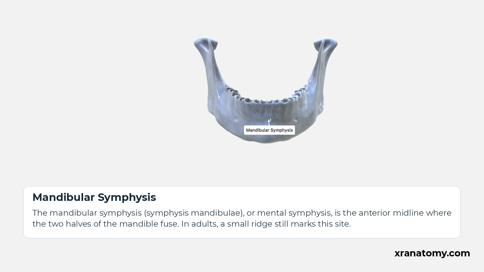
Mandibular Symphysis
The mandibular symphysis (symphysis mandibulae), or mental symphysis, is the anterior midline where the two halves of the mandible fuse. In adults, a small ridge still marks this site.
Illustration by XR Anatomy.
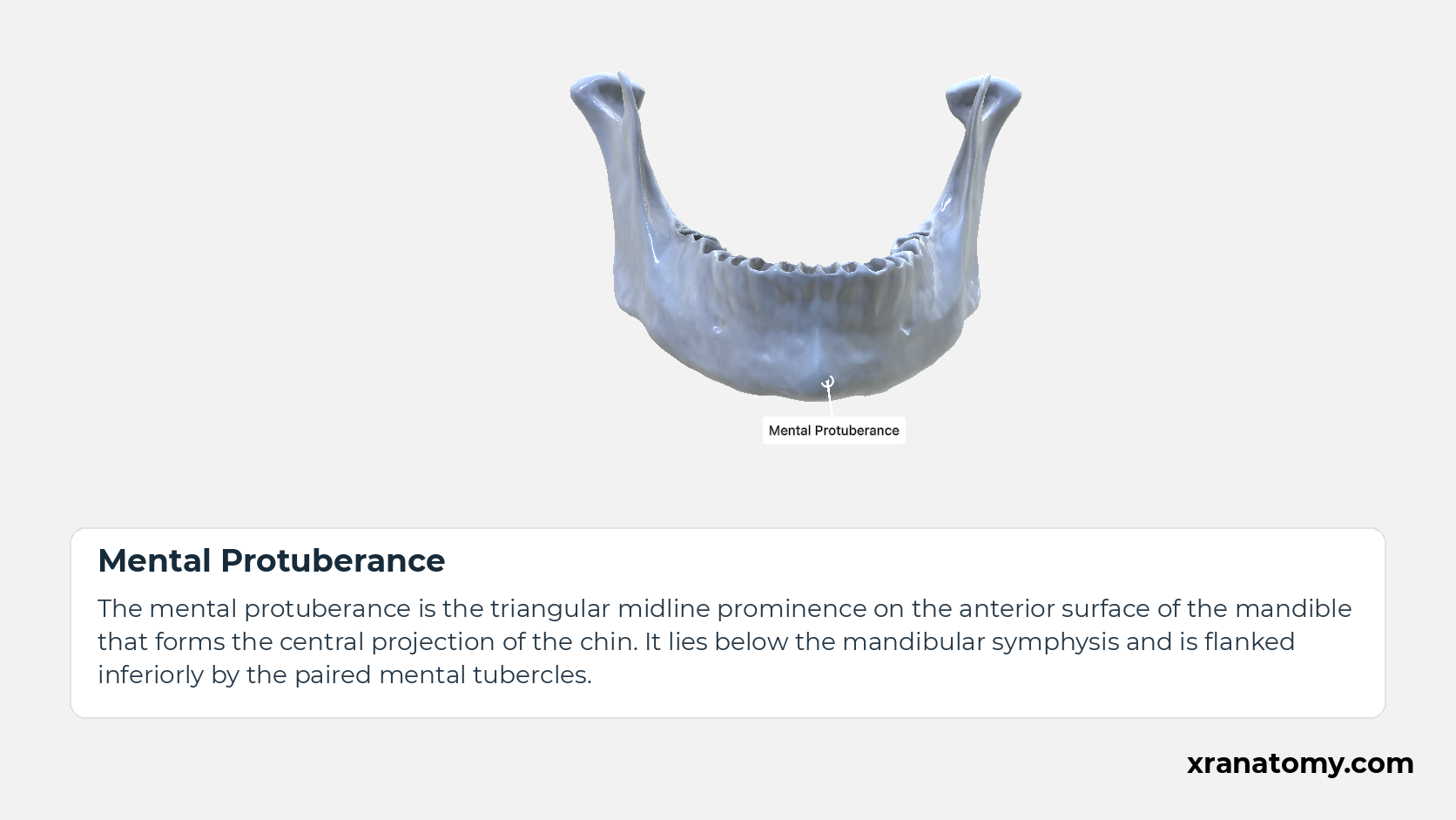
Mental Protuberance
The mental protuberance is the triangular midline prominence on the anterior surface of the mandible that forms the central projection of the chin. It lies below the mandibular symphysis and is flanked inferiorly by the paired mental tubercles.Show less
Illustration by XR Anatomy.
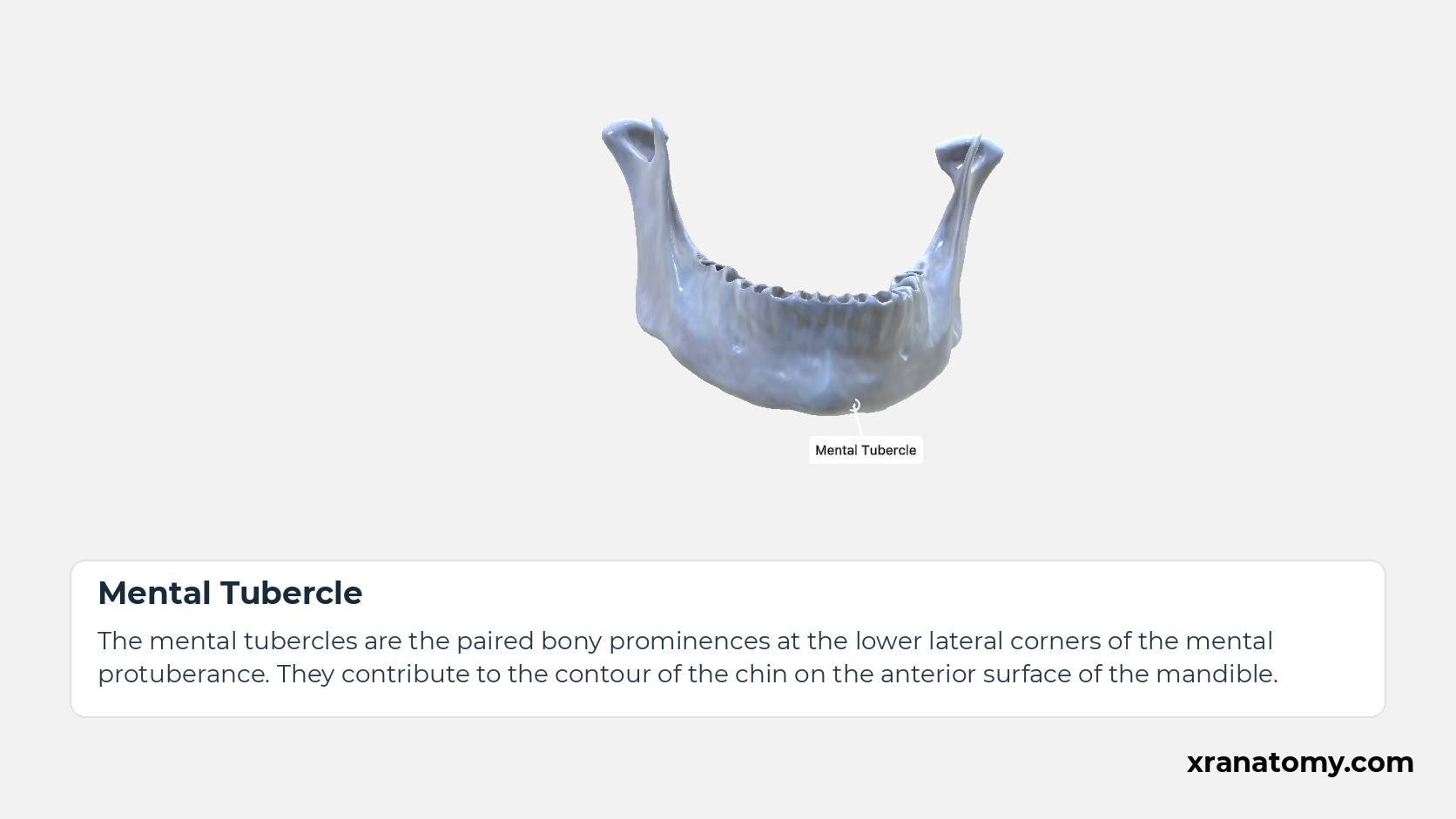
Mental Tubercle
The mental tubercles are the paired bony prominences at the lower lateral corners of the mental protuberance. They contribute to the contour of the chin on the anterior surface of the mandible.
Illustration by XR Anatomy.
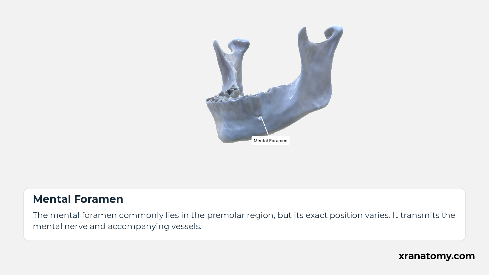
Mental Foramen
The mental foramen commonly lies in the premolar region, but its exact position varies. It transmits the mental nerve and accompanying vessels.
Illustration by XR Anatomy.
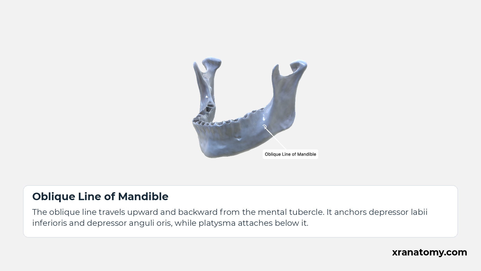
Oblique Line of Mandible
The oblique line travels upward and backward from the mental tubercle. It anchors depressor labii inferioris and depressor anguli oris, while platysma attaches below it.
Illustration by XR Anatomy.
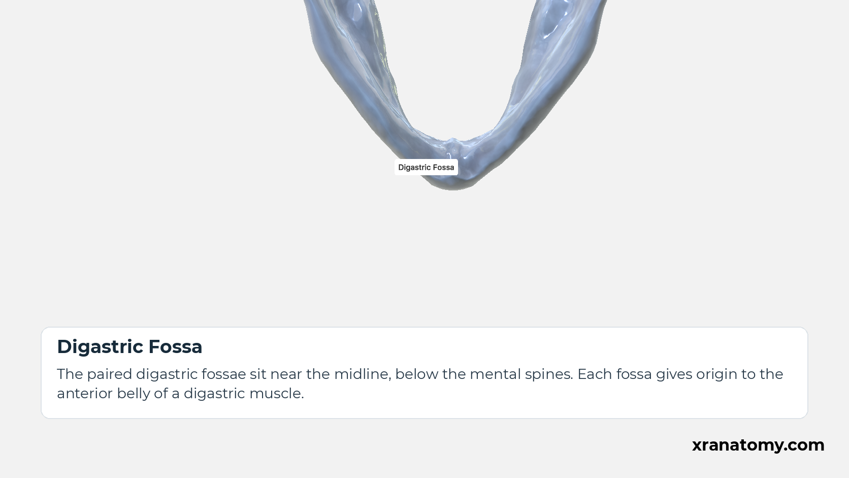
Digastric Fossa
The paired digastric fossae sit near the midline, below the mental spines. Each fossa gives origin to the anterior belly of a digastric muscle.
Illustration by XR Anatomy.
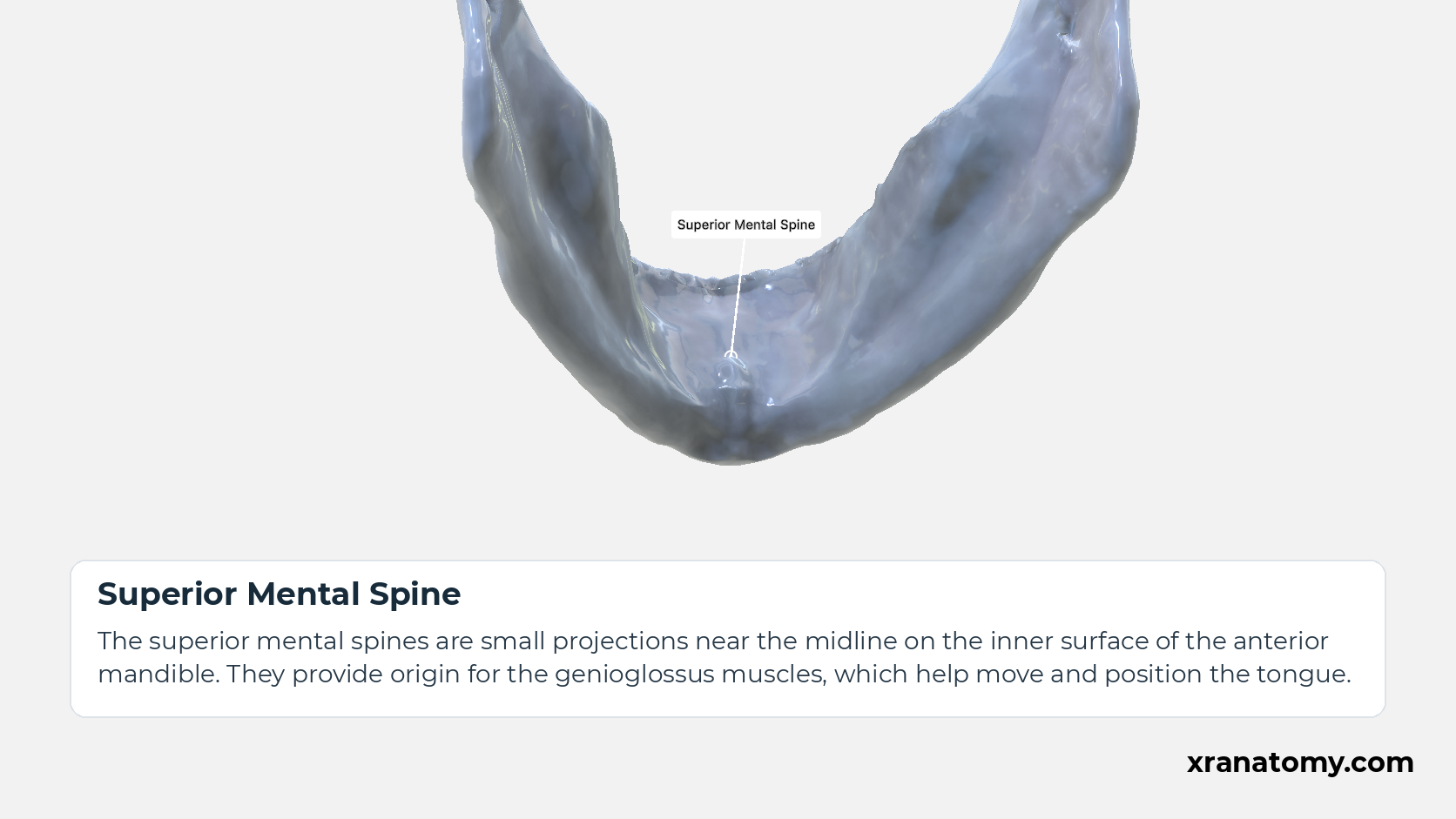
Superior Mental Spine
The superior mental spines are small projections near the midline on the inner surface of the anterior mandible. They provide origin for the genioglossus muscles, which help move and position the tongue.
Illustration by XR Anatomy.
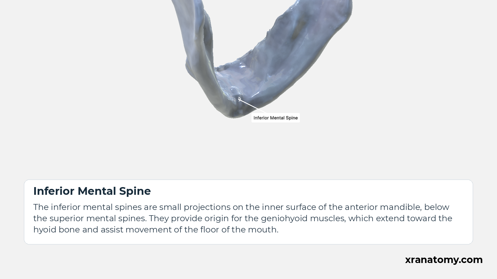
Inferior Mental Spine
The inferior mental spines are small projections on the inner surface of the anterior mandible, below the superior mental spines. They provide origin for the geniohyoid muscles, which extend toward the hyoid bone and assist movement of the floor of the mouth.Show less
Illustration by XR Anatomy.
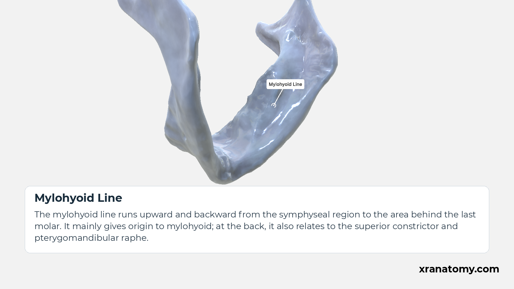
Mylohyoid Line
The mylohyoid line runs upward and backward from the symphyseal region to the area behind the last molar. It mainly gives origin to mylohyoid; at the back, it also relates to the superior constrictor and pterygomandibular raphe.
Illustration by XR Anatomy.
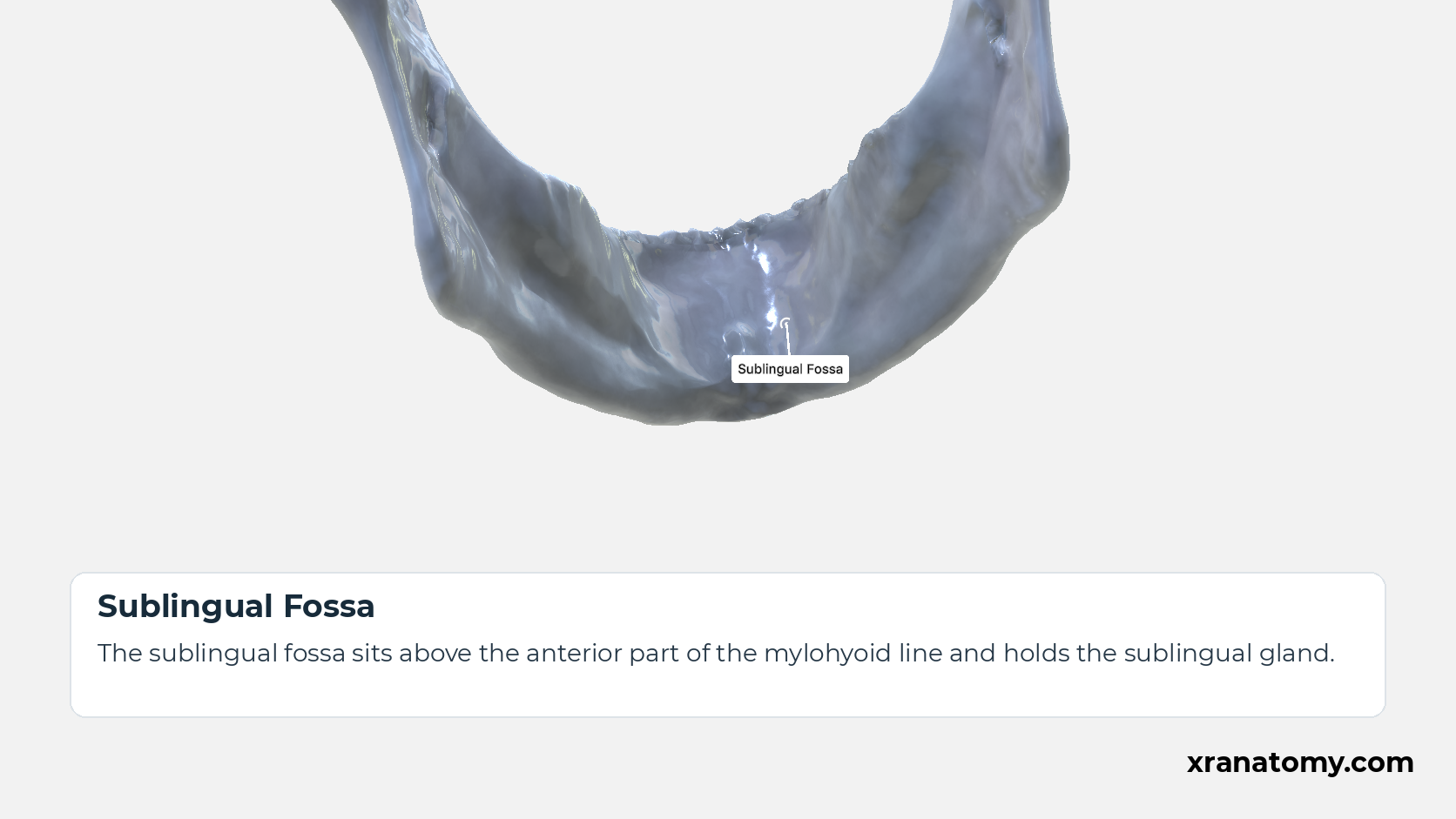
Sublingual Fossa
The sublingual fossa sits above the anterior part of the mylohyoid line and holds the sublingual gland.
Illustration by XR Anatomy.
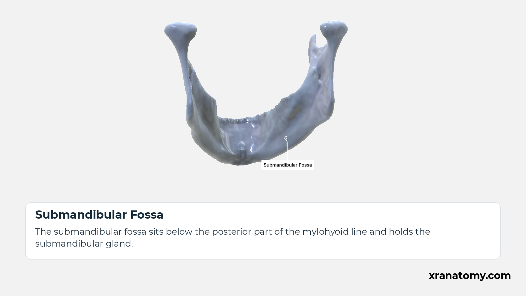
Submandibular Fossa
The submandibular fossa sits below the posterior part of the mylohyoid line and holds the submandibular gland.
Illustration by XR Anatomy.
Body of Mandible
The mandibular body is shaped like a horseshoe. It holds the tooth sockets and offers broad surfaces for muscle attachment.
Illustration by XR Anatomy.
Ramus of Mandible
Each ramus of the mandible is the upright posterior part of the lower jaw, rising from the body at the mandibular angle. Its upper end divides into the coronoid and condylar processes, separated by the mandibular notch; its surfaces provide major muscle attachments and passages for nerves and vessels.Show less
Illustration by XR Anatomy.
Mandible
The mandible is the unpaired movable lower jaw and carries the lower teeth. It is the largest and strongest bone of the face.
Illustration by XR Anatomy.
 ↺ drag to rotate · scroll to zoom
Base of the Mandible
Base of the Mandible
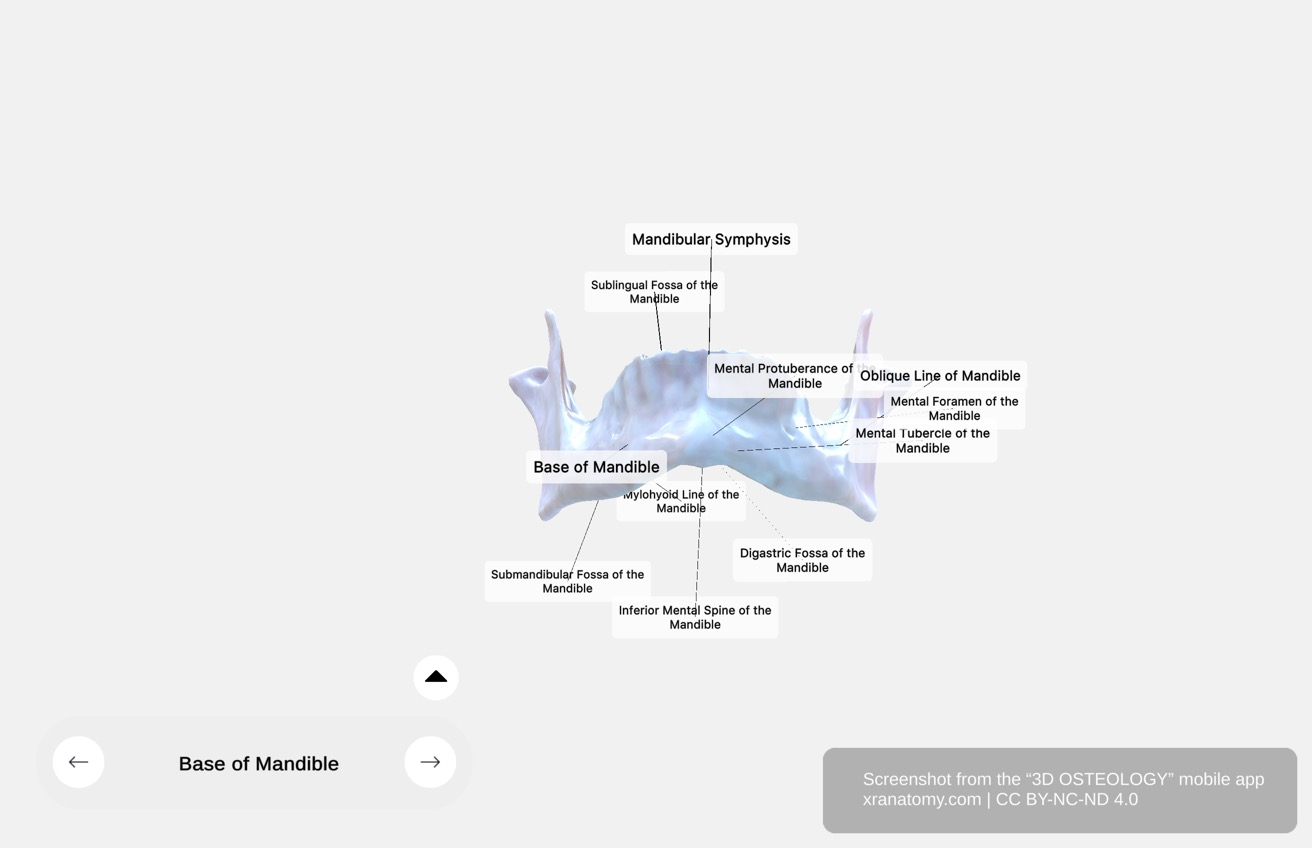
Drag or use the viewer controls to inspect the labelled landmarks around the base of the mandible.
Sourced from 3D Osteology.
The base is the inferior part of the mandibular body. Posteriorly, it continues into the lower border of each ramus.
External Surface of the Mandible
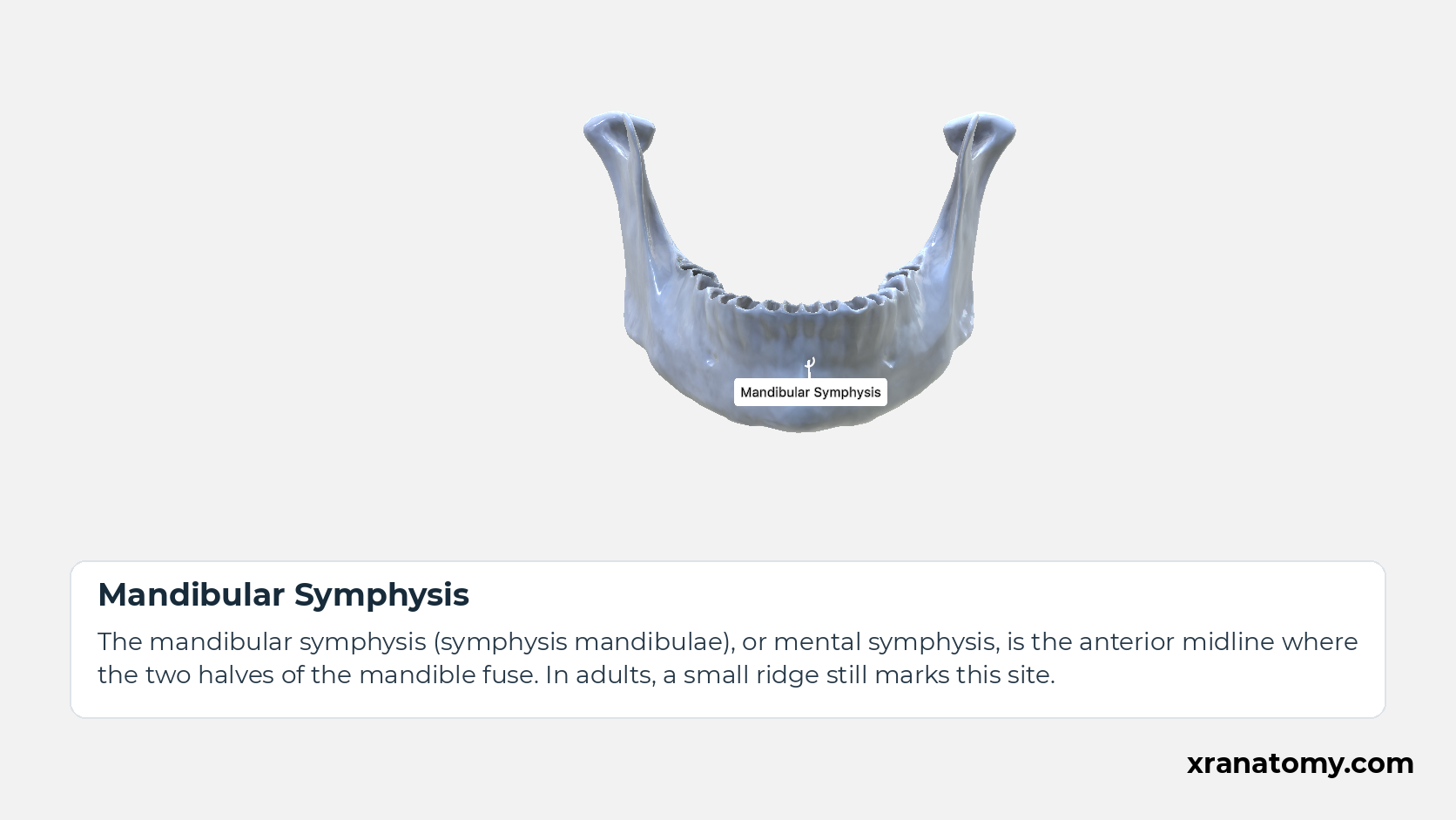
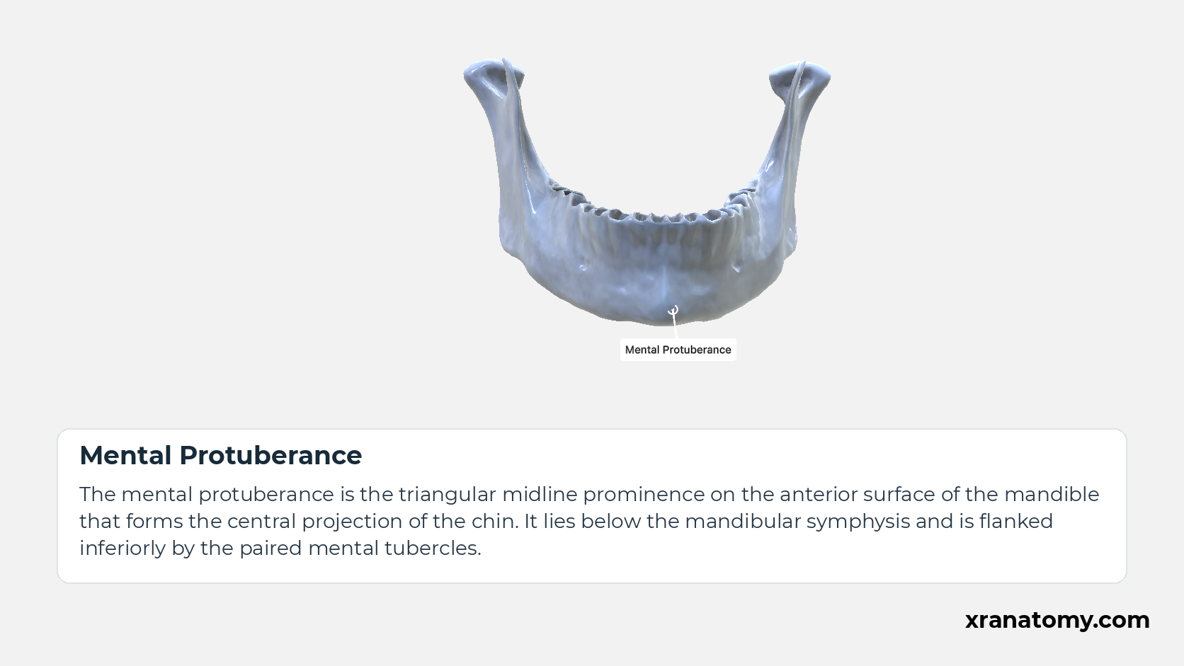
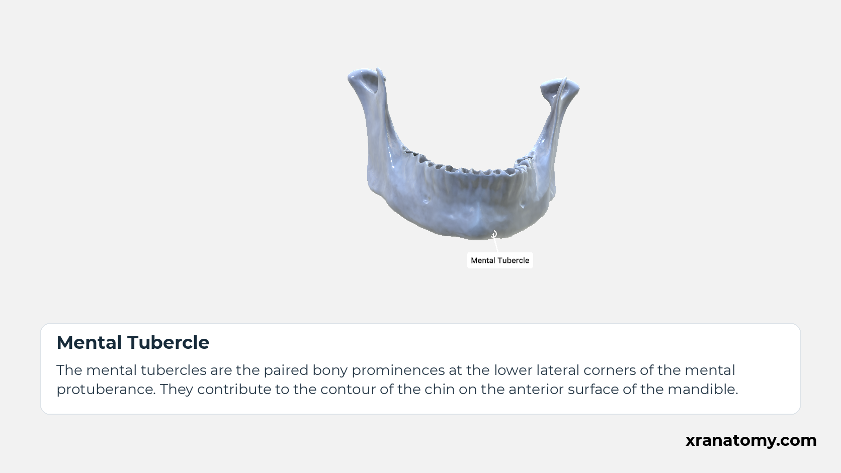
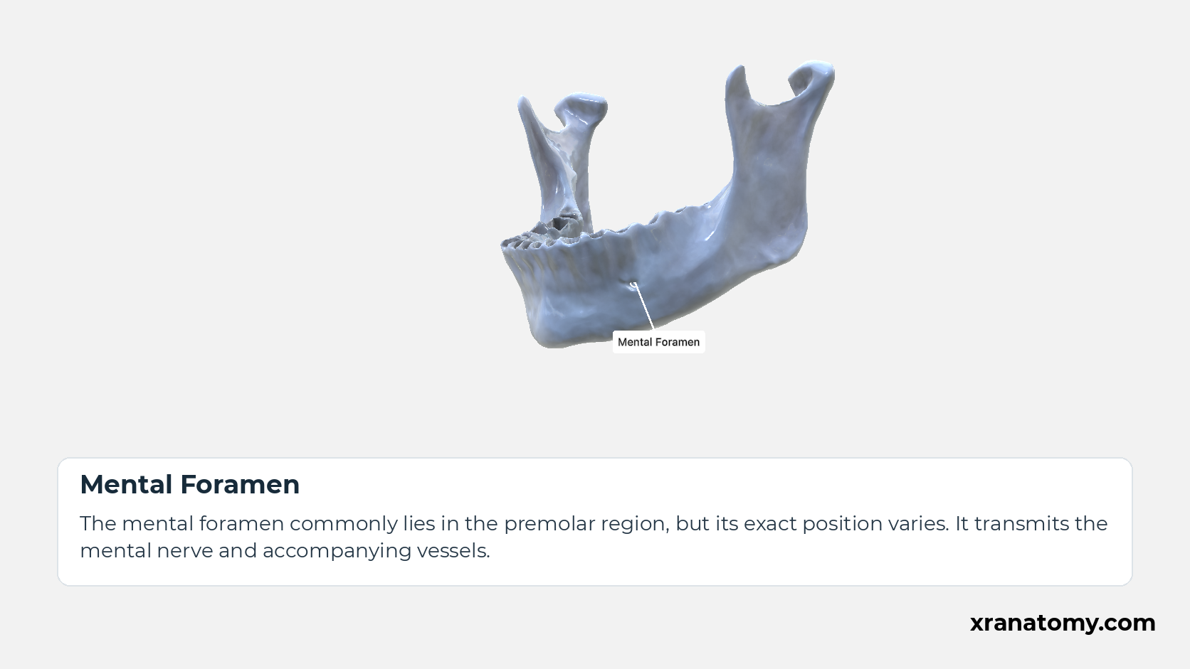
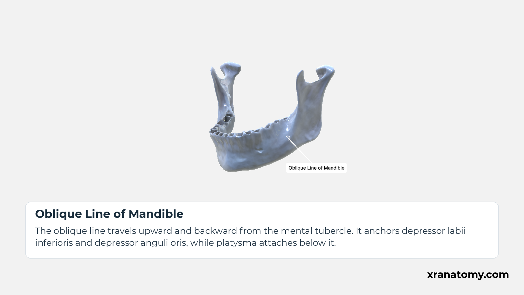
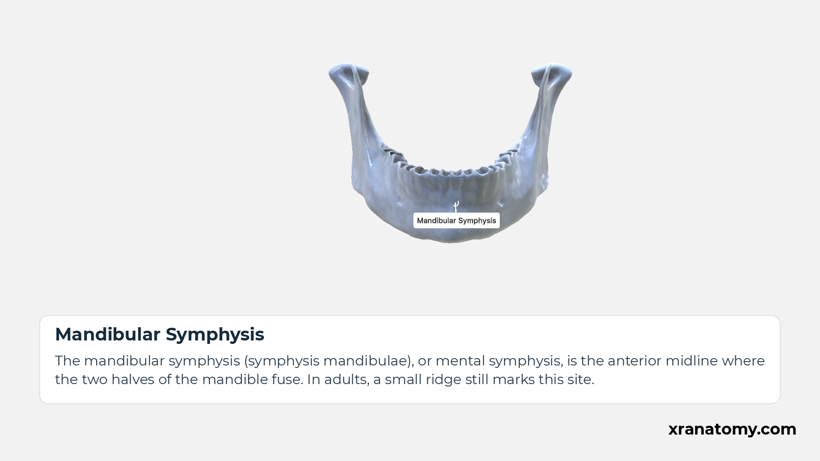
Mandibular Symphysis
The mandibular symphysis (symphysis mandibulae), or mental symphysis, is the anterior midline where the two halves of the mandible fuse. In adults, a small ridge still marks this site.
Study Mandibular Symphysis in this fixed image. Use the thumbnails to move between the 2D views. The image does not rotate.
Illustration by XR Anatomy.
Mental Protuberance
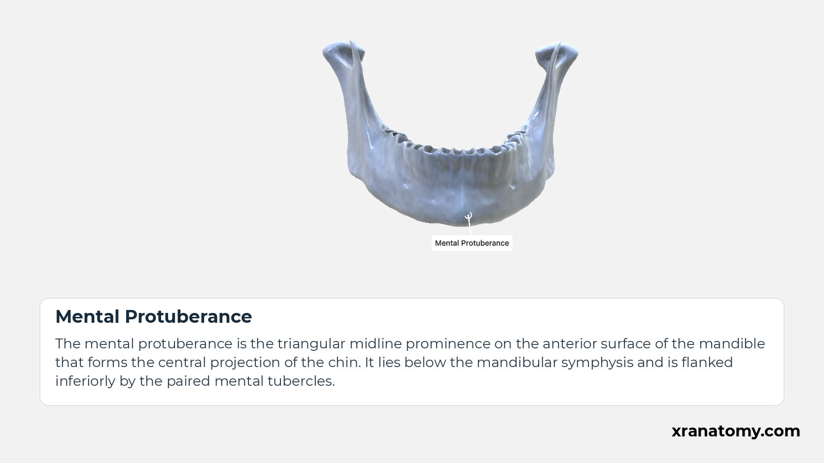
The mental protuberance is the triangular midline prominence on the anterior surface of the mandible that forms the central projection of the chin. It lies below the mandibular symphysis and is flanked inferiorly by the paired mental tubercles.Show less
Illustration by XR Anatomy.
Mental Tubercle
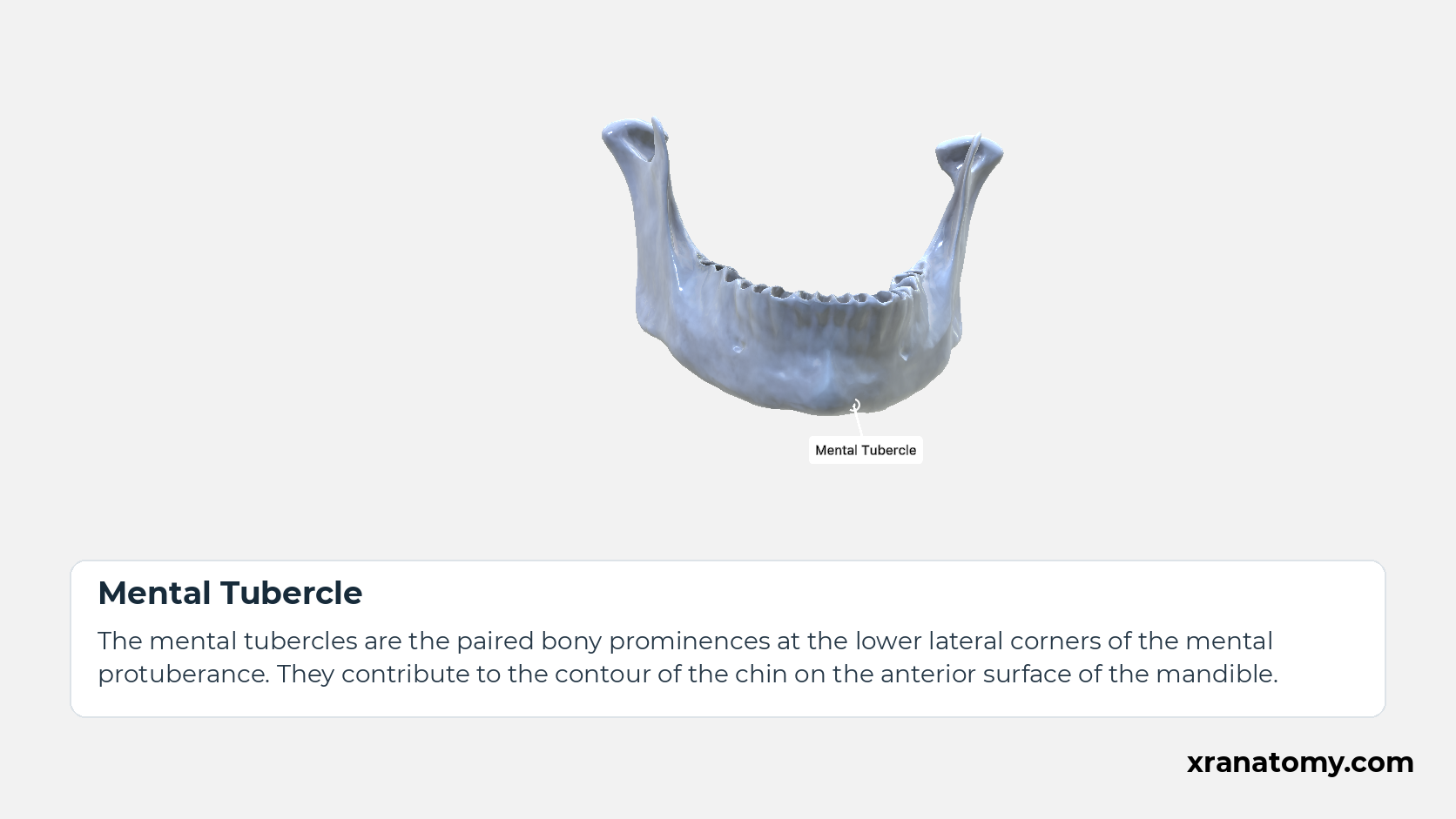
The mental tubercles are the paired bony prominences at the lower lateral corners of the mental protuberance. They contribute to the contour of the chin on the anterior surface of the mandible.
Illustration by XR Anatomy.
Mental Foramen
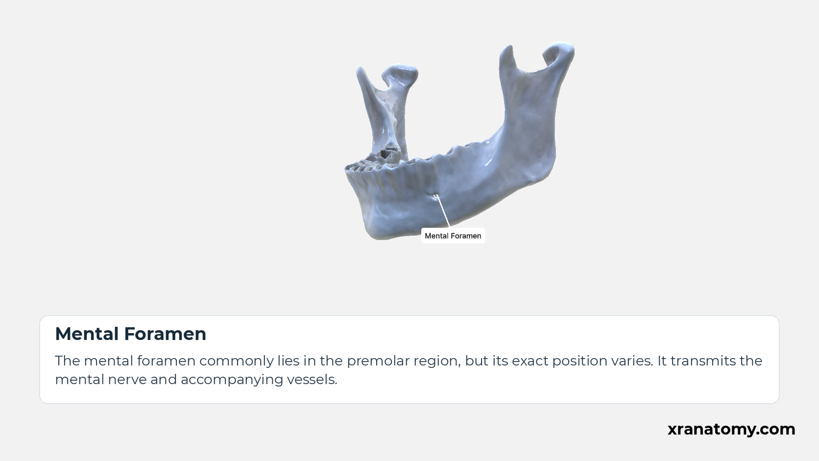
The mental foramen commonly lies in the premolar region, but its exact position varies. It transmits the mental nerve and accompanying vessels.
Illustration by XR Anatomy.
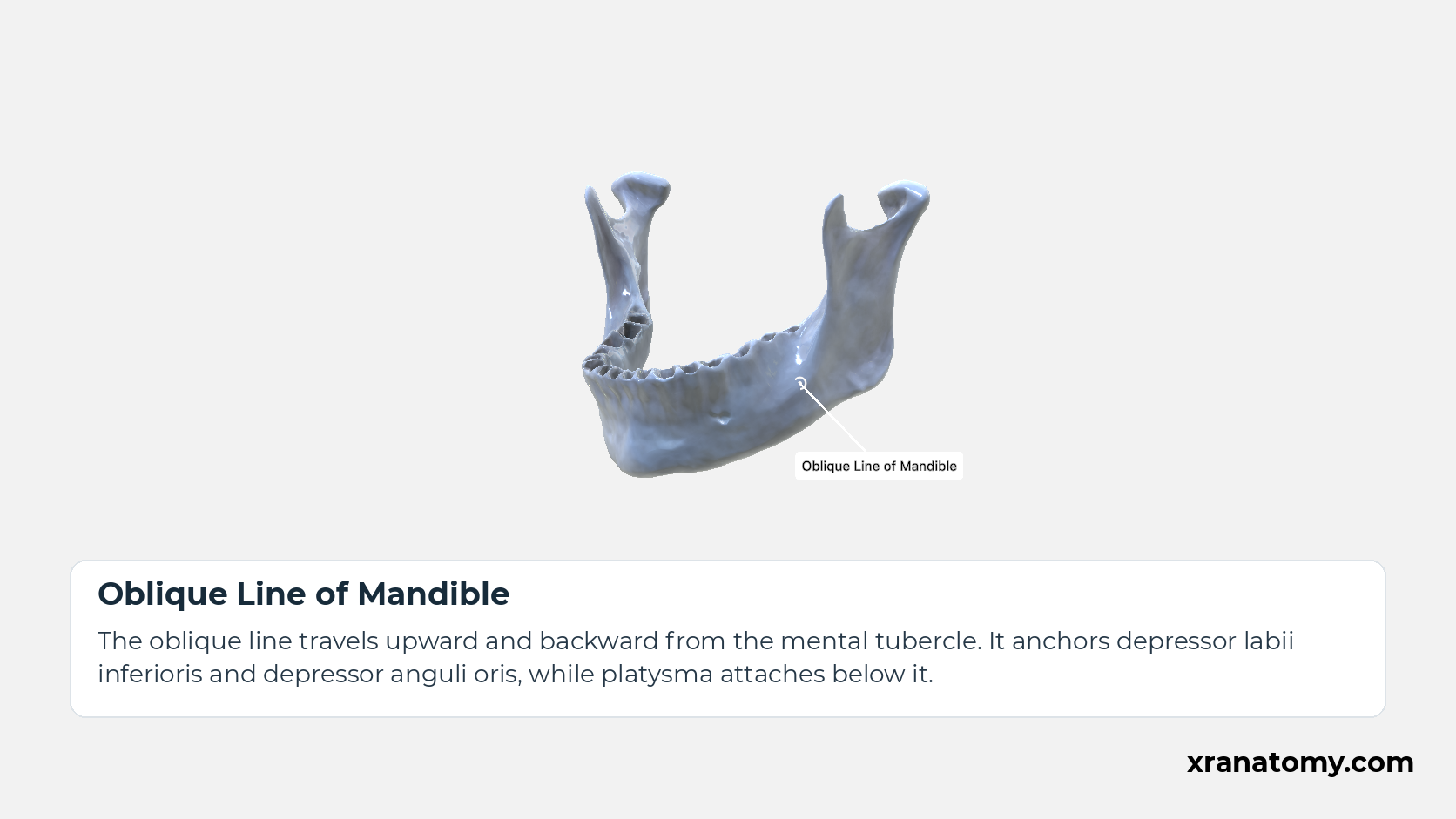
Oblique Line of Mandible
The oblique line travels upward and backward from the mental tubercle. It anchors depressor labii inferioris and depressor anguli oris, while platysma attaches below it.
Illustration by XR Anatomy.
Mandibular Symphysis
The mandibular symphysis (symphysis mandibulae), or mental symphysis, is the anterior midline where the two halves of the mandible fuse. In adults, a small ridge still marks this site.
Illustration by XR Anatomy.
Mental Protuberance
The mental protuberance is the triangular midline prominence on the anterior surface of the mandible that forms the central projection of the chin. It lies below the mandibular symphysis and is flanked inferiorly by the paired mental tubercles.Show less
Illustration by XR Anatomy.
Mental Tubercle
The mental tubercles are the paired bony prominences at the lower lateral corners of the mental protuberance. They contribute to the contour of the chin on the anterior surface of the mandible.
Illustration by XR Anatomy.
Mental Foramen
The mental foramen commonly lies in the premolar region, but its exact position varies. It transmits the mental nerve and accompanying vessels.
Illustration by XR Anatomy.
Oblique Line of Mandible
The oblique line travels upward and backward from the mental tubercle. It anchors depressor labii inferioris and depressor anguli oris, while platysma attaches below it.
Illustration by XR Anatomy.
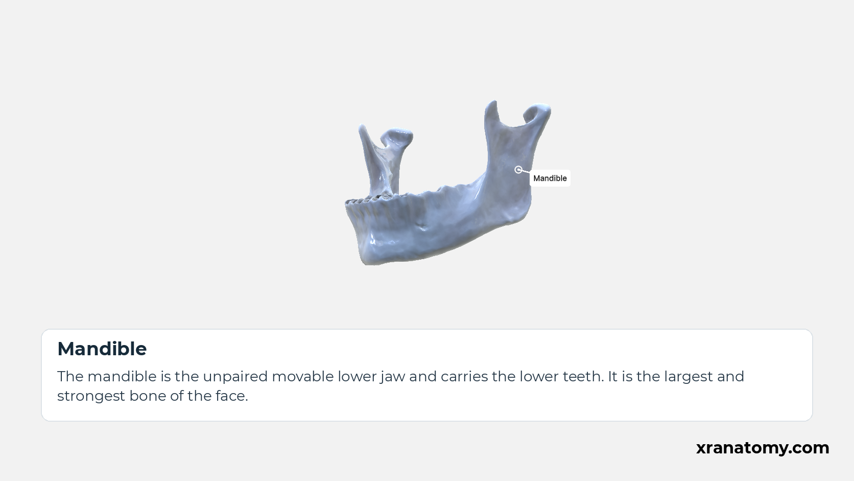
Mandible
The mandible is the unpaired movable lower jaw and carries the lower teeth. It is the largest and strongest bone of the face.
Illustration by XR Anatomy.
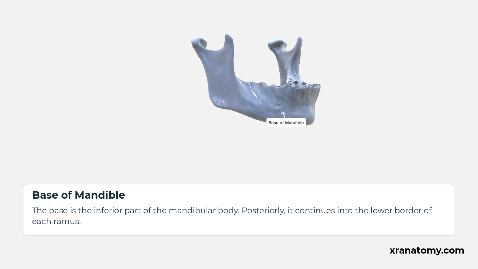
Base of Mandible
The base is the inferior part of the mandibular body. Posteriorly, it continues into the lower border of each ramus.
Illustration by XR Anatomy.
The external surface shows the mandibular symphysis, mental protuberance, paired mental tubercles, mental foramina, and oblique lines.
In depth · 5 sections · 1 min read5 more sections on this part5 sections open — reading In depth- Mandibular Symphysis
- Mental Protuberance
- Mental Tubercle
- Mental Foramen
- Oblique Line of Mandible
Mandibular Symphysis
The mandibular symphysis (symphysis mandibulae), or mental symphysis, is the anterior midline where the two halves of the mandible fuse. In adults, a small ridge still marks this site.
Mental Protuberance
Below the symphysis, the midline ridge splits around the triangular mental protuberance, which forms the central projection of the chin.
Mental Tubercle
The centre of the mental protuberance's base is depressed, while its sides rise as the paired mental tubercles.
Mental Foramen
The mental foramen commonly lies in the premolar region, but its exact position varies. It transmits the mental nerve and accompanying vessels.
Oblique Line of Mandible
The oblique line travels upward and backward from the mental tubercle. It anchors depressor labii inferioris and depressor anguli oris, while platysma attaches below it.
Internal Surface of the Mandible
Digastric Fossa
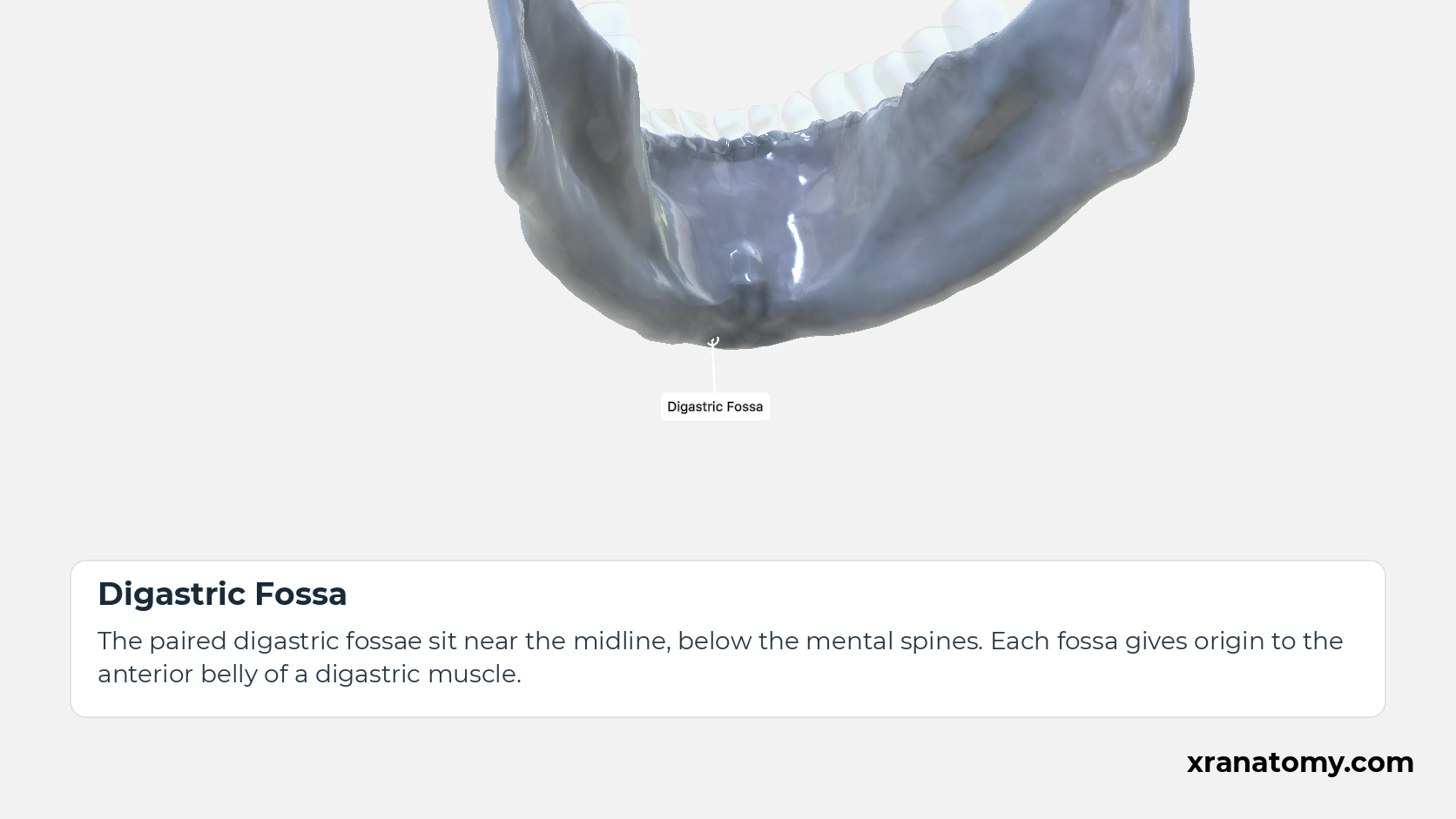
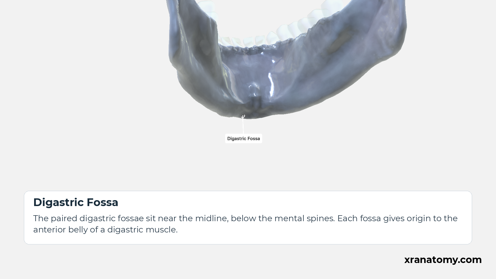
The paired digastric fossae sit near the midline, below the mental spines. Each fossa gives origin to the anterior belly of a digastric muscle.
Study Digastric Fossa in this fixed image. Use the thumbnails to move between the 2D views. The image does not rotate.
Illustration by XR Anatomy.
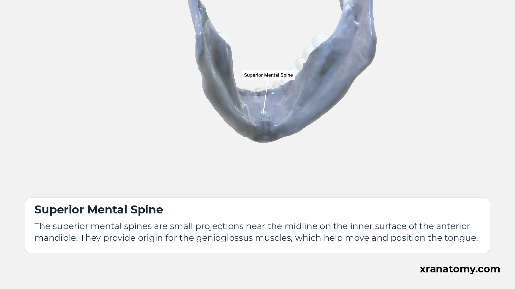
Superior Mental Spine
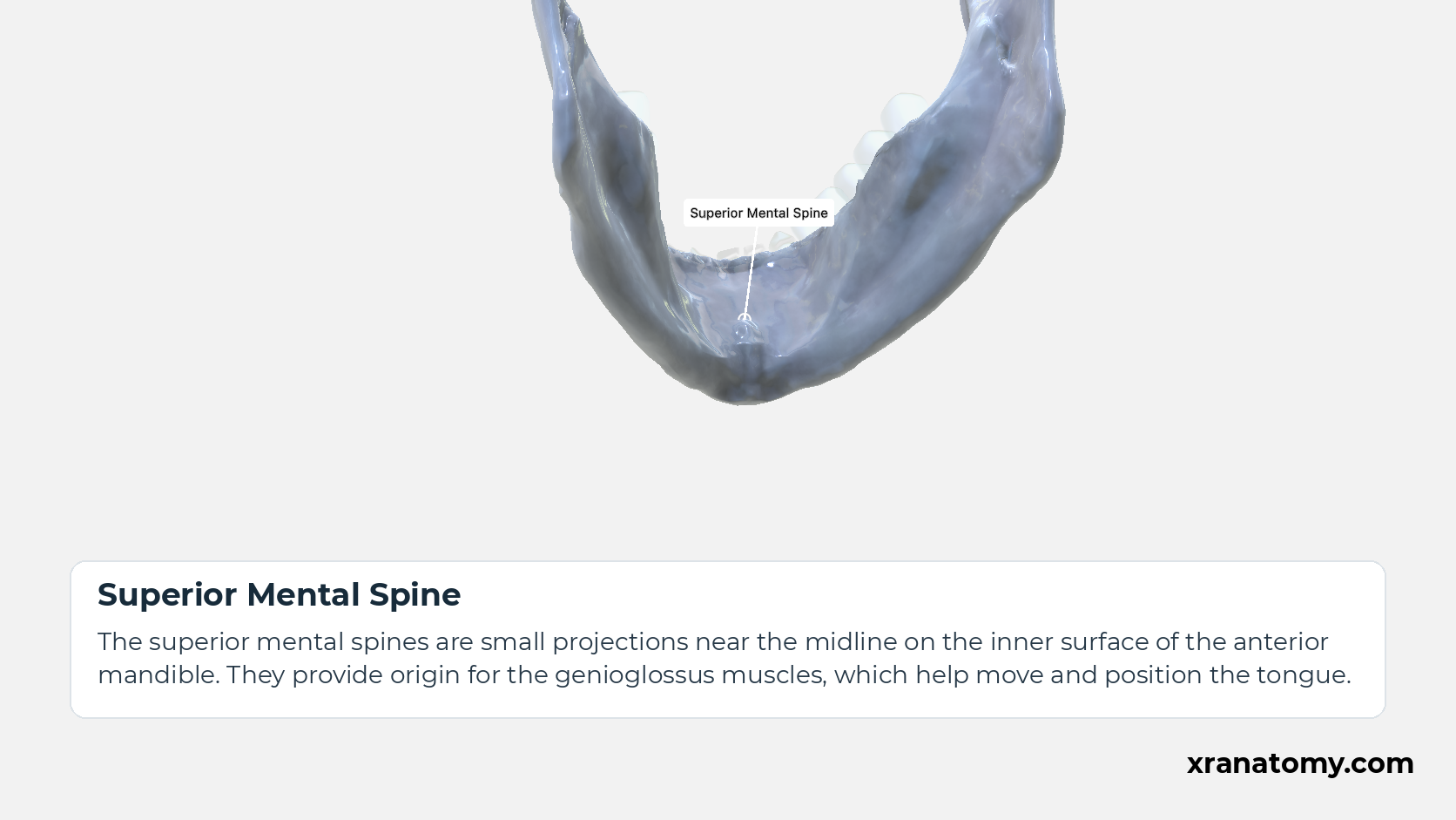
The superior mental spines are small projections near the midline on the inner surface of the anterior mandible. They provide origin for the genioglossus muscles, which help move and position the tongue.
Illustration by XR Anatomy.
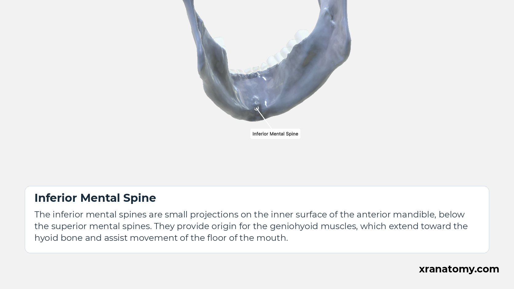
Inferior Mental Spine
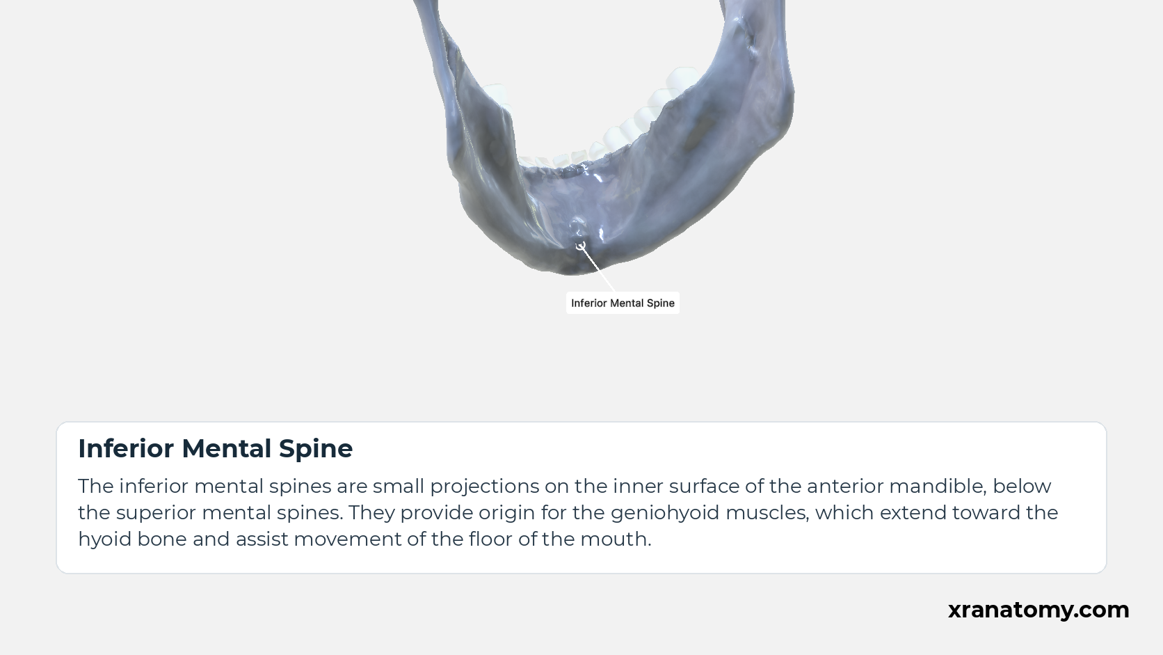
The inferior mental spines are small projections on the inner surface of the anterior mandible, below the superior mental spines. They provide origin for the geniohyoid muscles, which extend toward the hyoid bone and assist movement of the floor of the mouth.Show less
Illustration by XR Anatomy.
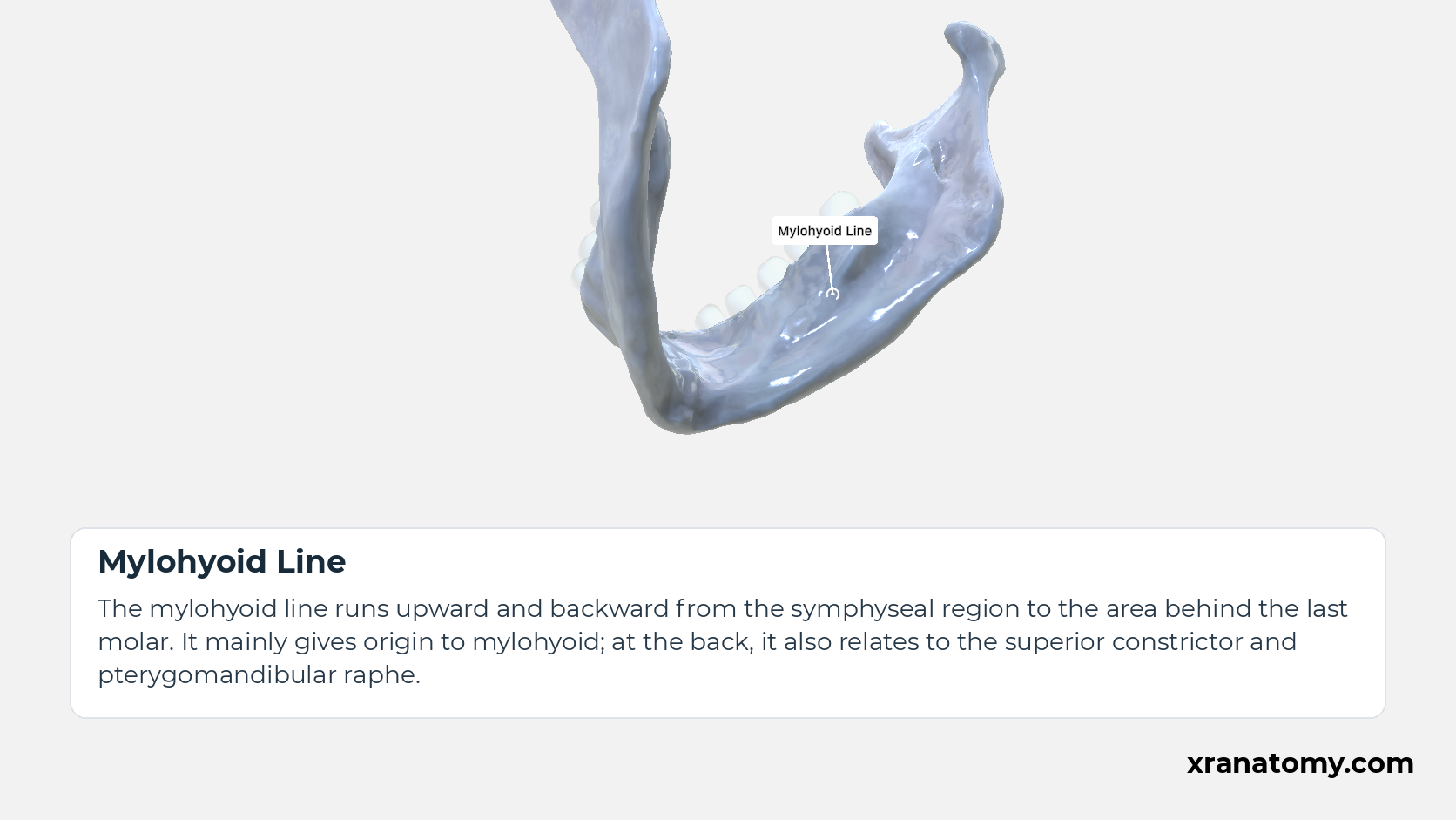
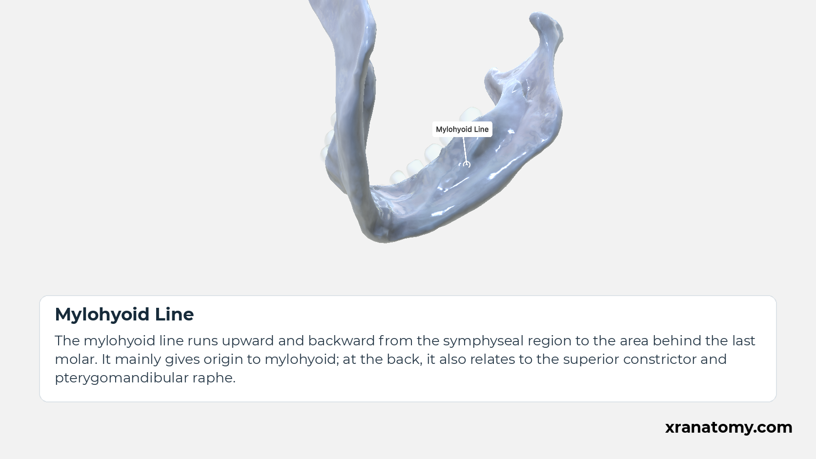
Mylohyoid Line
The mylohyoid line runs upward and backward from the symphyseal region to the area behind the last molar. It mainly gives origin to mylohyoid; at the back, it also relates to the superior constrictor and pterygomandibular raphe.
Illustration by XR Anatomy.
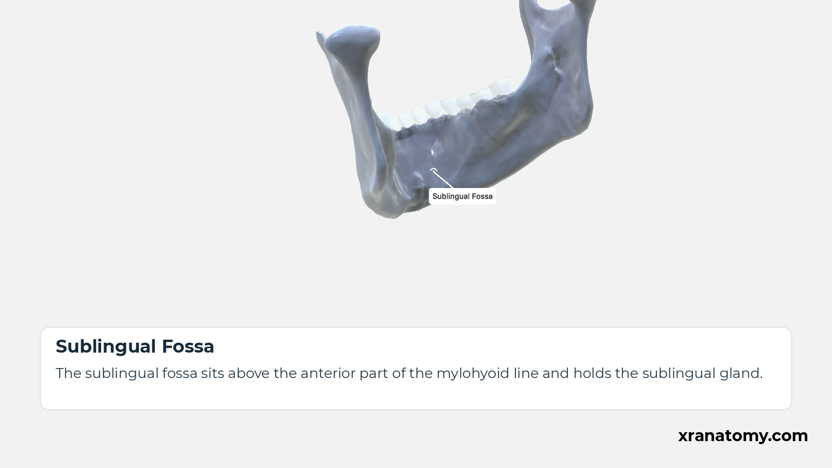
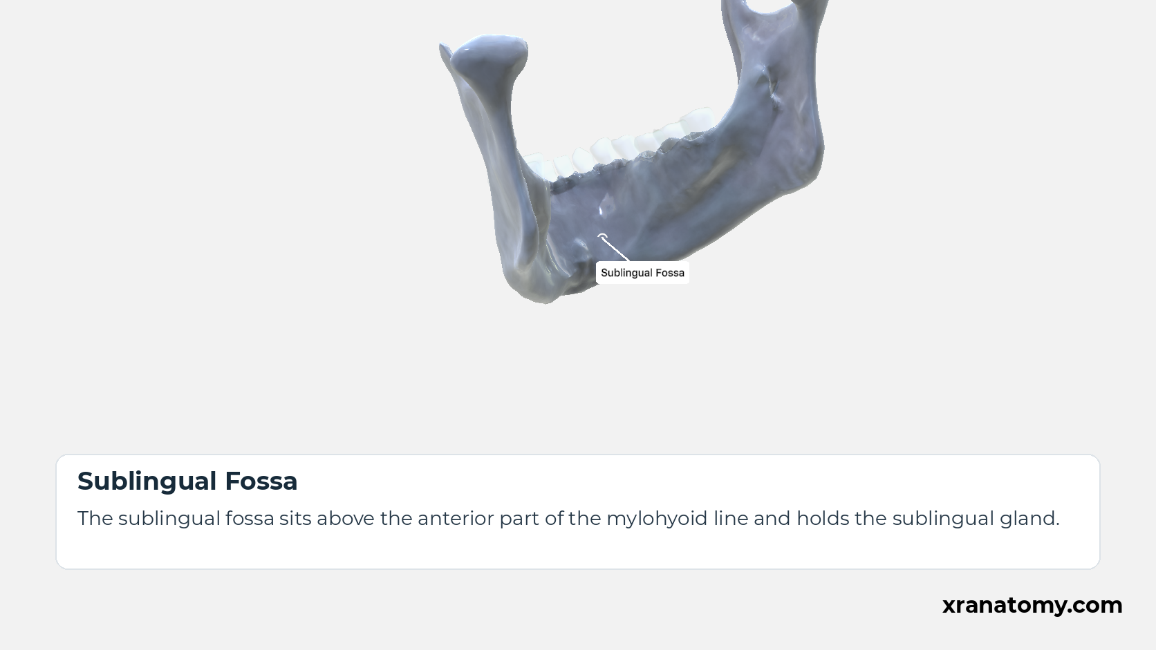
Sublingual Fossa
The sublingual fossa sits above the anterior part of the mylohyoid line and holds the sublingual gland.
Illustration by XR Anatomy.
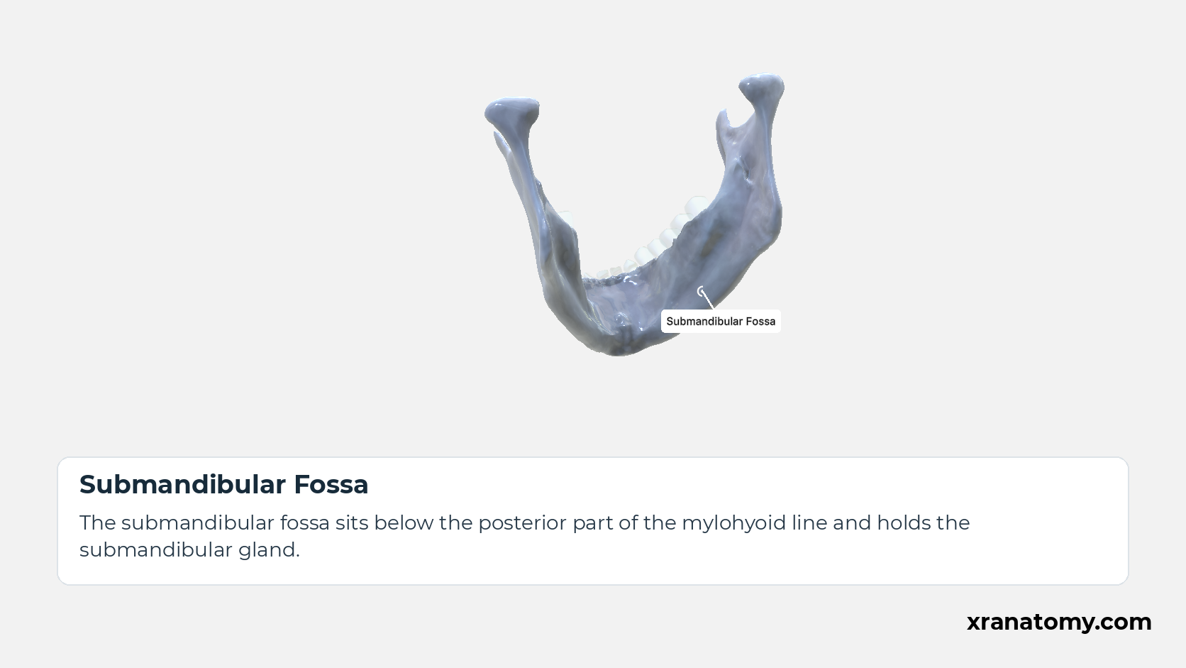
Submandibular Fossa
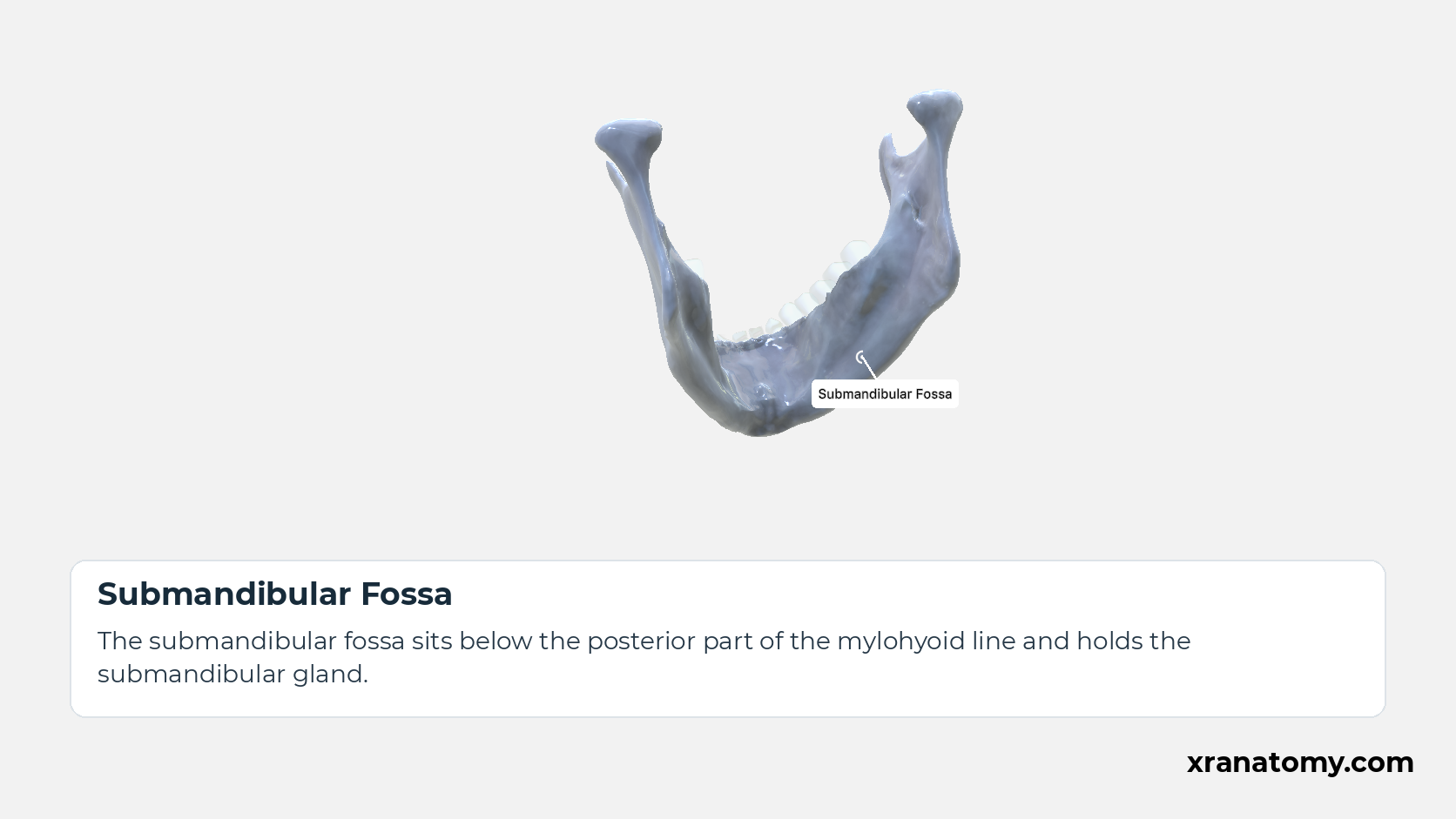
The submandibular fossa sits below the posterior part of the mylohyoid line and holds the submandibular gland.
Illustration by XR Anatomy.
Digastric Fossa
The paired digastric fossae sit near the midline, below the mental spines. Each fossa gives origin to the anterior belly of a digastric muscle.
Illustration by XR Anatomy.
Superior Mental Spine
The superior mental spines are small projections near the midline on the inner surface of the anterior mandible. They provide origin for the genioglossus muscles, which help move and position the tongue.
Illustration by XR Anatomy.
Inferior Mental Spine
The inferior mental spines are small projections on the inner surface of the anterior mandible, below the superior mental spines. They provide origin for the geniohyoid muscles, which extend toward the hyoid bone and assist movement of the floor of the mouth.Show less
Illustration by XR Anatomy.
Mylohyoid Line
The mylohyoid line runs upward and backward from the symphyseal region to the area behind the last molar. It mainly gives origin to mylohyoid; at the back, it also relates to the superior constrictor and pterygomandibular raphe.
Illustration by XR Anatomy.
Sublingual Fossa
The sublingual fossa sits above the anterior part of the mylohyoid line and holds the sublingual gland.
Illustration by XR Anatomy.
Submandibular Fossa
The submandibular fossa sits below the posterior part of the mylohyoid line and holds the submandibular gland.
Illustration by XR Anatomy.
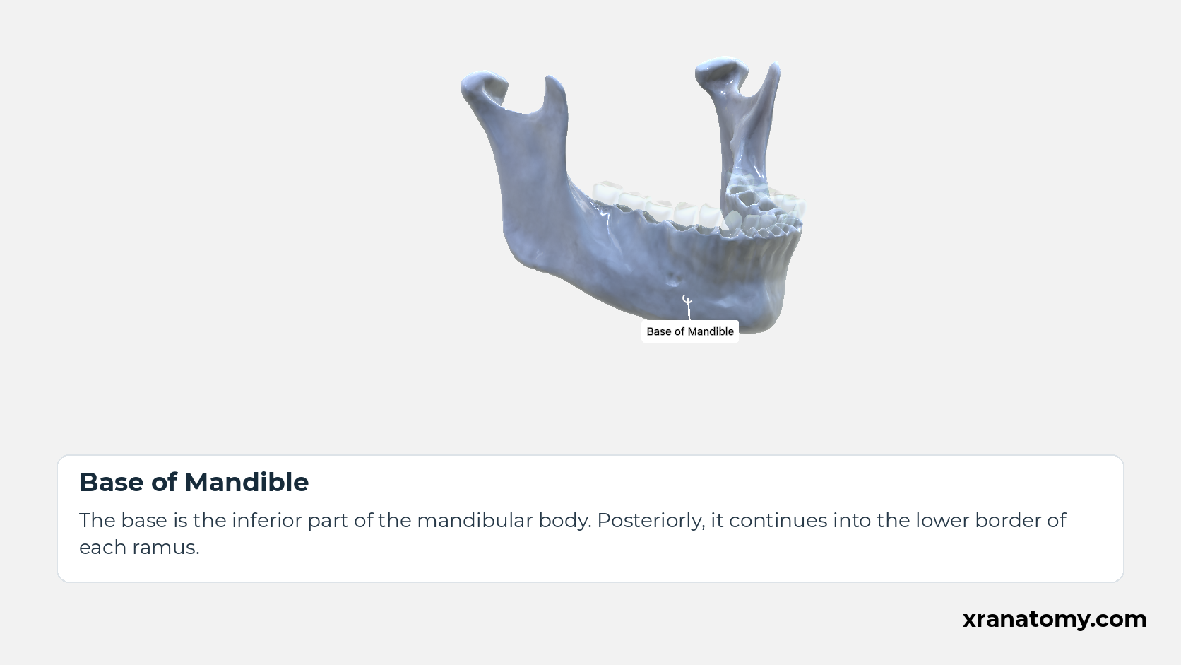
Base of Mandible
The base is the inferior part of the mandibular body. Posteriorly, it continues into the lower border of each ramus.
Illustration by XR Anatomy.
The internal surface carries the digastric fossae, superior mental spines, inferior mental spines, mylohyoid lines, sublingual fossae, and submandibular fossae.
In depth · 6 sections · 1 min read6 more sections on this part6 sections open — reading In depth- Digastric Fossa
- Superior Mental Spine
- Inferior Mental Spine
- Mylohyoid Line
- Sublingual Fossa
- Submandibular Fossa
Digastric Fossa
The paired digastric fossae sit near the midline, below the mental spines. Each fossa gives origin to the anterior belly of a digastric muscle.
Superior Mental Spine
The superior mental spines are the attachment sites where the genioglossus muscles originate.
Inferior Mental Spine
The inferior mental spines sit below the superior pair and give origin to the geniohyoid muscles.
Mylohyoid Line
The mylohyoid line runs upward and backward from the symphyseal region to the area behind the last molar. It mainly gives origin to mylohyoid; at the back, it also relates to the superior constrictor and pterygomandibular raphe.
Sublingual Fossa
The sublingual fossa sits above the anterior part of the mylohyoid line and holds the sublingual gland.
Submandibular Fossa
The submandibular fossa sits below the posterior part of the mylohyoid line and holds the submandibular gland.
Alveolar Part of Mandible
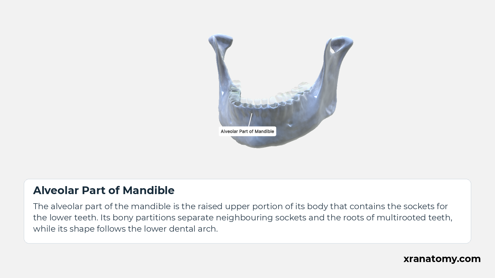
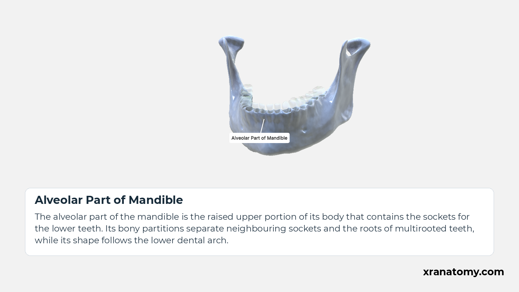
Alveolar Part of Mandible
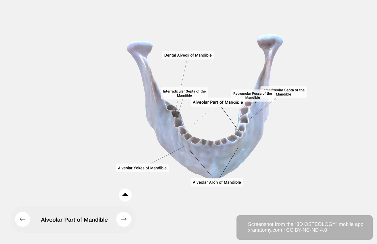
The alveolar part of the mandible is the raised upper portion of its body that contains the sockets for the lower teeth. Its bony partitions separate neighbouring sockets and the roots of multirooted teeth, while its shape follows the lower dental arch.Show less
Study Alveolar Part of Mandible in this fixed image. The image does not rotate.
Illustration by XR Anatomy.
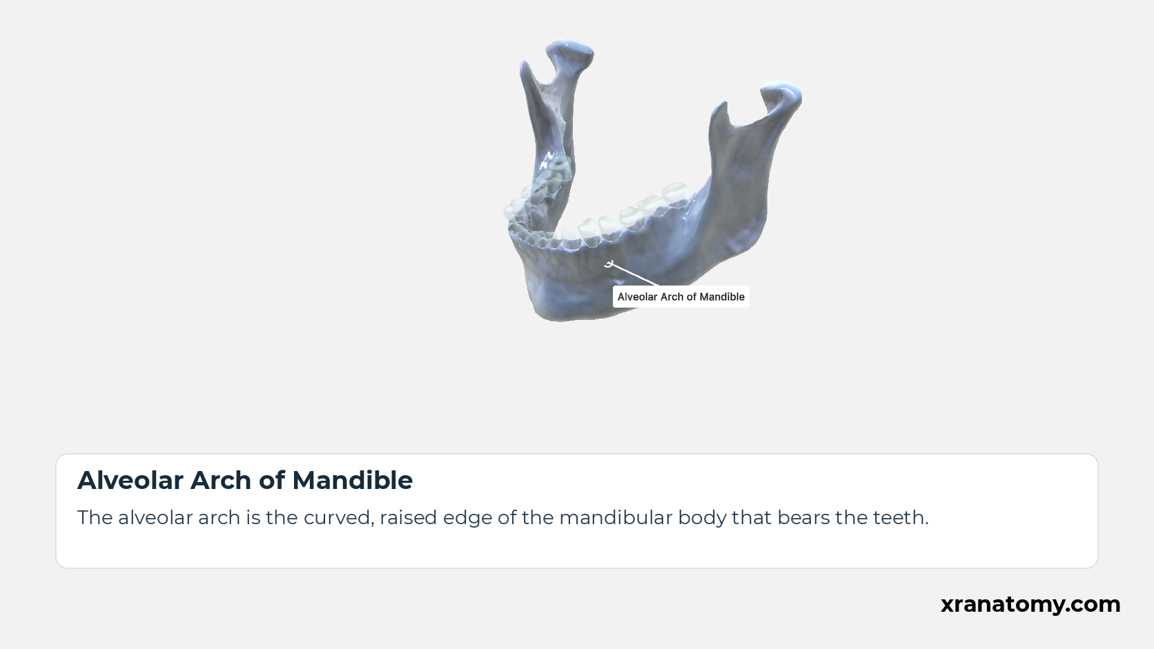
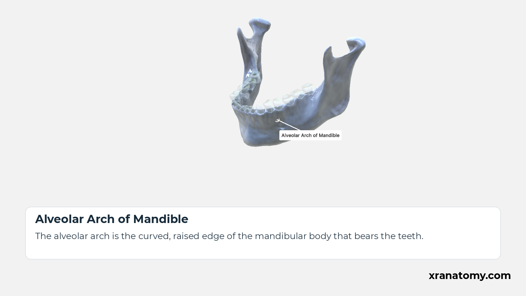
Alveolar Arch of Mandible
The alveolar arch is the curved, raised edge of the mandibular body that bears the teeth.
Illustration by XR Anatomy.
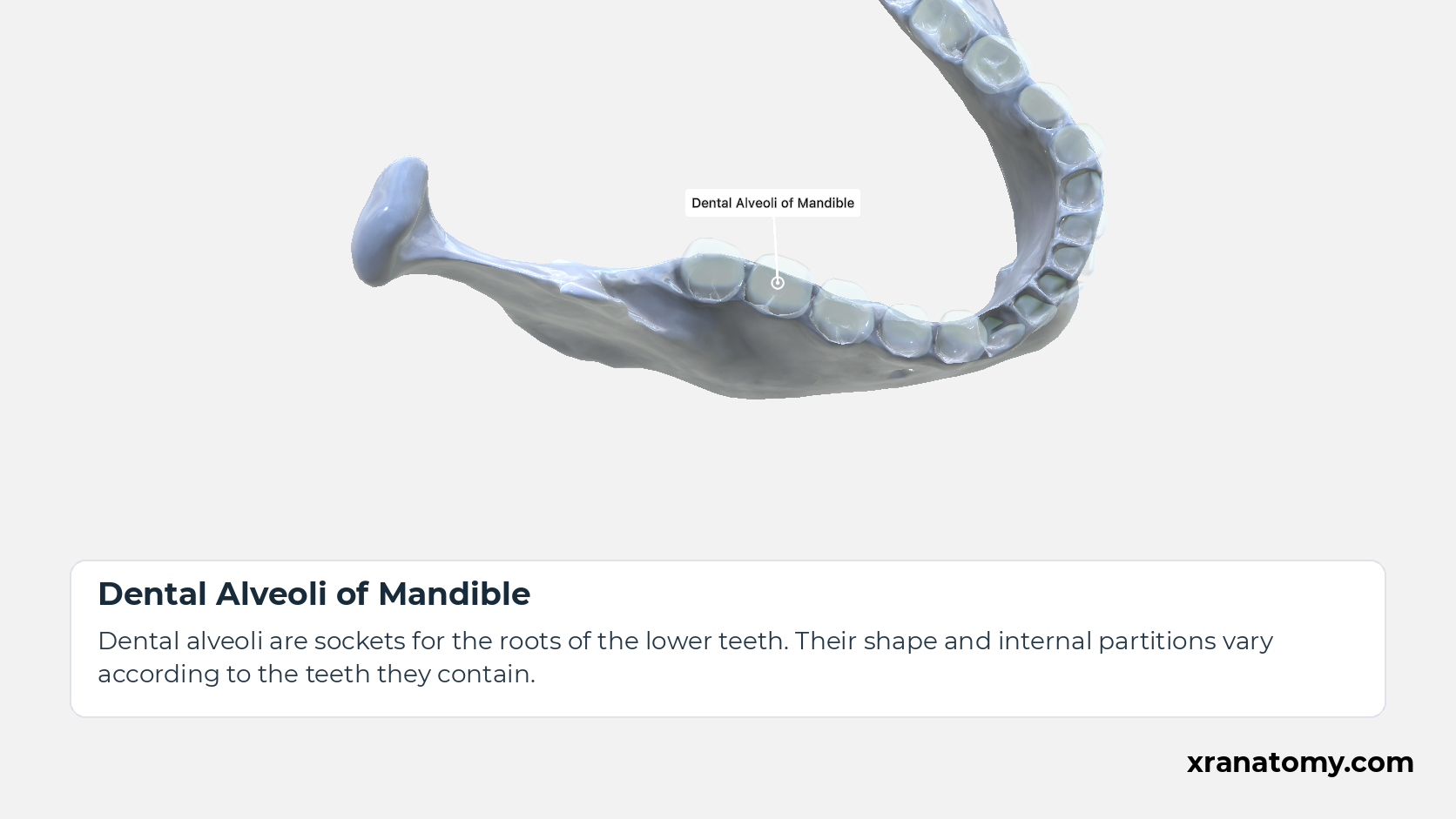
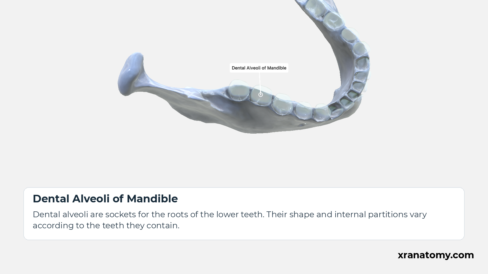
Dental Alveoli of Mandible
Dental alveoli are sockets for the roots of the lower teeth. Their shape and internal partitions vary according to the teeth they contain.
Illustration by XR Anatomy.
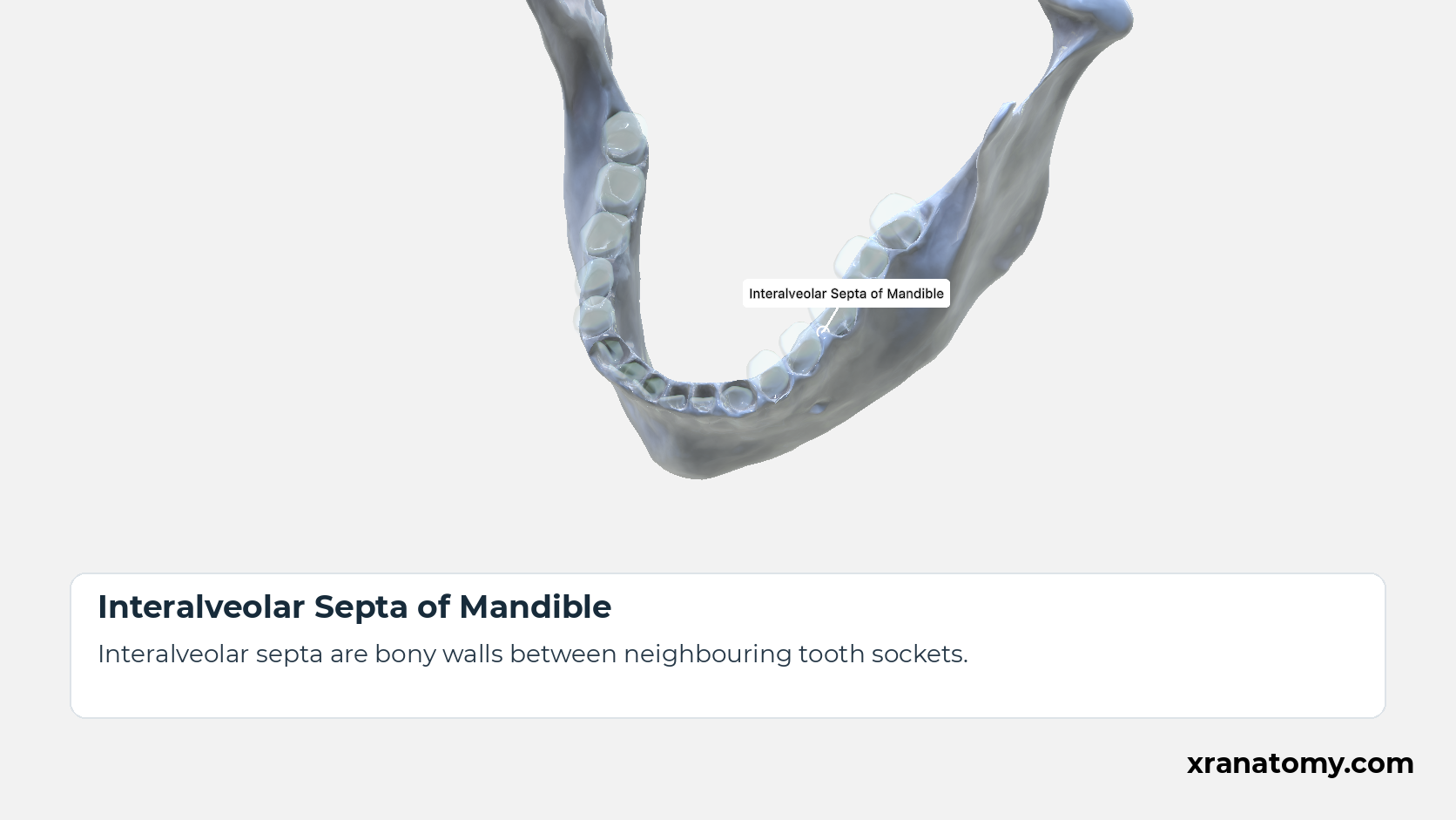
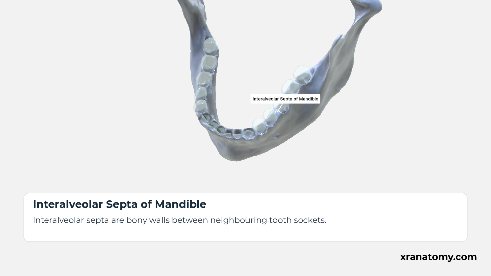
Interalveolar Septa of Mandible
Interalveolar septa are bony walls between neighbouring tooth sockets.
Illustration by XR Anatomy.
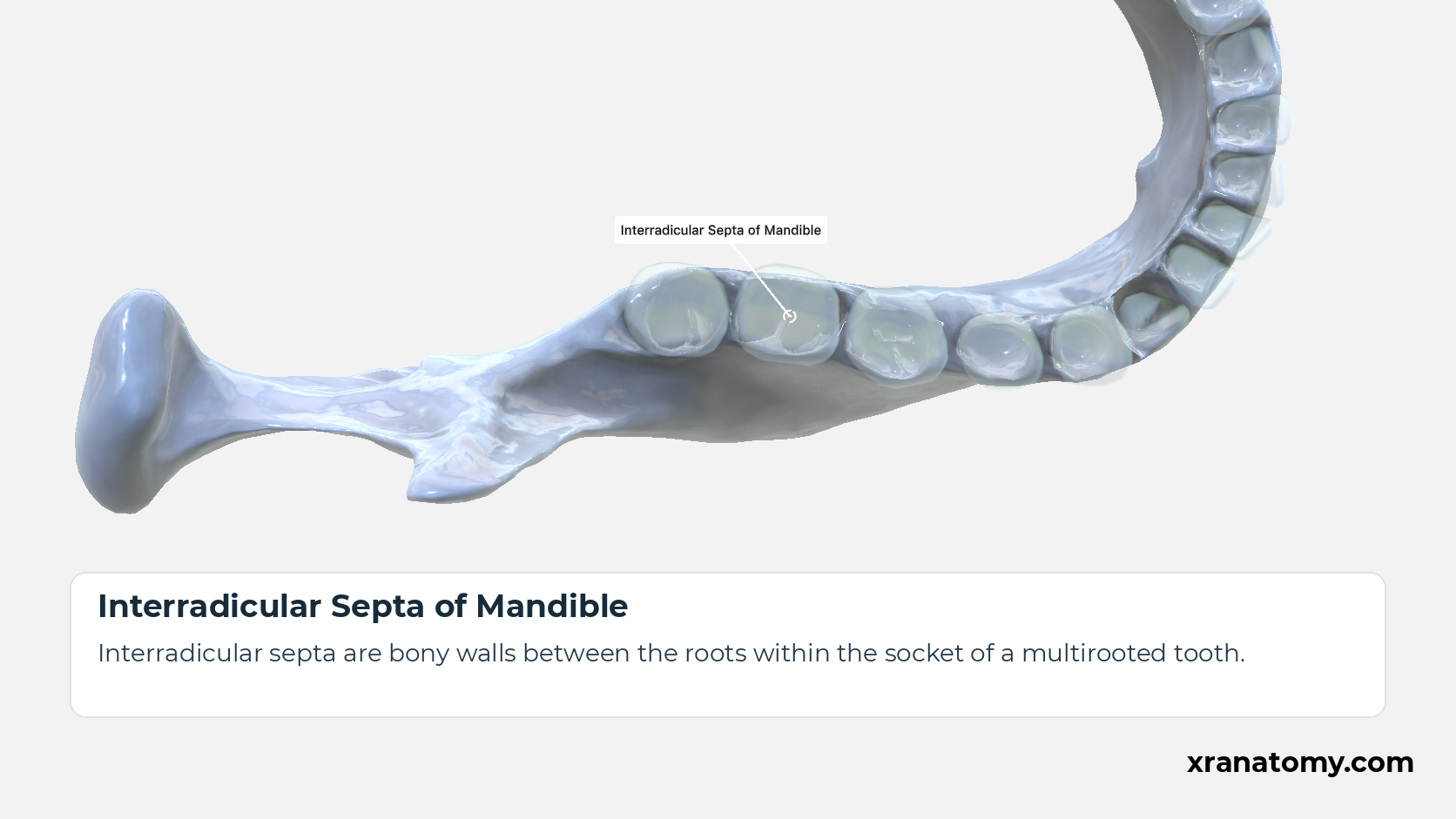
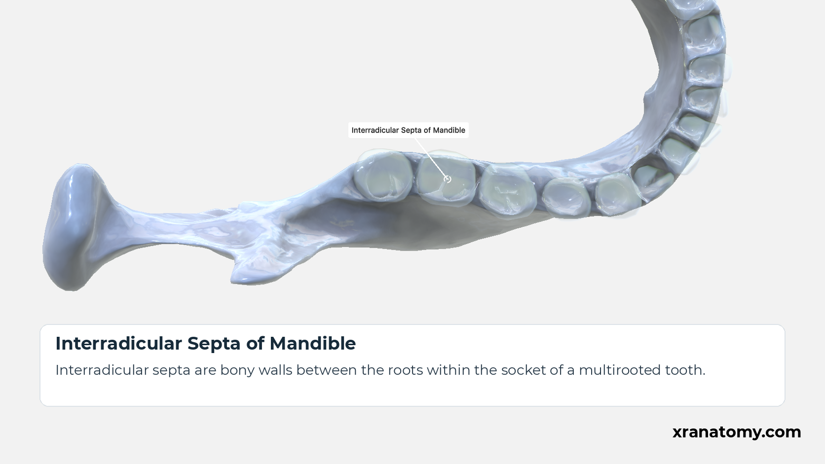
Interradicular Septa of Mandible
Interradicular septa are bony walls between the roots within the socket of a multirooted tooth.
Illustration by XR Anatomy.
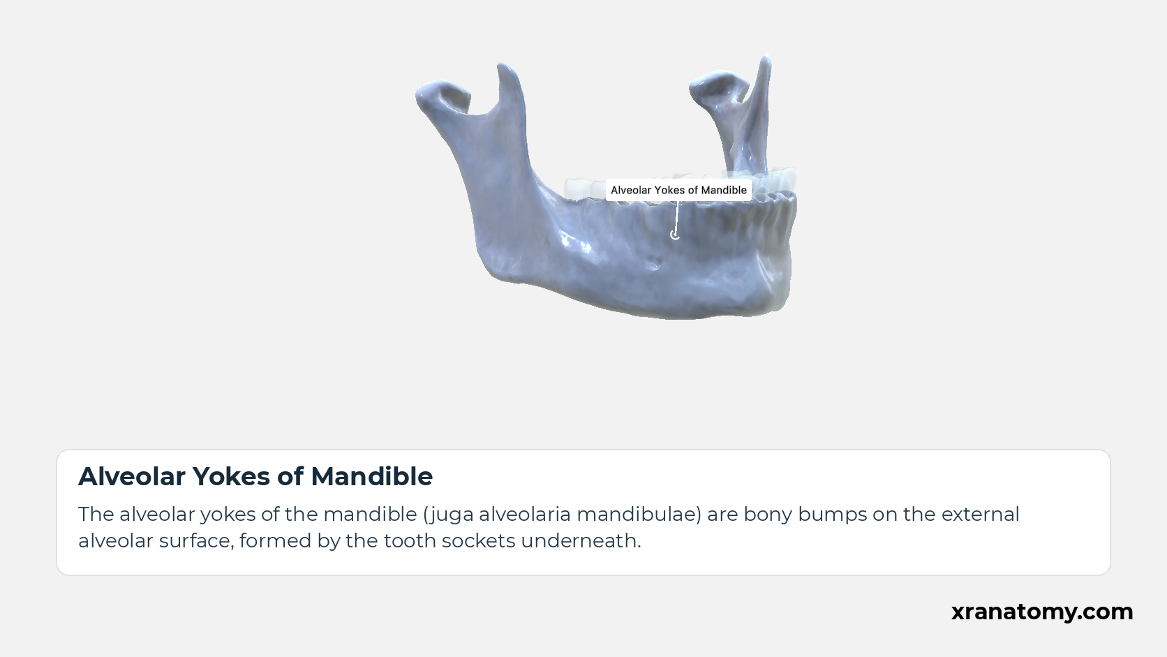
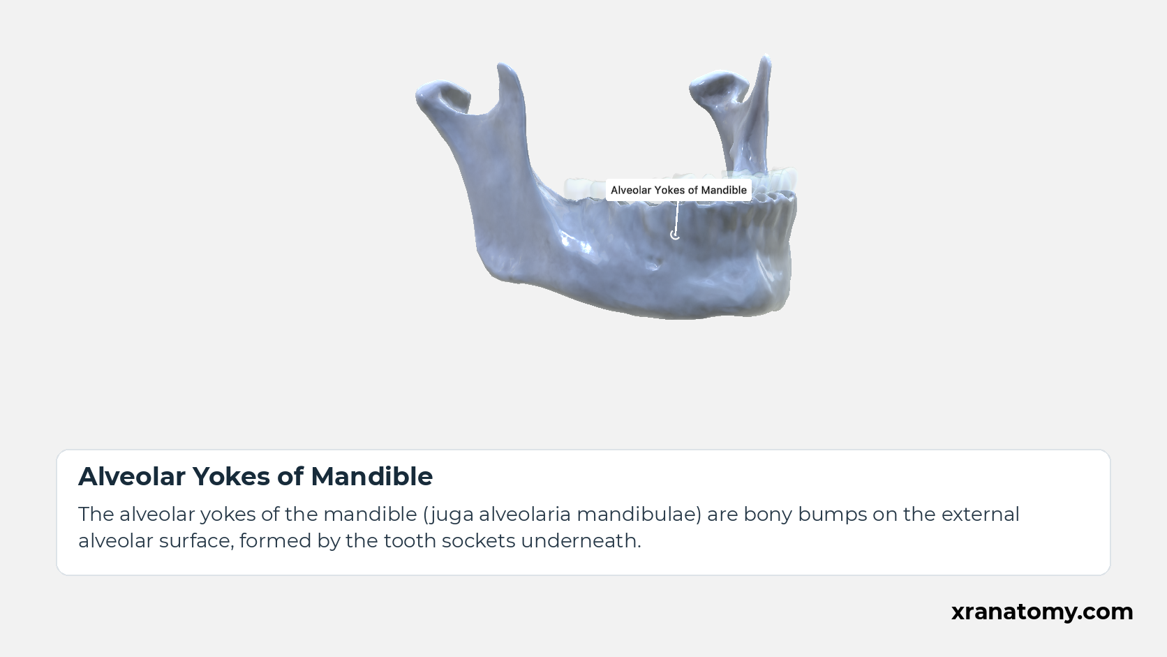
Alveolar Yokes of Mandible
The alveolar yokes of the mandible (juga alveolaria mandibulae) are bony bumps on the external alveolar surface, formed by the tooth sockets underneath.
Illustration by XR Anatomy.
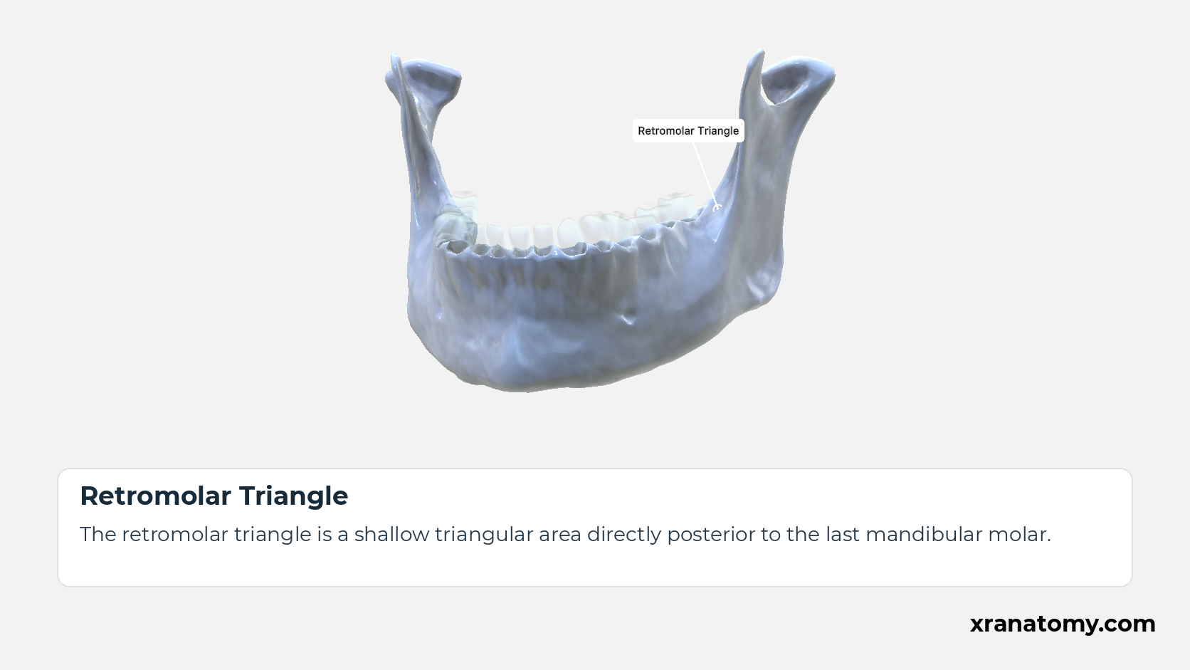
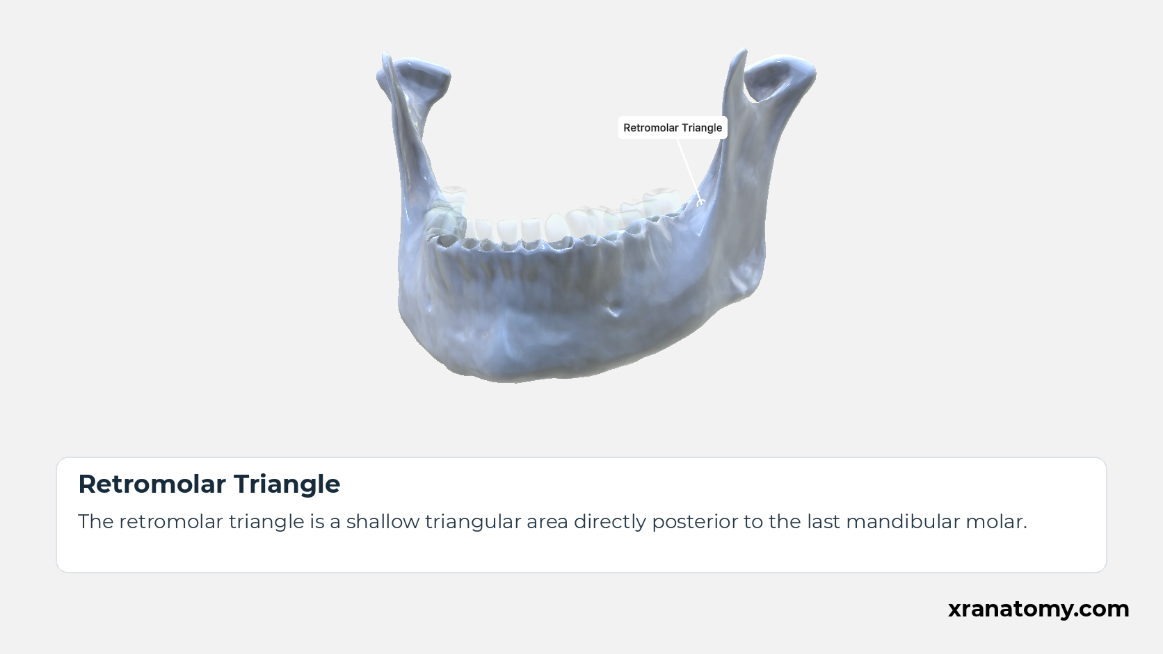
Retromolar Triangle
The retromolar triangle is a shallow triangular area directly posterior to the last mandibular molar.
Illustration by XR Anatomy.
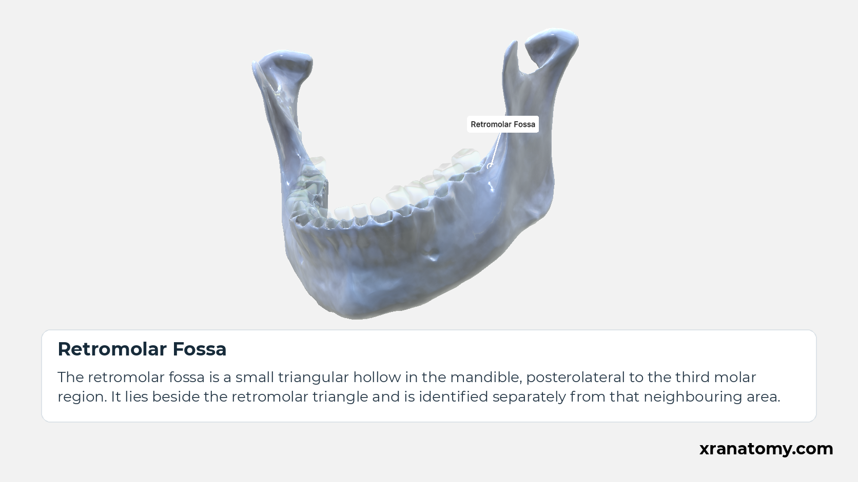
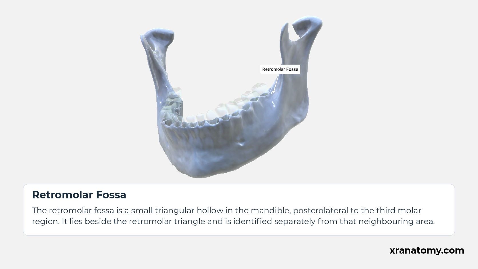
Retromolar Fossa
The retromolar fossa is a small triangular hollow in the mandible, posterolateral to the third molar region. It lies beside the retromolar triangle and is identified separately from that neighbouring area.
Illustration by XR Anatomy.
Alveolar Part of Mandible
The alveolar part of the mandible is the raised upper portion of its body that contains the sockets for the lower teeth. Its bony partitions separate neighbouring sockets and the roots of multirooted teeth, while its shape follows the lower dental arch.Show less
Illustration by XR Anatomy.
Alveolar Arch of Mandible
The alveolar arch is the curved, raised edge of the mandibular body that bears the teeth.
Illustration by XR Anatomy.
Dental Alveoli of Mandible
Dental alveoli are sockets for the roots of the lower teeth. Their shape and internal partitions vary according to the teeth they contain.
Illustration by XR Anatomy.
Interalveolar Septa of Mandible
Interalveolar septa are bony walls between neighbouring tooth sockets.
Illustration by XR Anatomy.
Interradicular Septa of Mandible
Interradicular septa are bony walls between the roots within the socket of a multirooted tooth.
Illustration by XR Anatomy.
Alveolar Yokes of Mandible
The alveolar yokes of the mandible (juga alveolaria mandibulae) are bony bumps on the external alveolar surface, formed by the tooth sockets underneath.
Illustration by XR Anatomy.
Retromolar Triangle
The retromolar triangle is a shallow triangular area directly posterior to the last mandibular molar.
Illustration by XR Anatomy.
Retromolar Fossa
The retromolar fossa is a small triangular hollow in the mandible, posterolateral to the third molar region. It lies beside the retromolar triangle and is identified separately from that neighbouring area.
Illustration by XR Anatomy.
Body of Mandible
The mandibular body is shaped like a horseshoe. It holds the tooth sockets and offers broad surfaces for muscle attachment.
Illustration by XR Anatomy.
 ↺ drag to rotate · scroll to zoom
Alveolar Part of the Mandible
Alveolar Part of the Mandible
Drag or use the viewer controls to inspect each labelled subdivision of the alveolar part.
Sourced from 3D Osteology.
The alveolar part is the raised superior portion of the body that anchors the lower teeth. Its landmarks include the alveolar arch, dental alveoli, interalveolar septa, interradicular septa, alveolar yokes, retromolar triangle, and retromolar fossa.
In depth · 7 sections · 1 min read7 more sections on this part7 sections open — reading In depth- Alveolar Arch of Mandible
- Dental Alveoli of Mandible
- Interalveolar Septa of Mandible
- Interradicular Septa of Mandible
- Alveolar Yokes of Mandible
- Retromolar Triangle
- Retromolar Fossa
Alveolar Arch of Mandible
The alveolar arch is the curved, raised edge of the mandibular body that bears the teeth.
Dental Alveoli of Mandible
Dental alveoli are sockets for the roots of the lower teeth. Their shape and internal partitions vary according to the teeth they contain.
Interalveolar Septa of Mandible
Interalveolar septa are bony walls between neighbouring tooth sockets.
Interradicular Septa of Mandible
Interradicular septa are bony walls between the roots within the socket of a multirooted tooth.
Alveolar Yokes of Mandible
The alveolar yokes of the mandible (juga alveolaria mandibulae) are bony bumps on the external alveolar surface, formed by the tooth sockets underneath.
Retromolar Triangle
The retromolar triangle is a shallow triangular area directly posterior to the last mandibular molar.
Retromolar Fossa
The retromolar fossa (fossa retromolaris) is a triangular depression behind the last molar in the mandibular alveolar part. This region is also commonly called the retromolar triangle.
Practice · Active recall · 5 questions Recall: Body of Mandible
The mandibular body is shaped like a .
horseshoe
The mandibular body is shaped like a horseshoe. It holds the tooth sockets and offers broad surfaces for muscle attachment.
Review this sectionHow well did you know it? · press 1–4
The mandibular body has a lower base and a raised part.
alveolar
The body has a lower base and a raised alveolar part. Landmarks on its outer and inner surfaces relate to the chin, glands, muscles, teeth, and neurovascular pathways.
Review this sectionHow well did you know it? · press 1–4
The raised alveolar part of the mandibular body bears the .
teeth
The base is the lower part of the body, while the alveolar part is the raised upper part that bears the teeth.
Review this sectionHow well did you know it? · press 1–4
The base is the part of the mandibular body.
inferior
The base is the inferior part of the mandibular body. Posteriorly, it continues into the lower border of each ramus.
Review this sectionHow well did you know it? · press 1–4
The alveolar part of the mandible anchors the teeth.
lower
The alveolar part is the raised superior portion of the body that anchors the lower teeth. Its landmarks include the alveolar arch, dental alveoli, interalveolar septa, interradicular septa, alveolar yokes, retromolar triangle, and retromolar fossa.
Review this sectionHow well did you know it? · press 1–4
Nice work — revisit in a few days.
Ramus of Mandible
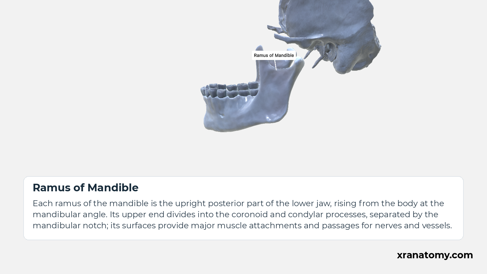
Ramus of Mandible
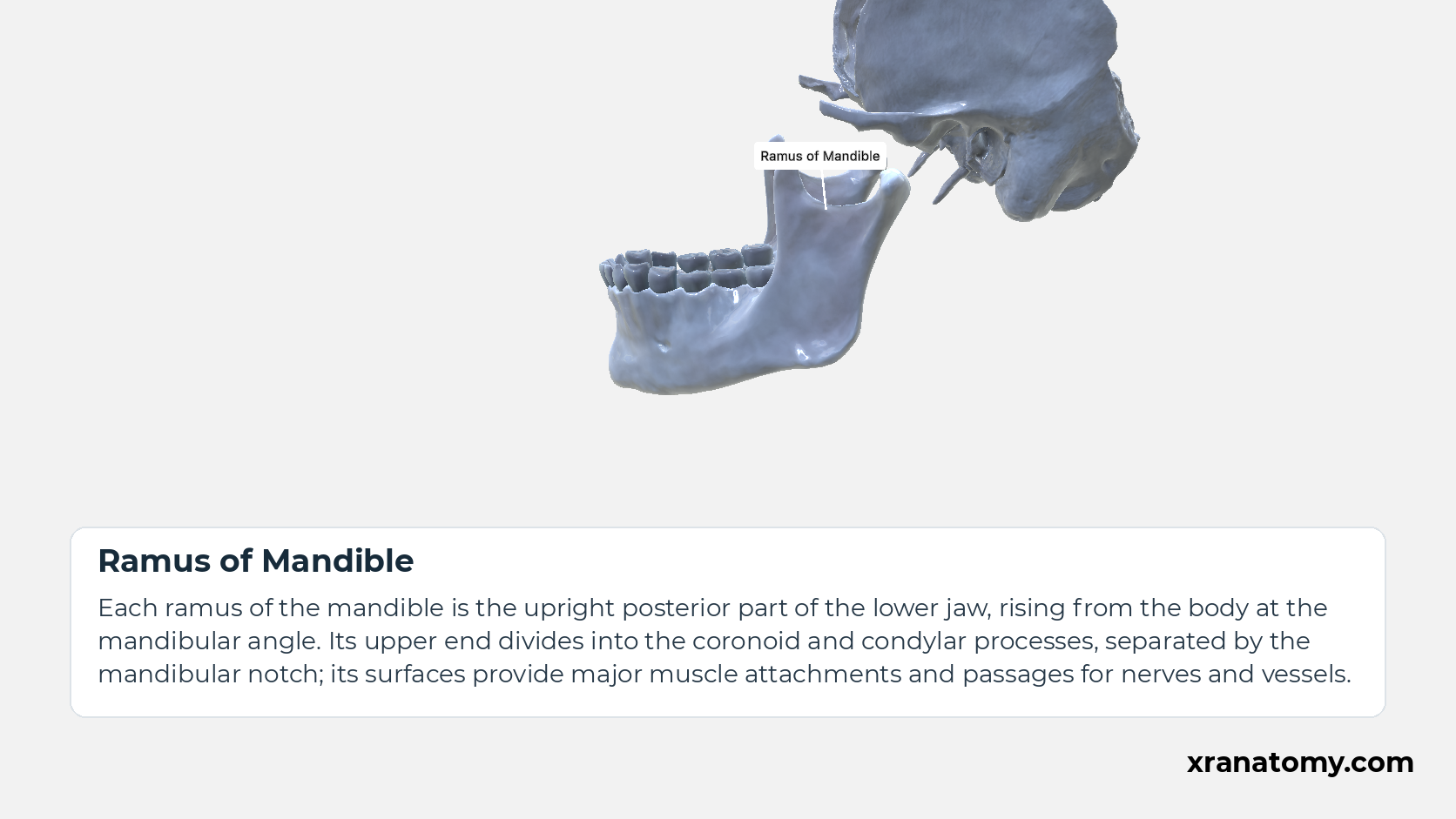
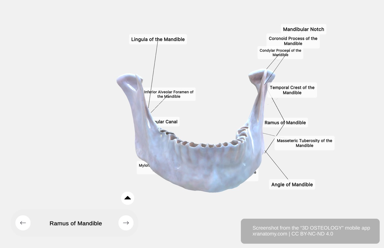
Each ramus of the mandible is the upright posterior part of the lower jaw, rising from the body at the mandibular angle. Its upper end divides into the coronoid and condylar processes, separated by the mandibular notch; its surfaces provide major muscle attachments and passages for nerves and vessels.Show less
Study Ramus of Mandible in this fixed image. The image does not rotate.
Illustration by XR Anatomy.
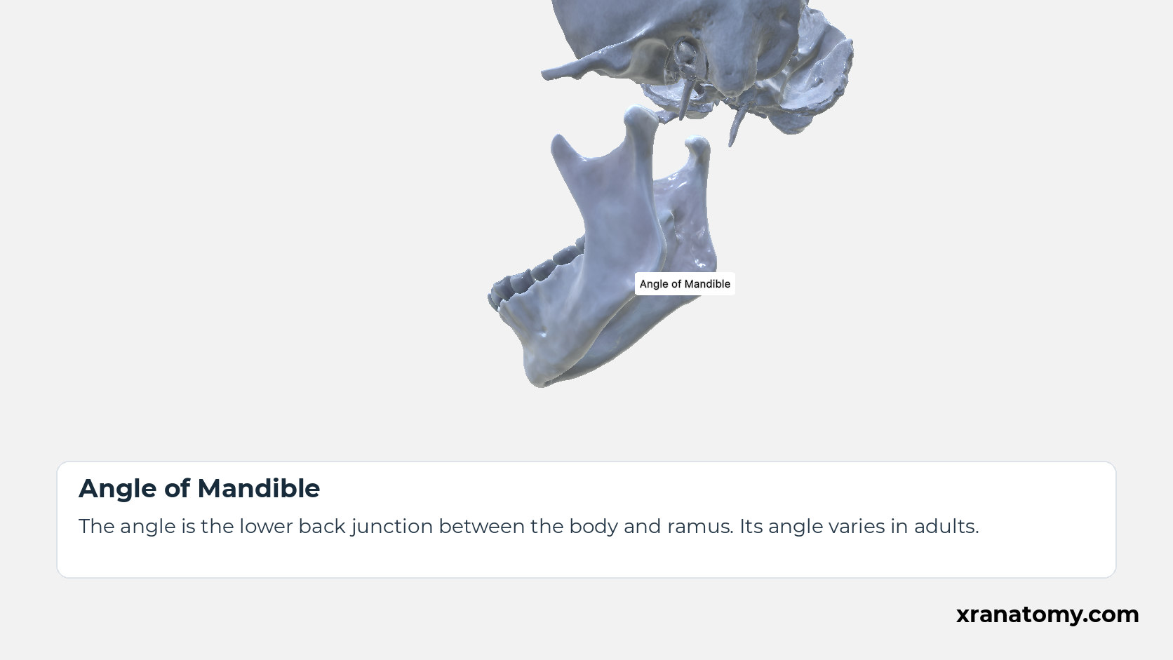
Angle of Mandible
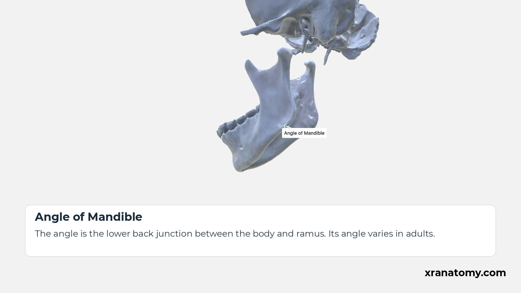
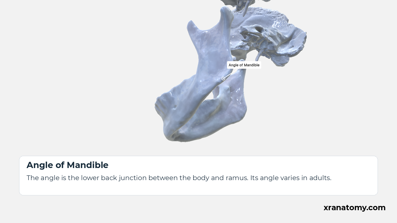
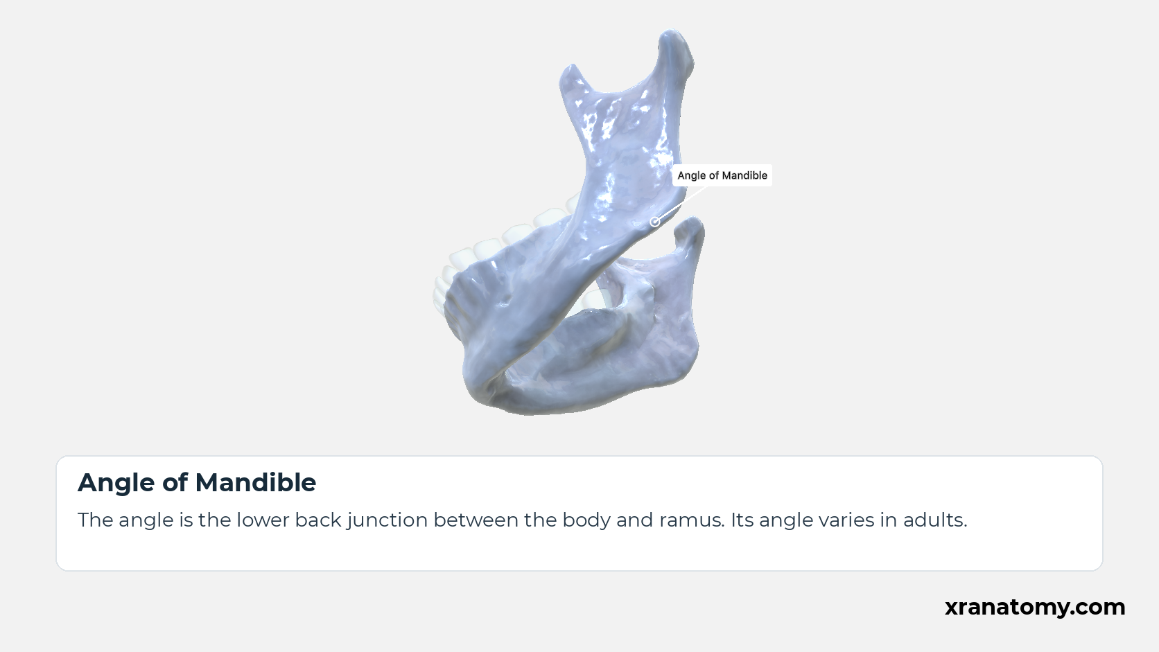
The angle is the lower back junction between the body and ramus. Its angle varies in adults.
Illustration by XR Anatomy.
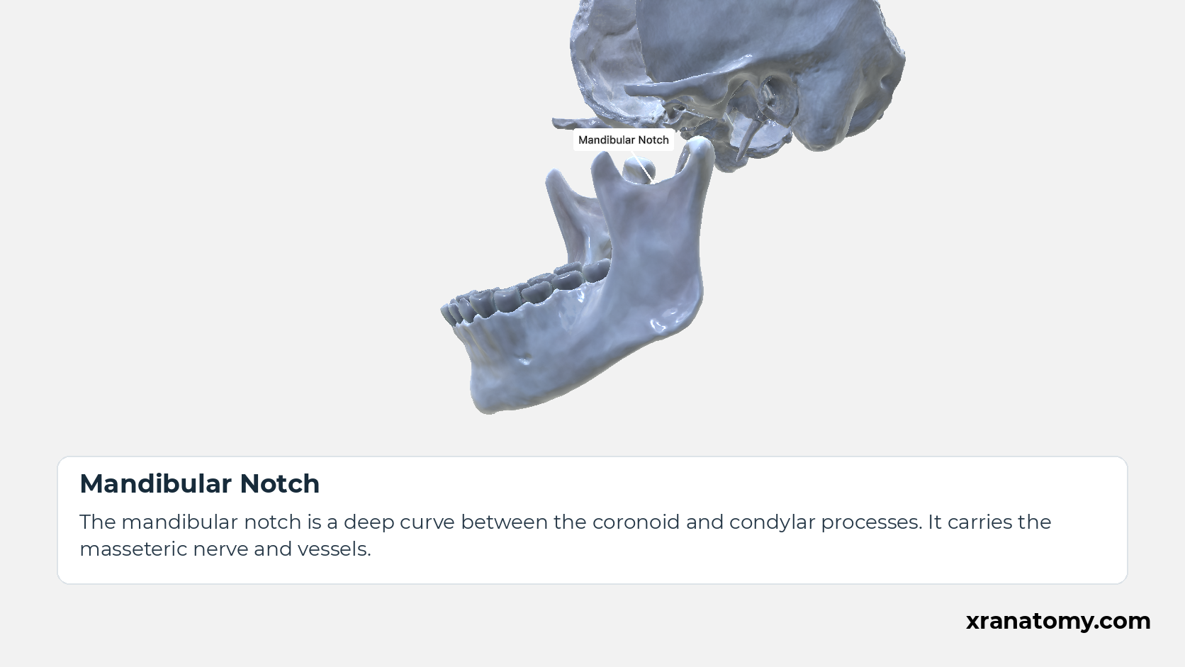
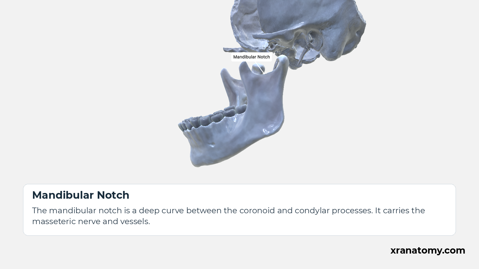
Mandibular Notch
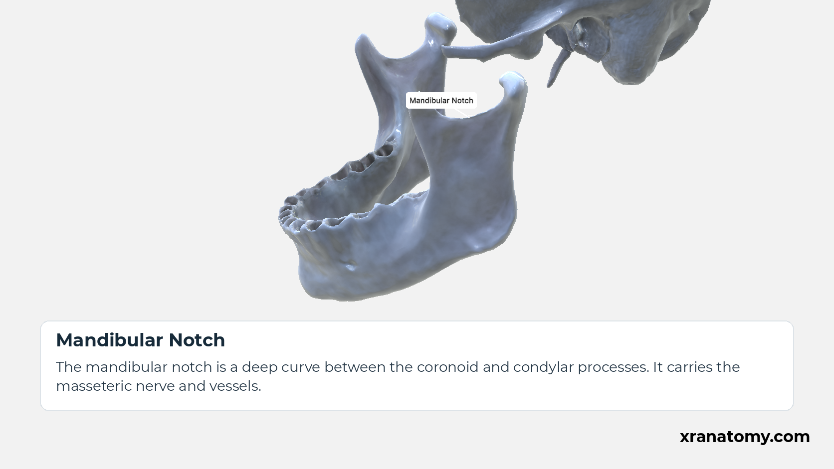
The mandibular notch is a deep curve between the coronoid and condylar processes. It carries the masseteric nerve and vessels.
Illustration by XR Anatomy.
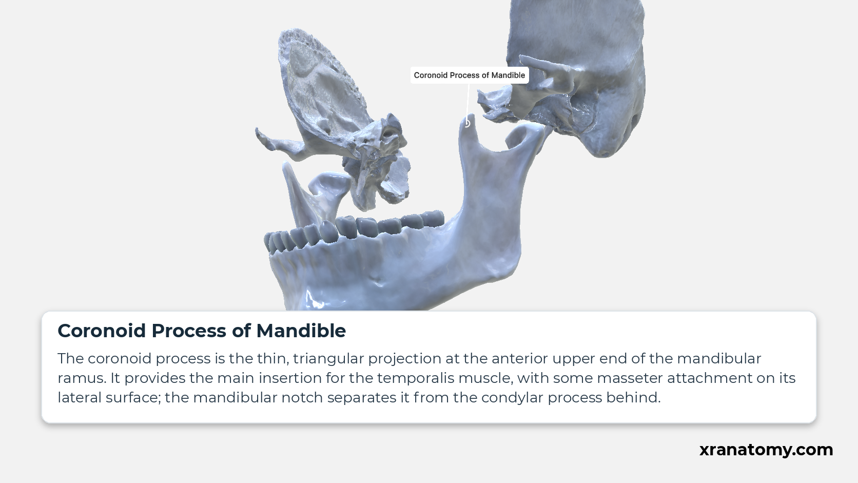
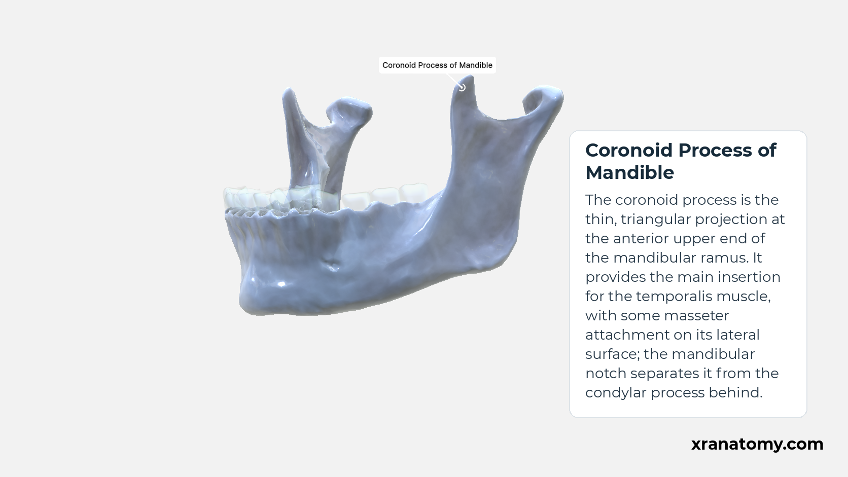
Coronoid Process of Mandible
The coronoid process is the thin, triangular projection at the anterior upper end of the mandibular ramus. It provides the main insertion for the temporalis muscle, with some masseter attachment on its lateral surface; the mandibular notch separates it from the condylar process behind.Show less
Illustration by XR Anatomy.
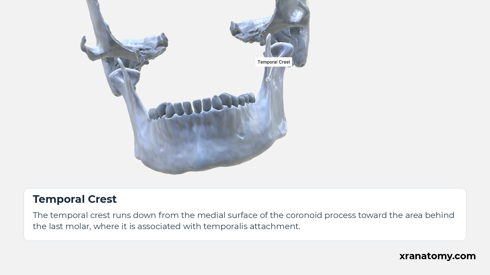
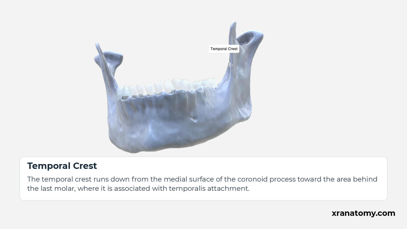
Temporal Crest
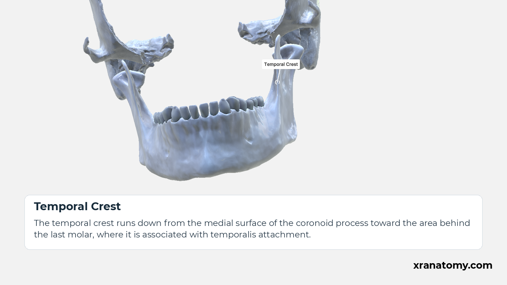
The temporal crest runs down from the medial surface of the coronoid process toward the area behind the last molar, where it is associated with temporalis attachment.
Illustration by XR Anatomy.
Ramus of Mandible
Each ramus of the mandible is the upright posterior part of the lower jaw, rising from the body at the mandibular angle. Its upper end divides into the coronoid and condylar processes, separated by the mandibular notch; its surfaces provide major muscle attachments and passages for nerves and vessels.Show less
Illustration by XR Anatomy.
Angle of Mandible
The angle is the lower back junction between the body and ramus. Its angle varies in adults.
Illustration by XR Anatomy.
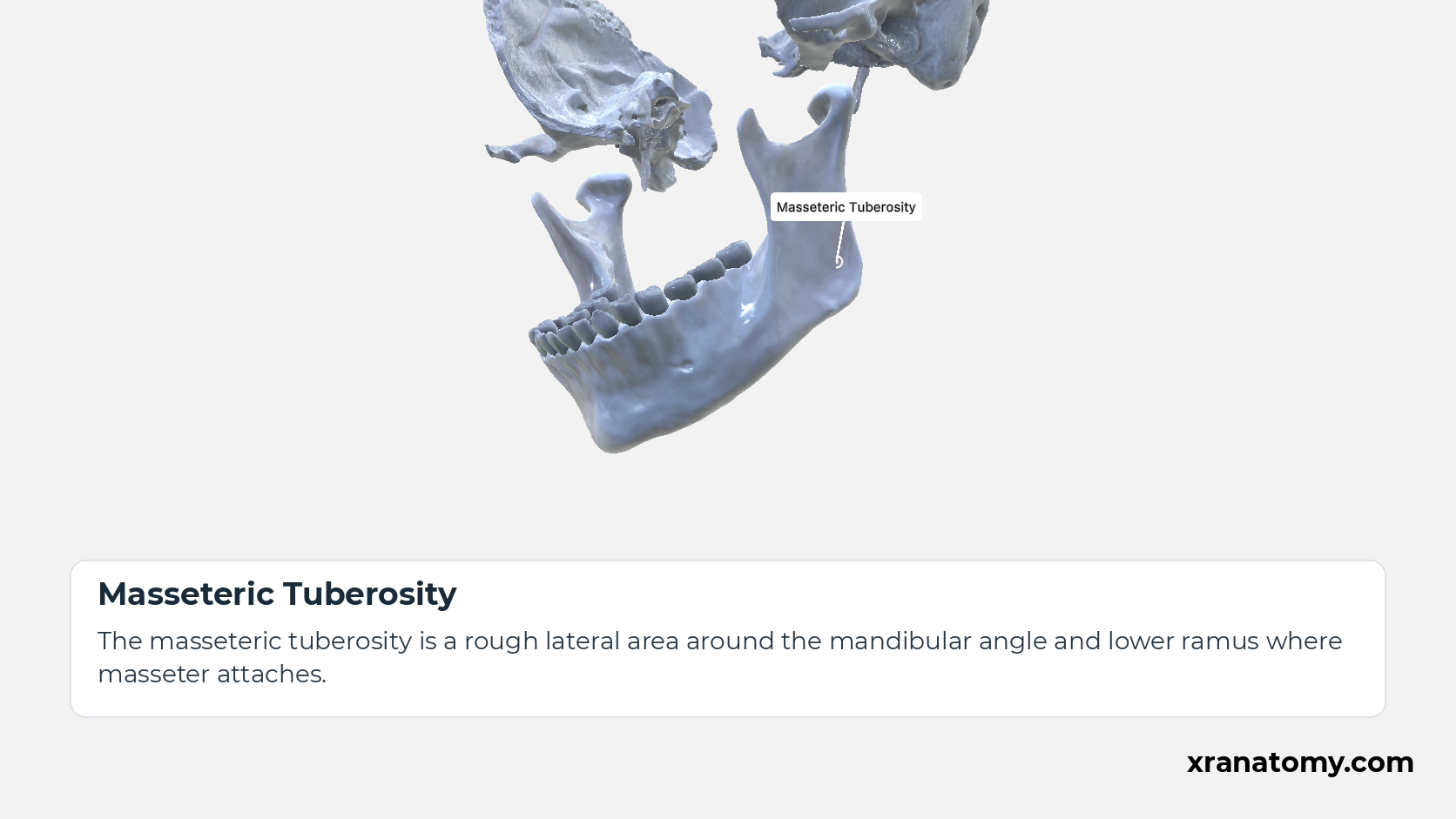
Masseteric Tuberosity
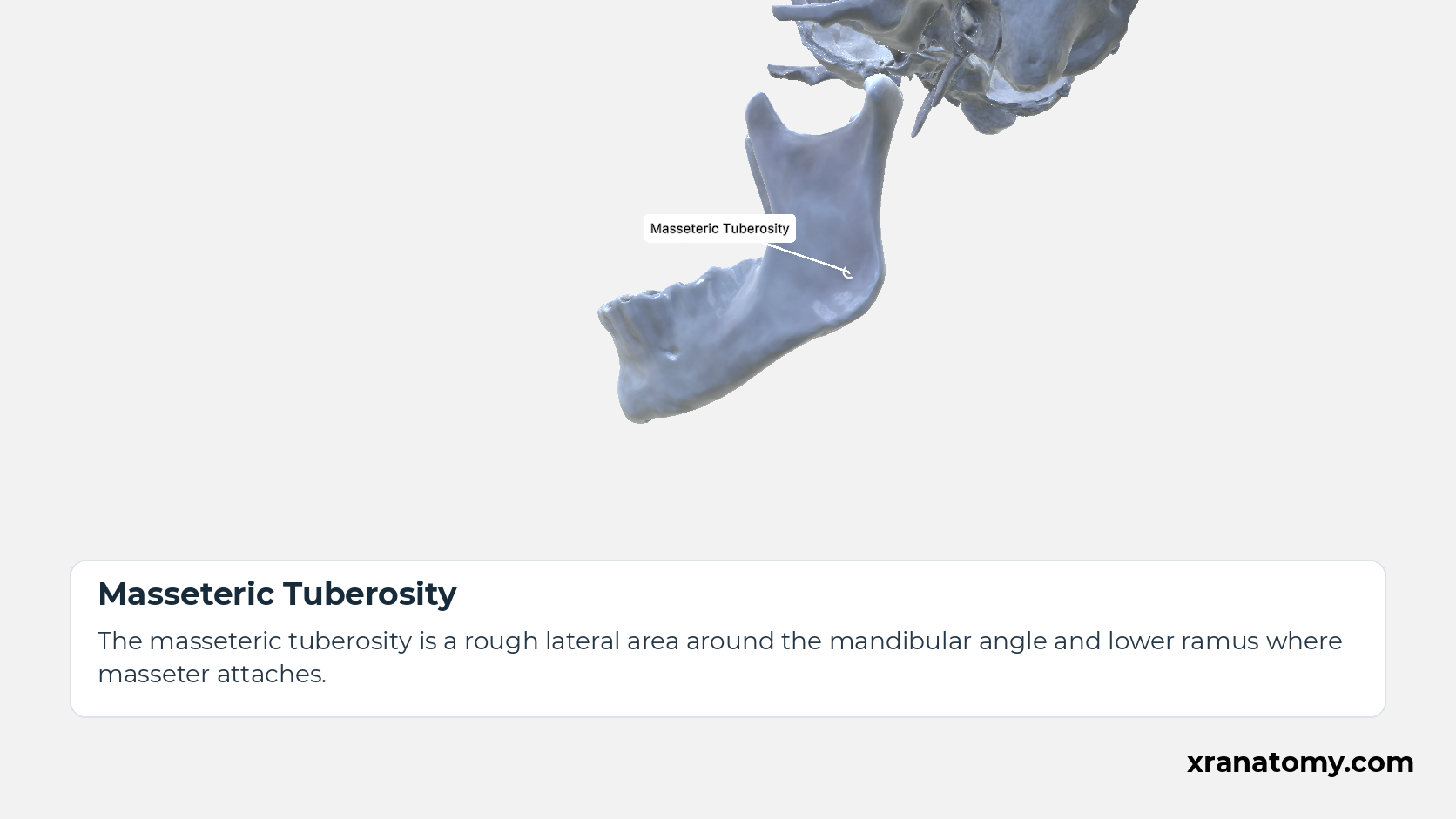
The masseteric tuberosity is a rough lateral area around the mandibular angle and lower ramus where masseter attaches.
Illustration by XR Anatomy.
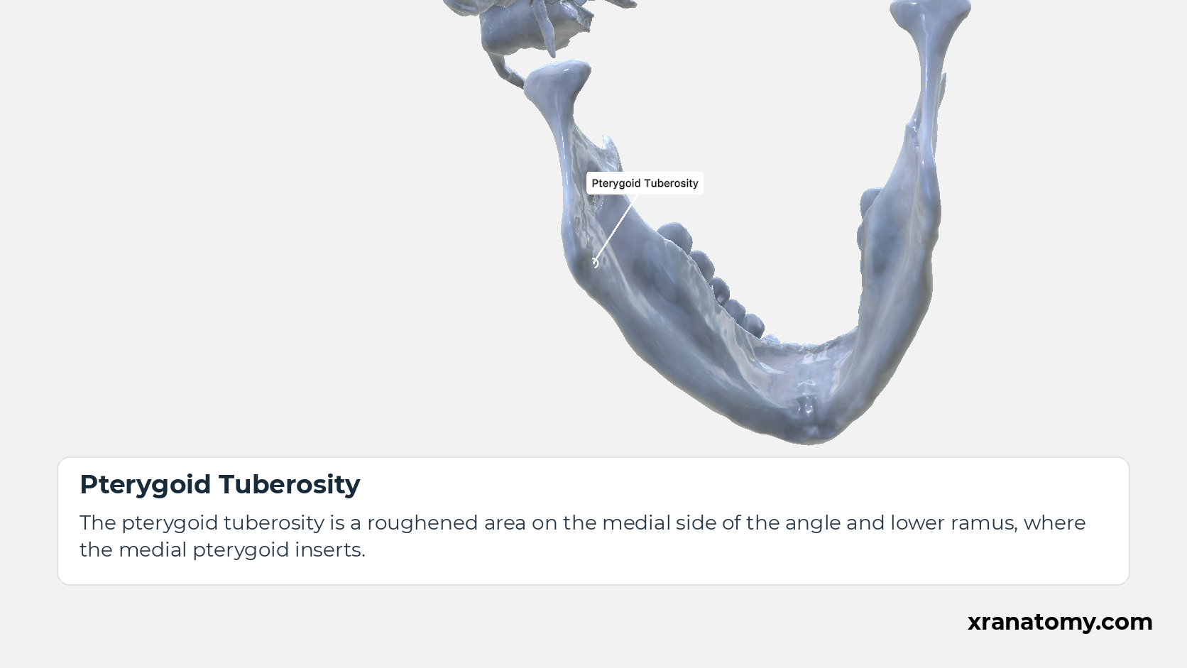
Pterygoid Tuberosity
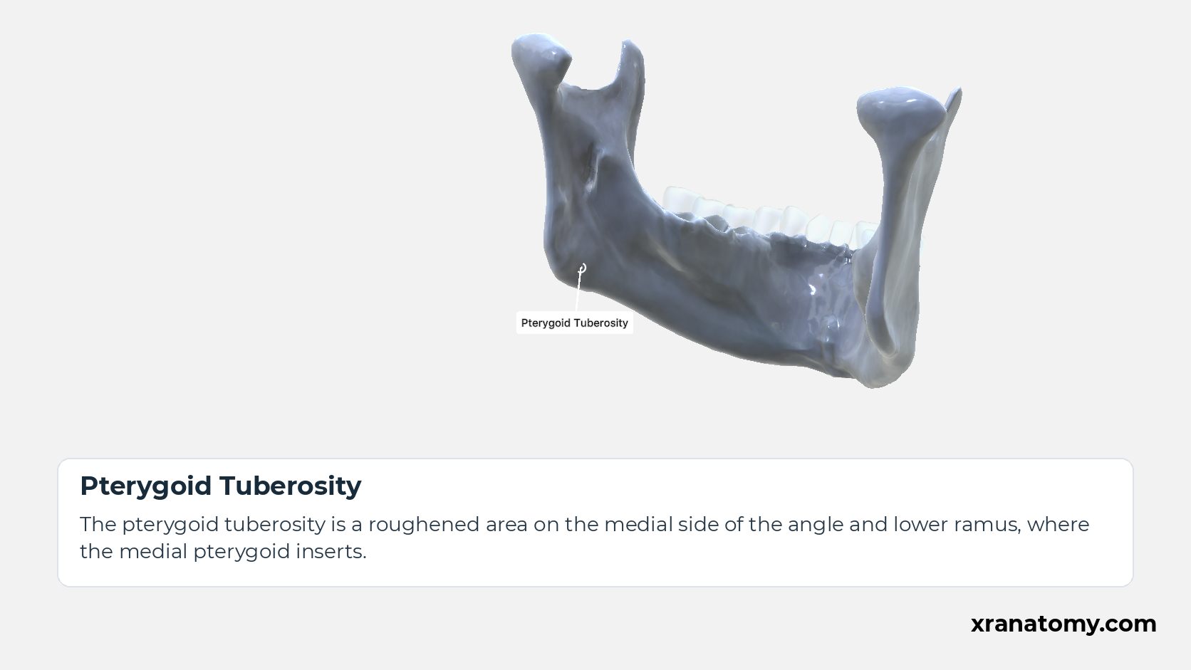
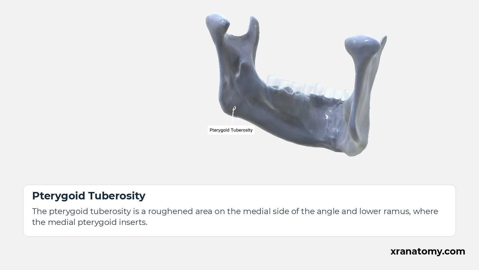
The pterygoid tuberosity is a roughened area on the medial side of the angle and lower ramus, where the medial pterygoid inserts.
Illustration by XR Anatomy.
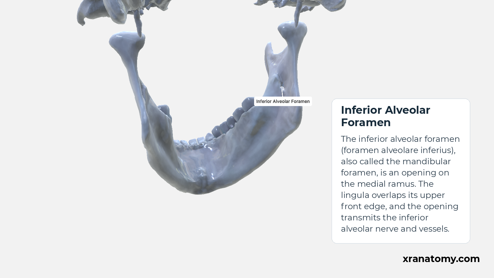
Inferior Alveolar Foramen
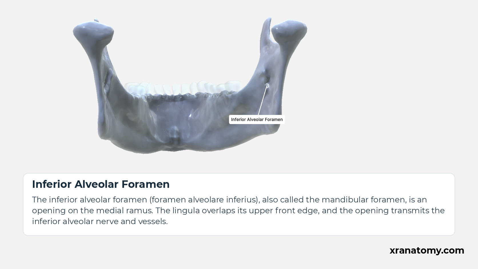
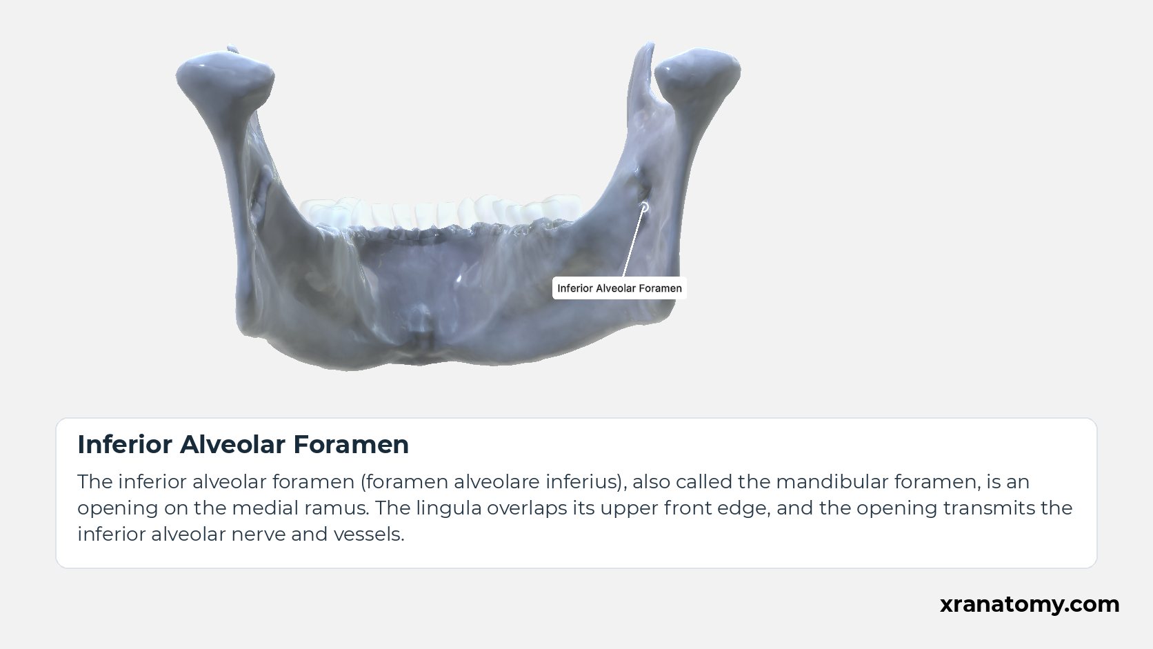
The inferior alveolar foramen (foramen alveolare inferius), also called the mandibular foramen, is an opening on the medial ramus. The lingula overlaps its upper front edge, and the opening transmits the inferior alveolar nerve and vessels.
Illustration by XR Anatomy.
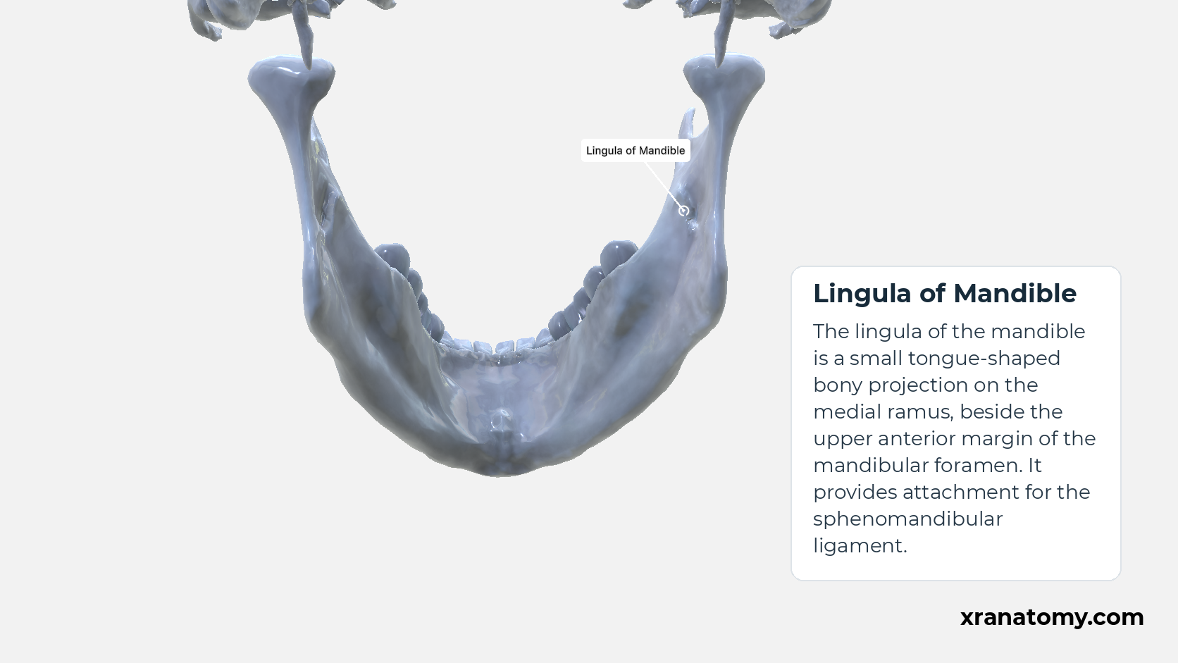
Lingula of Mandible
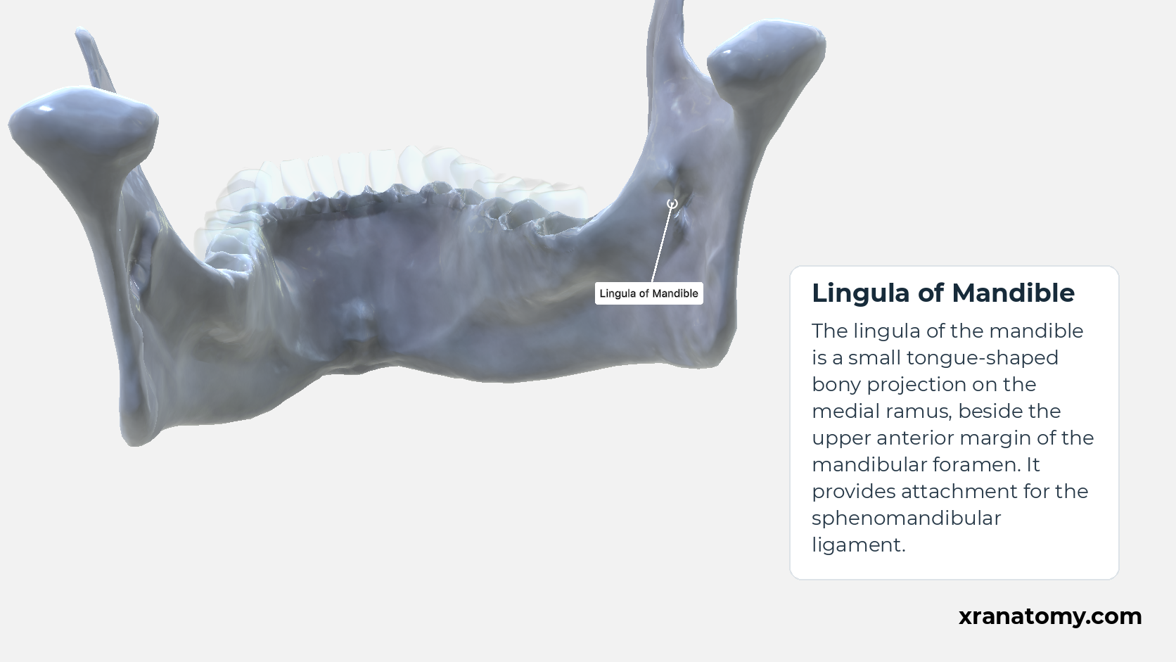
The lingula of the mandible is a small tongue-shaped bony projection on the medial ramus, beside the upper anterior margin of the mandibular foramen. It provides attachment for the sphenomandibular ligament.
Illustration by XR Anatomy.
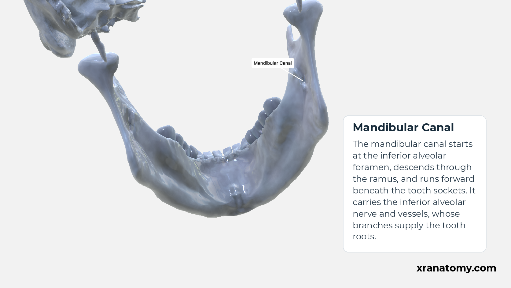
Mandibular Canal
The mandibular canal starts at the inferior alveolar foramen, descends through the ramus, and runs forward beneath the tooth sockets. It carries the inferior alveolar nerve and vessels, whose branches supply the tooth roots.
Illustration by XR Anatomy.
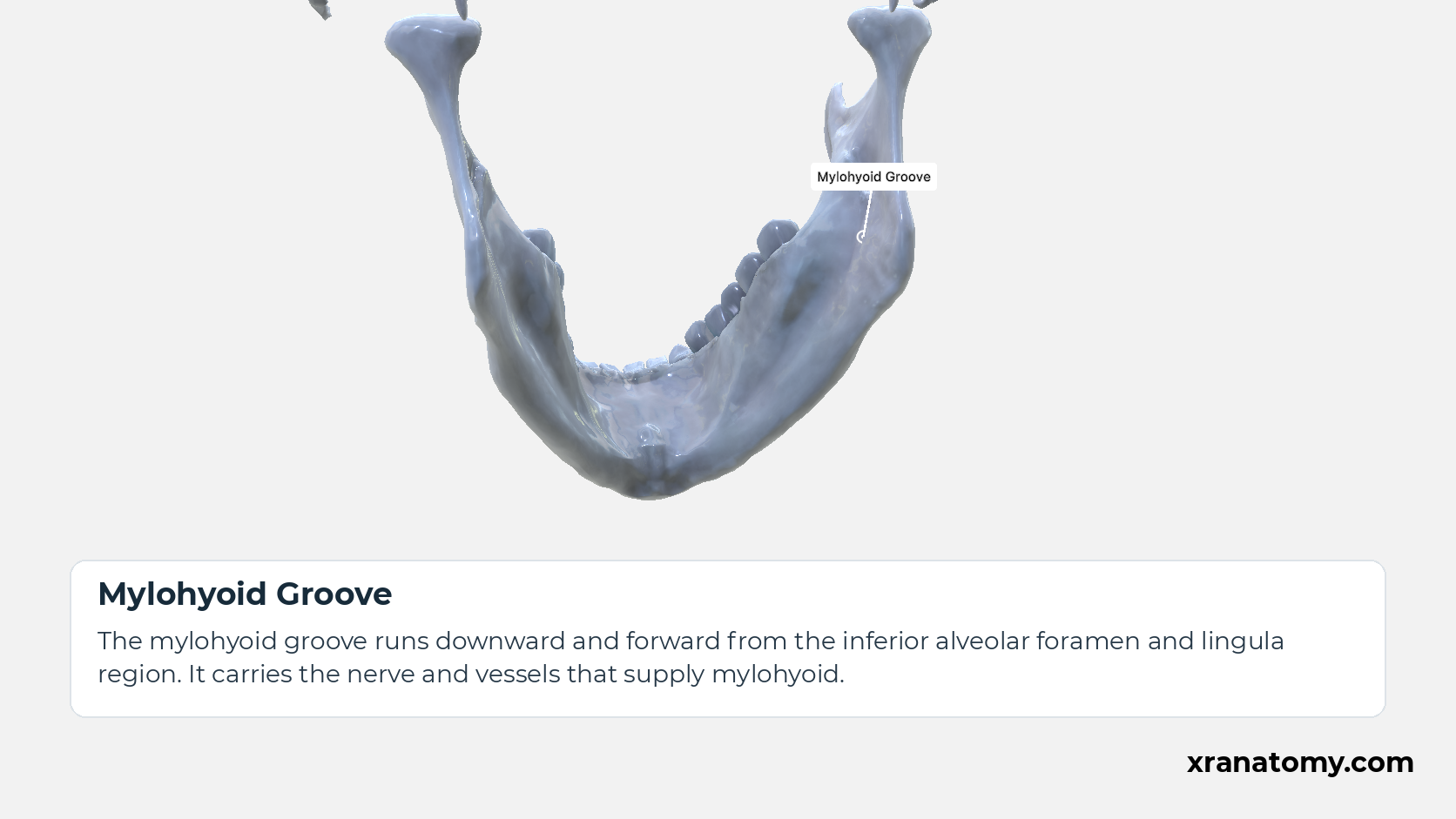
Mylohyoid Groove
The mylohyoid groove runs downward and forward from the inferior alveolar foramen and lingula region. It carries the nerve and vessels that supply mylohyoid.
Illustration by XR Anatomy.
Mandibular Notch
The mandibular notch is a deep curve between the coronoid and condylar processes. It carries the masseteric nerve and vessels.
Illustration by XR Anatomy.
Coronoid Process of Mandible
The coronoid process is the thin, triangular projection at the anterior upper end of the mandibular ramus. It provides the main insertion for the temporalis muscle, with some masseter attachment on its lateral surface; the mandibular notch separates it from the condylar process behind.Show less
Illustration by XR Anatomy.
Temporal Crest
The temporal crest runs down from the medial surface of the coronoid process toward the area behind the last molar, where it is associated with temporalis attachment.
Illustration by XR Anatomy.
Condylar Process
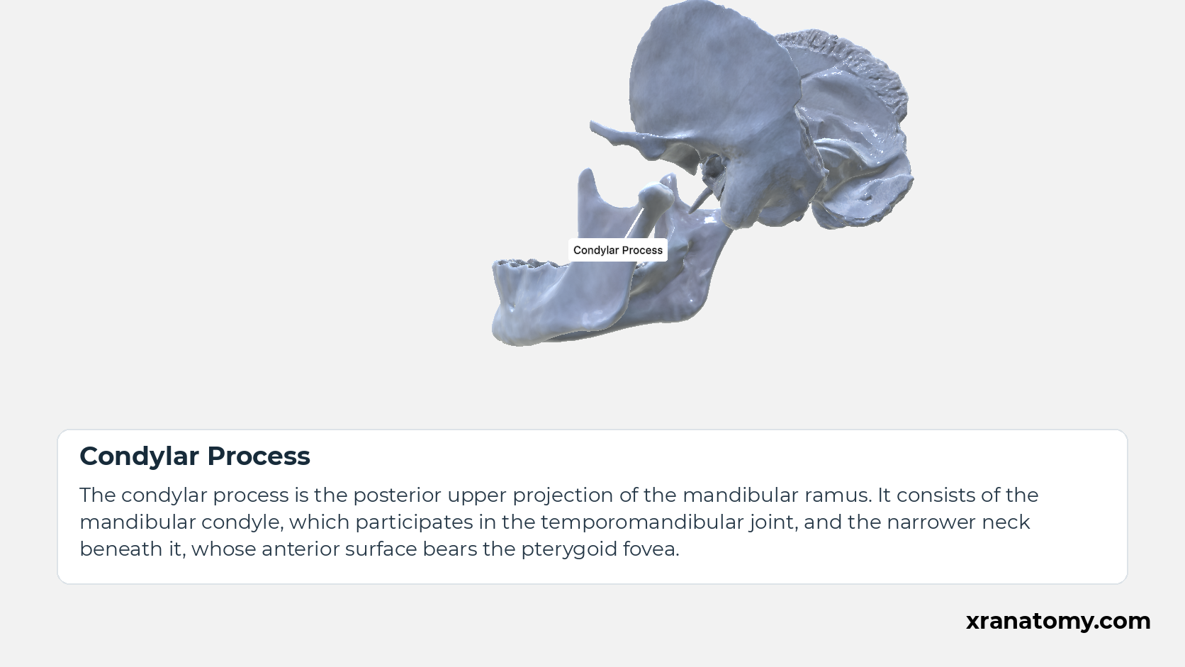
The condylar process is the posterior upper projection of the mandibular ramus. It consists of the mandibular condyle, which participates in the temporomandibular joint, and the narrower neck beneath it, whose anterior surface bears the pterygoid fovea.Show less
Illustration by XR Anatomy.
Mandibular Condyle
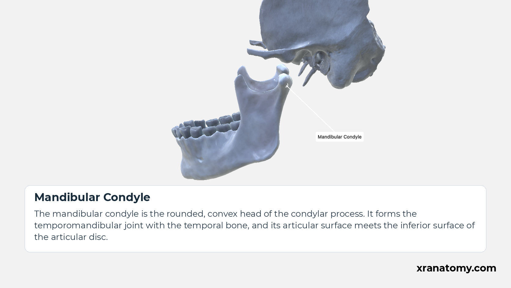
The mandibular condyle is the rounded, convex head of the condylar process. It forms the temporomandibular joint with the temporal bone, and its articular surface meets the inferior surface of the articular disc.
Illustration by XR Anatomy.
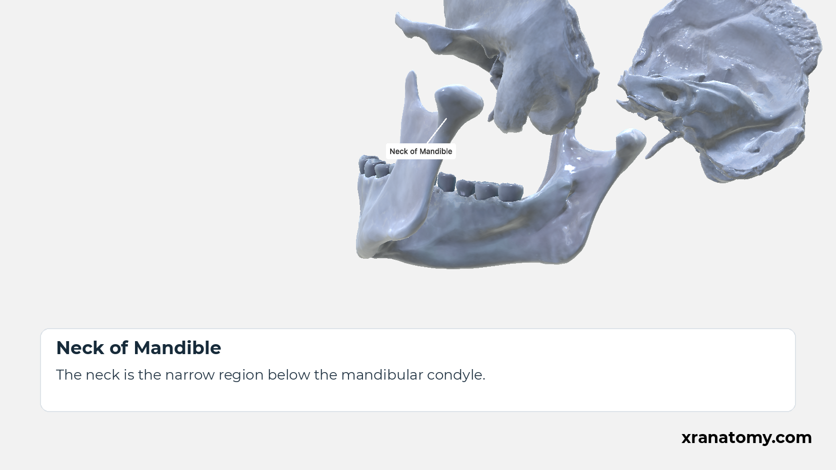
Neck of Mandible
The neck is the narrow region below the mandibular condyle.
Illustration by XR Anatomy.
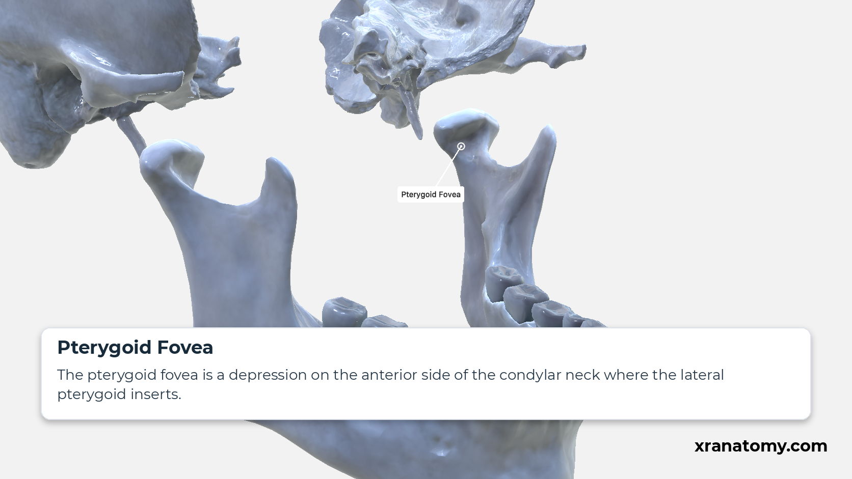
Pterygoid Fovea
The pterygoid fovea is a depression on the anterior side of the condylar neck where the lateral pterygoid inserts.
Illustration by XR Anatomy.
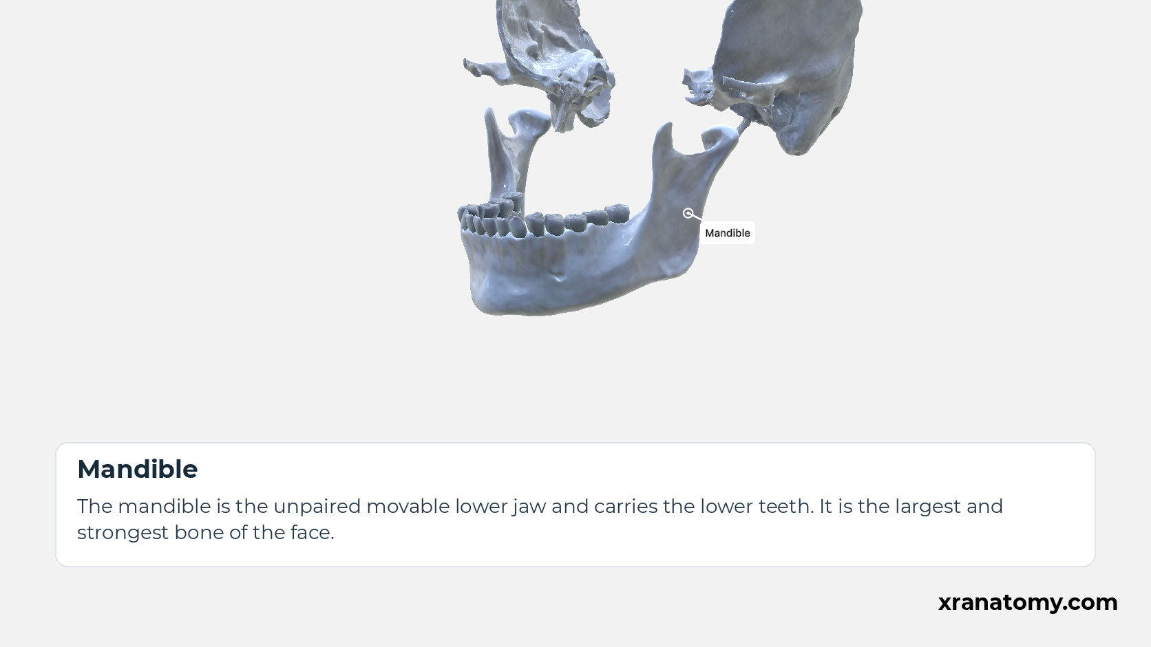
Mandible
The mandible is the unpaired movable lower jaw and carries the lower teeth. It is the largest and strongest bone of the face.
Illustration by XR Anatomy.
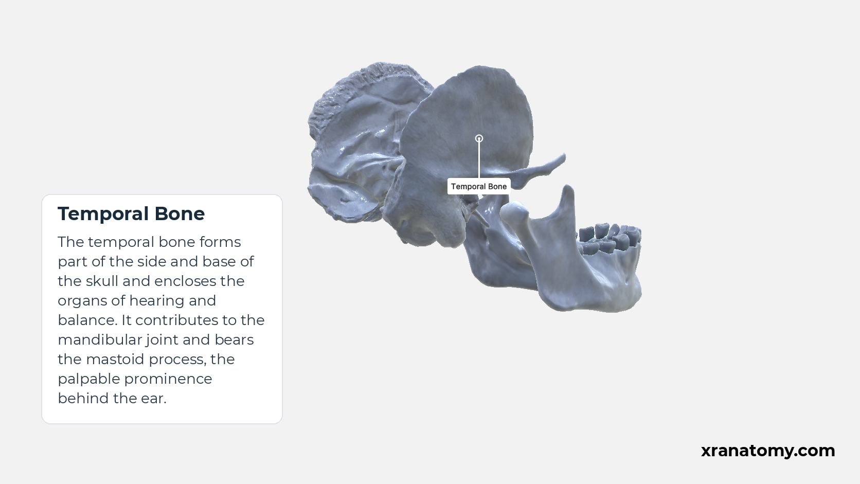
Temporal Bone
The temporal bone forms part of the side and base of the skull and encloses the organs of hearing and balance. It contributes to the mandibular joint and bears the mastoid process, the palpable prominence behind the ear.
Illustration by XR Anatomy.
Body of Mandible
The mandibular body is shaped like a horseshoe. It holds the tooth sockets and offers broad surfaces for muscle attachment.
Illustration by XR Anatomy.
 ↺ drag to rotate · scroll to zoom
Ramus of the Mandible
Ramus of the Mandible
Drag or use the viewer controls to inspect the labelled ramus landmarks from different sides.
Sourced from 3D Osteology.
Each ramus rises from the posterior part of the body at the angle. Its main features are the lateral surface, medial surface, inferior border, mandibular notch, coronoid process, and condylar process.
Lateral Surface
Masseteric Tuberosity
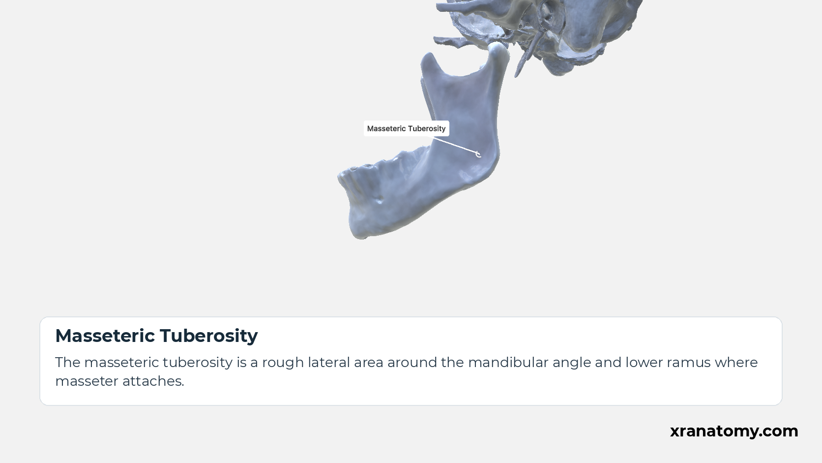
The masseteric tuberosity is a rough lateral area around the mandibular angle and lower ramus where masseter attaches.
Study Masseteric Tuberosity in this fixed image. The image does not rotate.
Illustration by XR Anatomy.
Mandible
The mandible is the unpaired movable lower jaw and carries the lower teeth. It is the largest and strongest bone of the face.
Illustration by XR Anatomy.
Ramus of Mandible
Each ramus of the mandible is the upright posterior part of the lower jaw, rising from the body at the mandibular angle. Its upper end divides into the coronoid and condylar processes, separated by the mandibular notch; its surfaces provide major muscle attachments and passages for nerves and vessels.Show less
Illustration by XR Anatomy.
Masseteric Tuberosity
The masseteric tuberosity is a rough lateral area around the mandibular angle and lower ramus where masseter attaches.
Illustration by XR Anatomy.
Angle of Mandible
The angle is the lower back junction between the body and ramus. Its angle varies in adults.
Illustration by XR Anatomy.
Mandibular Notch
The mandibular notch is a deep curve between the coronoid and condylar processes. It carries the masseteric nerve and vessels.
Illustration by XR Anatomy.
Coronoid Process of Mandible
The coronoid process is the thin, triangular projection at the anterior upper end of the mandibular ramus. It provides the main insertion for the temporalis muscle, with some masseter attachment on its lateral surface; the mandibular notch separates it from the condylar process behind.Show less
Illustration by XR Anatomy.
Condylar Process
The condylar process is the posterior upper projection of the mandibular ramus. It consists of the mandibular condyle, which participates in the temporomandibular joint, and the narrower neck beneath it, whose anterior surface bears the pterygoid fovea.Show less
Illustration by XR Anatomy.
The masseter attaches across much of the lateral surface of the ramus. Lower oblique ridges mark the masseteric tuberosity, while deeper fibres also attach to the upper ramus and coronoid process.
In depth · 1 min read Masseteric Tuberosity
The masseteric tuberosity is a rough lateral area around the mandibular angle and lower ramus where masseter attaches.
Medial Surface
Pterygoid Tuberosity
The pterygoid tuberosity is a roughened area on the medial side of the angle and lower ramus, where the medial pterygoid inserts.
Study Pterygoid Tuberosity in this fixed image. Use the thumbnails to move between the 2D views. The image does not rotate.
Illustration by XR Anatomy.
Inferior Alveolar Foramen
The inferior alveolar foramen (foramen alveolare inferius), also called the mandibular foramen, is an opening on the medial ramus. The lingula overlaps its upper front edge, and the opening transmits the inferior alveolar nerve and vessels.
Illustration by XR Anatomy.
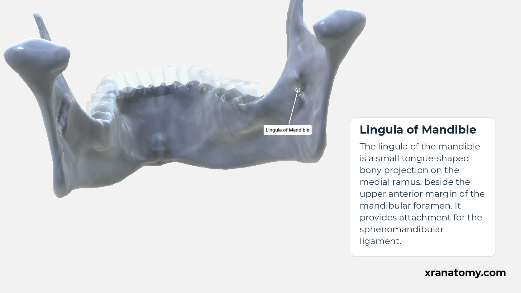
Lingula of Mandible
The lingula of the mandible is a small tongue-shaped bony projection on the medial ramus, beside the upper anterior margin of the mandibular foramen. It provides attachment for the sphenomandibular ligament.
Illustration by XR Anatomy.
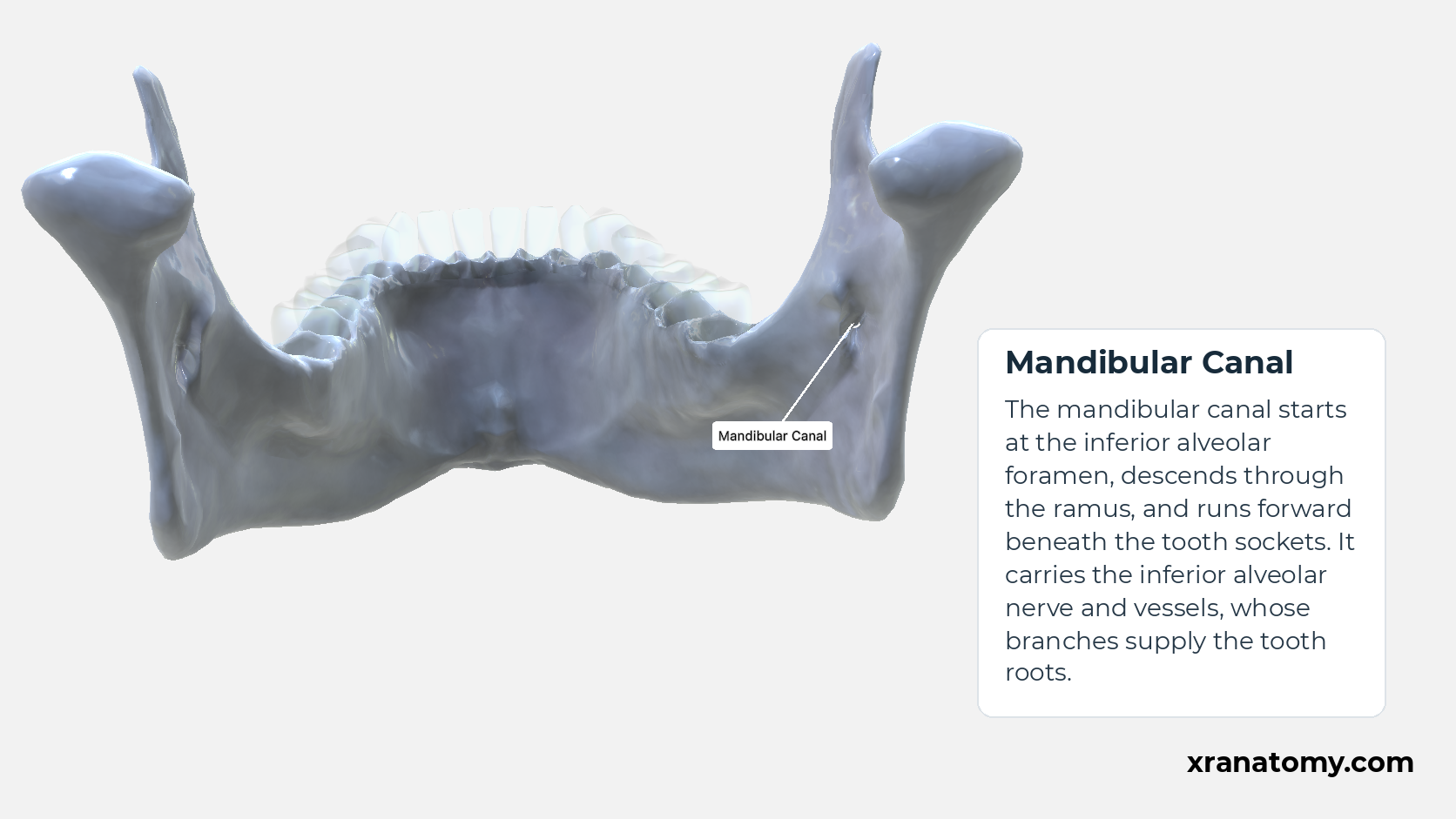
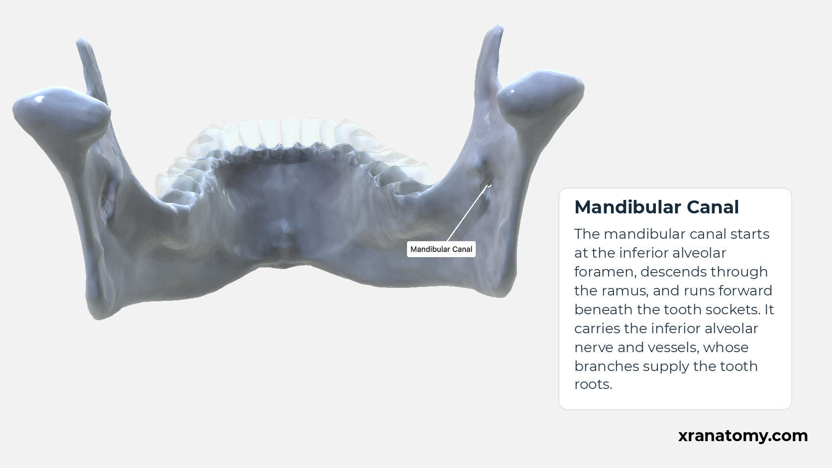
Mandibular Canal
The mandibular canal starts at the inferior alveolar foramen, descends through the ramus, and runs forward beneath the tooth sockets. It carries the inferior alveolar nerve and vessels, whose branches supply the tooth roots.
Illustration by XR Anatomy.
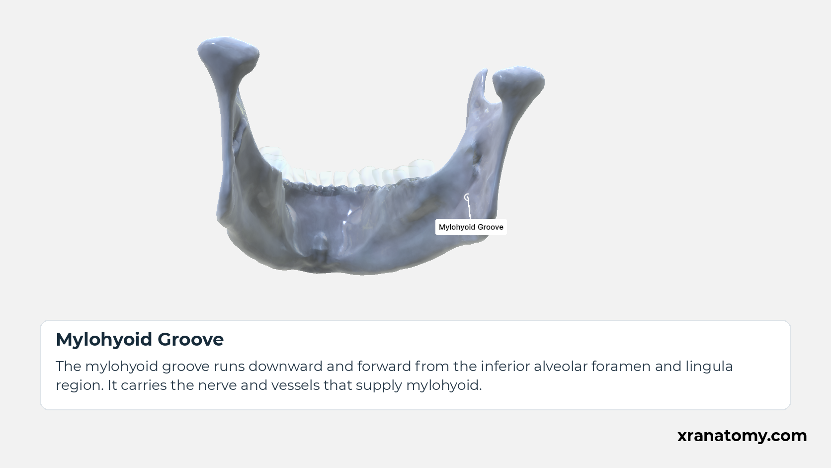
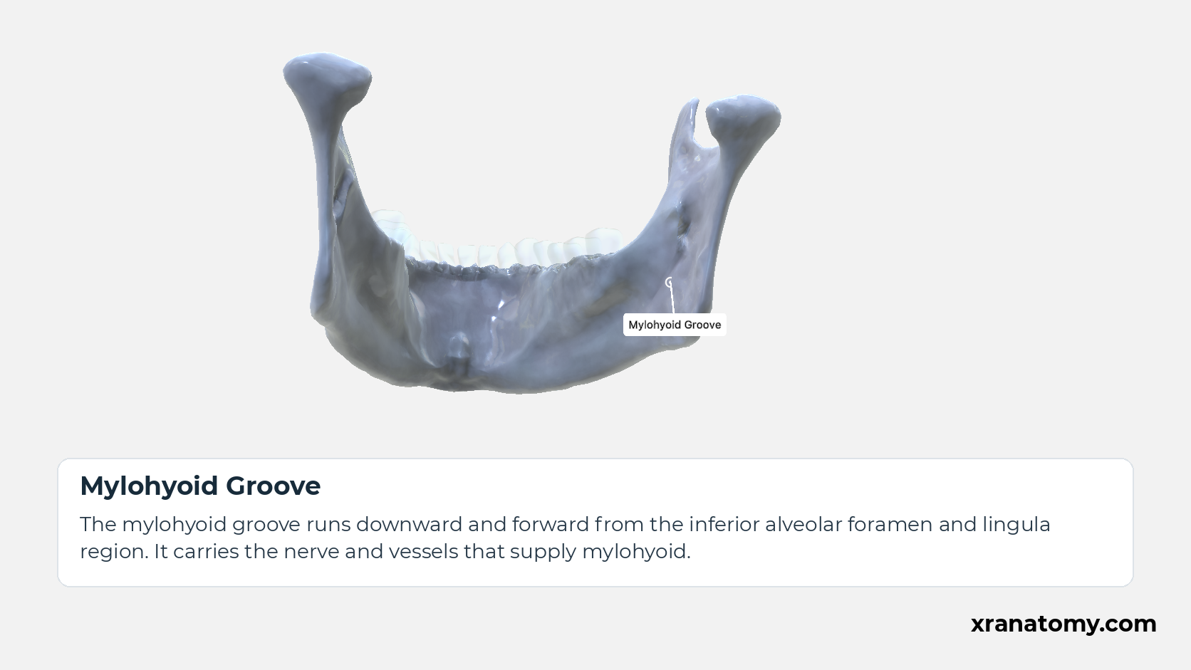
Mylohyoid Groove
The mylohyoid groove runs downward and forward from the inferior alveolar foramen and lingula region. It carries the nerve and vessels that supply mylohyoid.
Illustration by XR Anatomy.
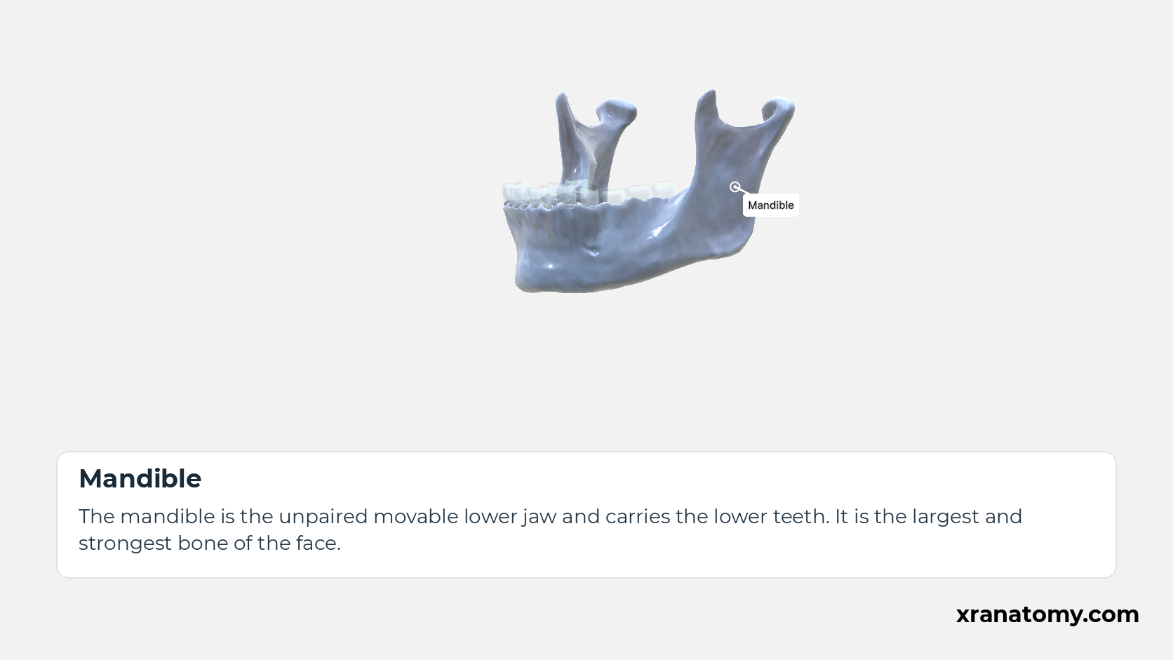
Mandible
The mandible is the unpaired movable lower jaw and carries the lower teeth. It is the largest and strongest bone of the face.
Illustration by XR Anatomy.
Ramus of Mandible
Each ramus of the mandible is the upright posterior part of the lower jaw, rising from the body at the mandibular angle. Its upper end divides into the coronoid and condylar processes, separated by the mandibular notch; its surfaces provide major muscle attachments and passages for nerves and vessels.Show less
Illustration by XR Anatomy.
Pterygoid Tuberosity
The pterygoid tuberosity is a roughened area on the medial side of the angle and lower ramus, where the medial pterygoid inserts.
Illustration by XR Anatomy.
Inferior Alveolar Foramen
The inferior alveolar foramen (foramen alveolare inferius), also called the mandibular foramen, is an opening on the medial ramus. The lingula overlaps its upper front edge, and the opening transmits the inferior alveolar nerve and vessels.
Illustration by XR Anatomy.
Lingula of Mandible
The lingula of the mandible is a small tongue-shaped bony projection on the medial ramus, beside the upper anterior margin of the mandibular foramen. It provides attachment for the sphenomandibular ligament.
Illustration by XR Anatomy.
Mylohyoid Groove
The mylohyoid groove runs downward and forward from the inferior alveolar foramen and lingula region. It carries the nerve and vessels that supply mylohyoid.
Illustration by XR Anatomy.
Mandibular Canal
The mandibular canal starts at the inferior alveolar foramen, descends through the ramus, and runs forward beneath the tooth sockets. It carries the inferior alveolar nerve and vessels, whose branches supply the tooth roots.
Illustration by XR Anatomy.
Temporal Crest
The temporal crest runs down from the medial surface of the coronoid process toward the area behind the last molar, where it is associated with temporalis attachment.
Illustration by XR Anatomy.
Coronoid Process of Mandible
The coronoid process is the thin, triangular projection at the anterior upper end of the mandibular ramus. It provides the main insertion for the temporalis muscle, with some masseter attachment on its lateral surface; the mandibular notch separates it from the condylar process behind.Show less
Illustration by XR Anatomy.
Angle of Mandible
The angle is the lower back junction between the body and ramus. Its angle varies in adults.
Illustration by XR Anatomy.
The medial surface has the inferior alveolar foramen, together with its lingula, canal, and mylohyoid groove. The lower part also bears the pterygoid tuberosity.
In depth · 5 sections · 1 min read5 more sections on this part5 sections open — reading In depth- Pterygoid Tuberosity
- Inferior Alveolar Foramen
- Mylohyoid Groove
- Inferior Border
- Mandibular Notch
Pterygoid Tuberosity
The pterygoid tuberosity is a roughened area on the medial side of the angle and lower ramus, where the medial pterygoid inserts.
Inferior Alveolar Foramen
The inferior alveolar foramen (foramen alveolare inferius), also called the mandibular foramen, is an opening on the medial ramus. The lingula overlaps its upper front edge, and the opening transmits the inferior alveolar nerve and vessels.
Lingula of Mandible
The lingula projects beside the inferior alveolar foramen and anchors the sphenomandibular ligament.
Mandibular Canal
The mandibular canal starts at the inferior alveolar foramen, descends through the ramus, and runs forward beneath the tooth sockets. It carries the inferior alveolar nerve and vessels, whose branches supply the tooth roots.
Mylohyoid Groove
The mylohyoid groove runs downward and forward from the inferior alveolar foramen and lingula region. It carries the nerve and vessels that supply mylohyoid.
In depth · continued · 2 more sections- Inferior Border
- Mandibular Notch
Inferior Border
The inferior border is thick, fairly straight, and continuous with the lower border of the mandibular body.
Mandibular Notch
The mandibular notch is a deep curve between the coronoid and condylar processes. It carries the masseteric nerve and vessels.
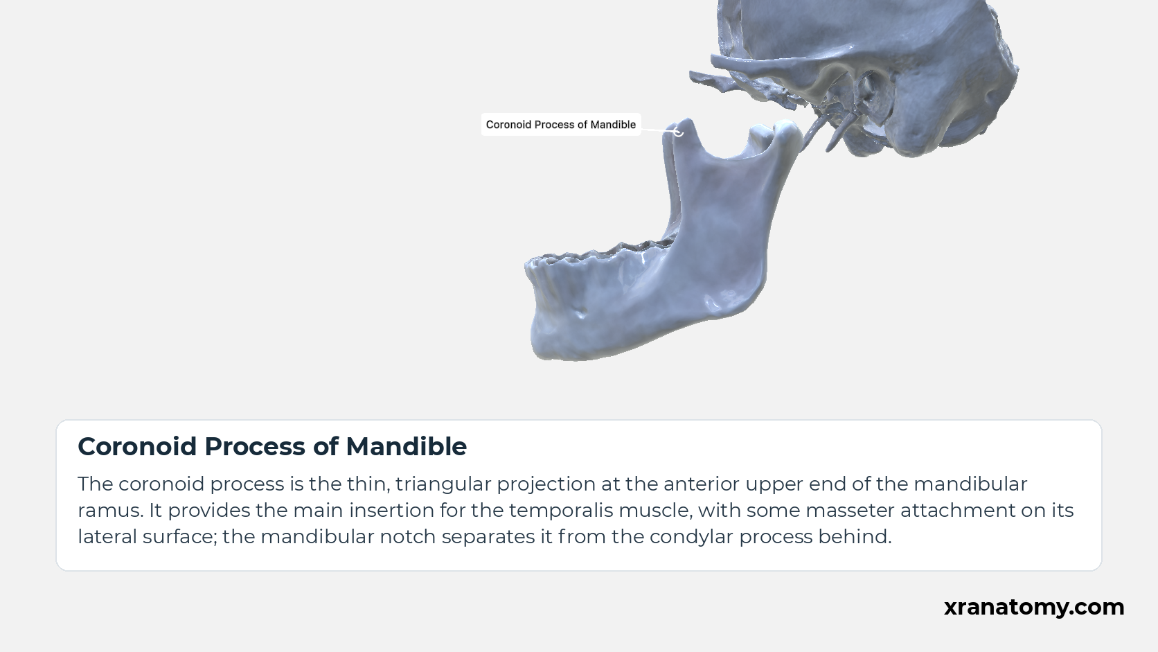
Coronoid Process of Mandible
The coronoid process is thin, triangular, and flattened side to side. Most of the temporalis inserts on it, and masseter also reaches its lateral surface. The medial side bears the temporal crest.
In depth · 1 min read Temporal Crest
The temporal crest runs down from the medial surface of the coronoid process toward the area behind the last molar, where it is associated with temporalis attachment.
Condylar Process
Condylar Process
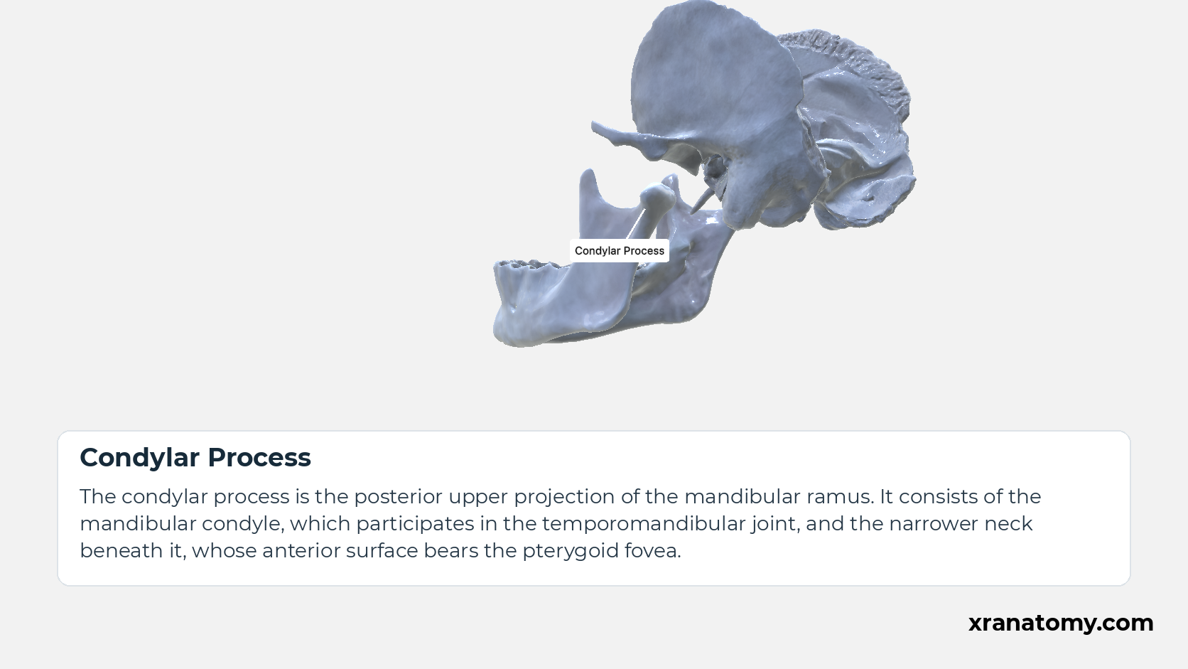
The condylar process is the posterior upper projection of the mandibular ramus. It consists of the mandibular condyle, which participates in the temporomandibular joint, and the narrower neck beneath it, whose anterior surface bears the pterygoid fovea.Show less
Study Condylar Process in this fixed image. The image does not rotate.
Illustration by XR Anatomy.
Mandibular Condyle
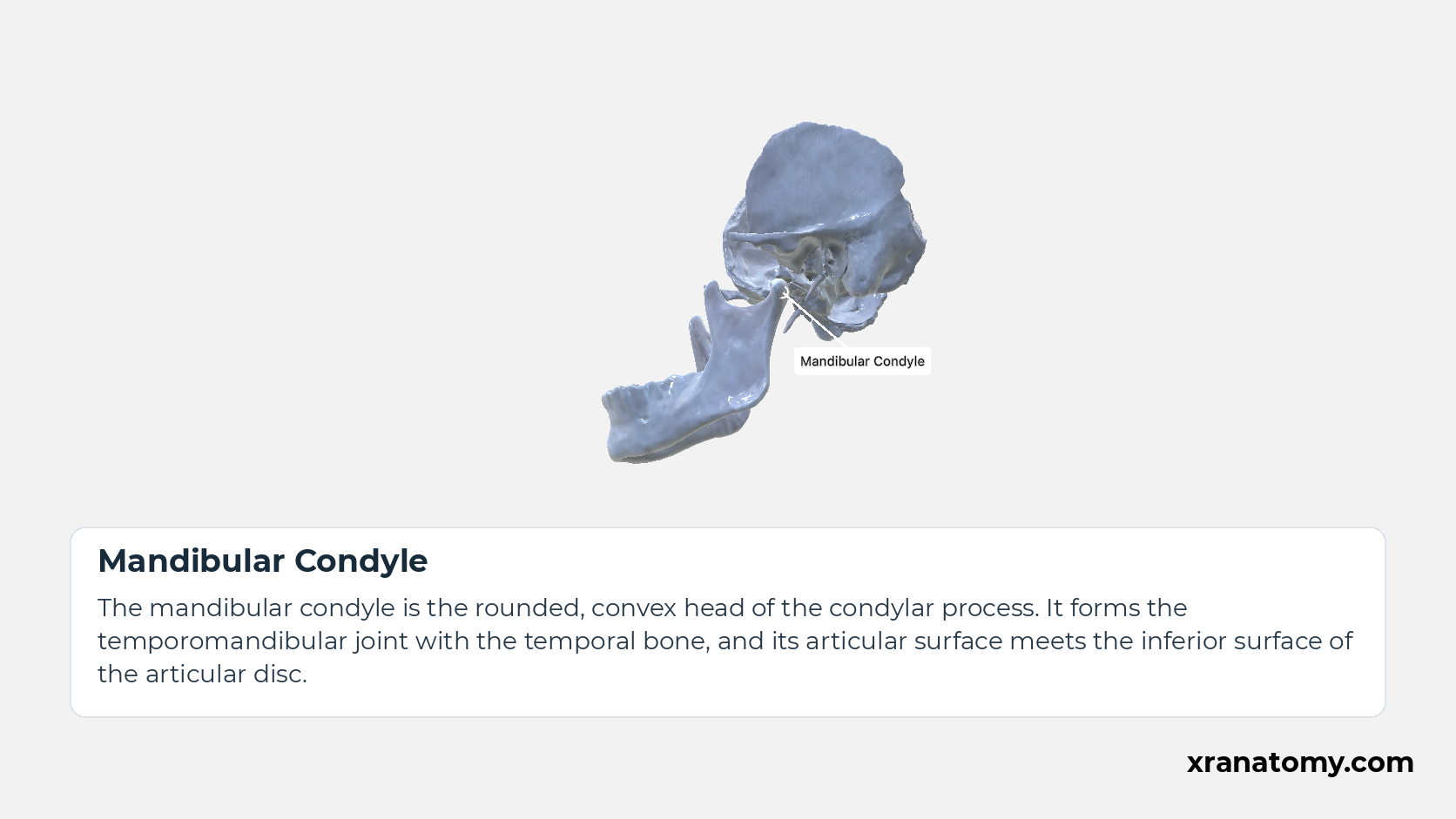
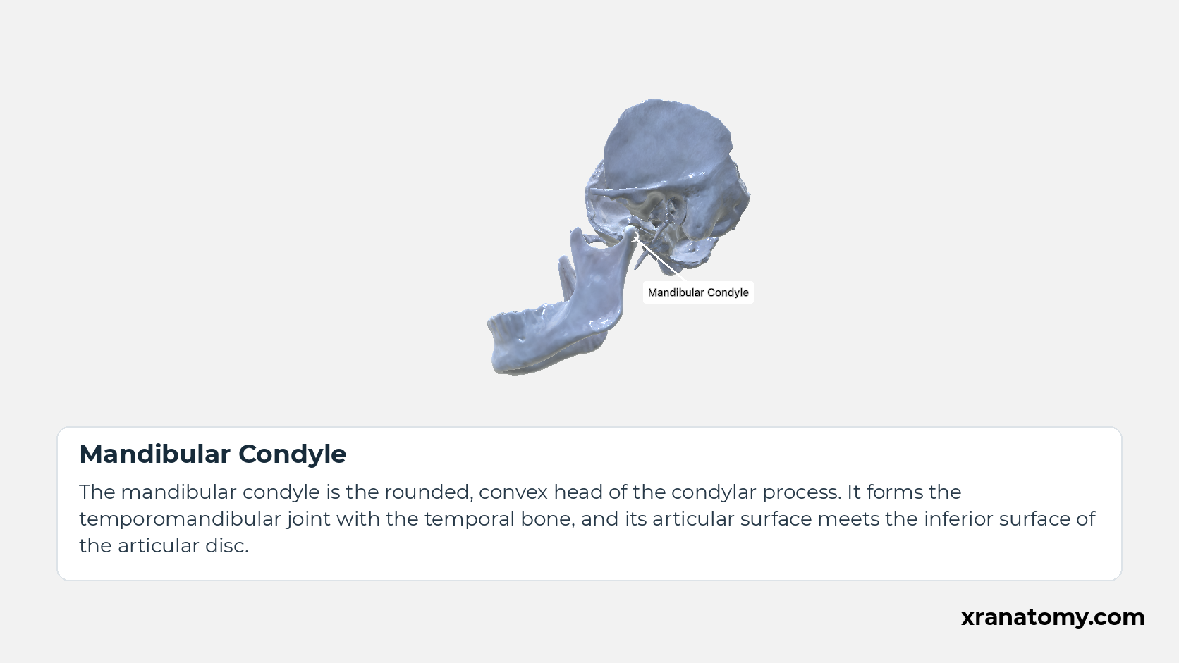
The mandibular condyle is the rounded, convex head of the condylar process. It forms the temporomandibular joint with the temporal bone, and its articular surface meets the inferior surface of the articular disc.
Illustration by XR Anatomy.
Neck of Mandible
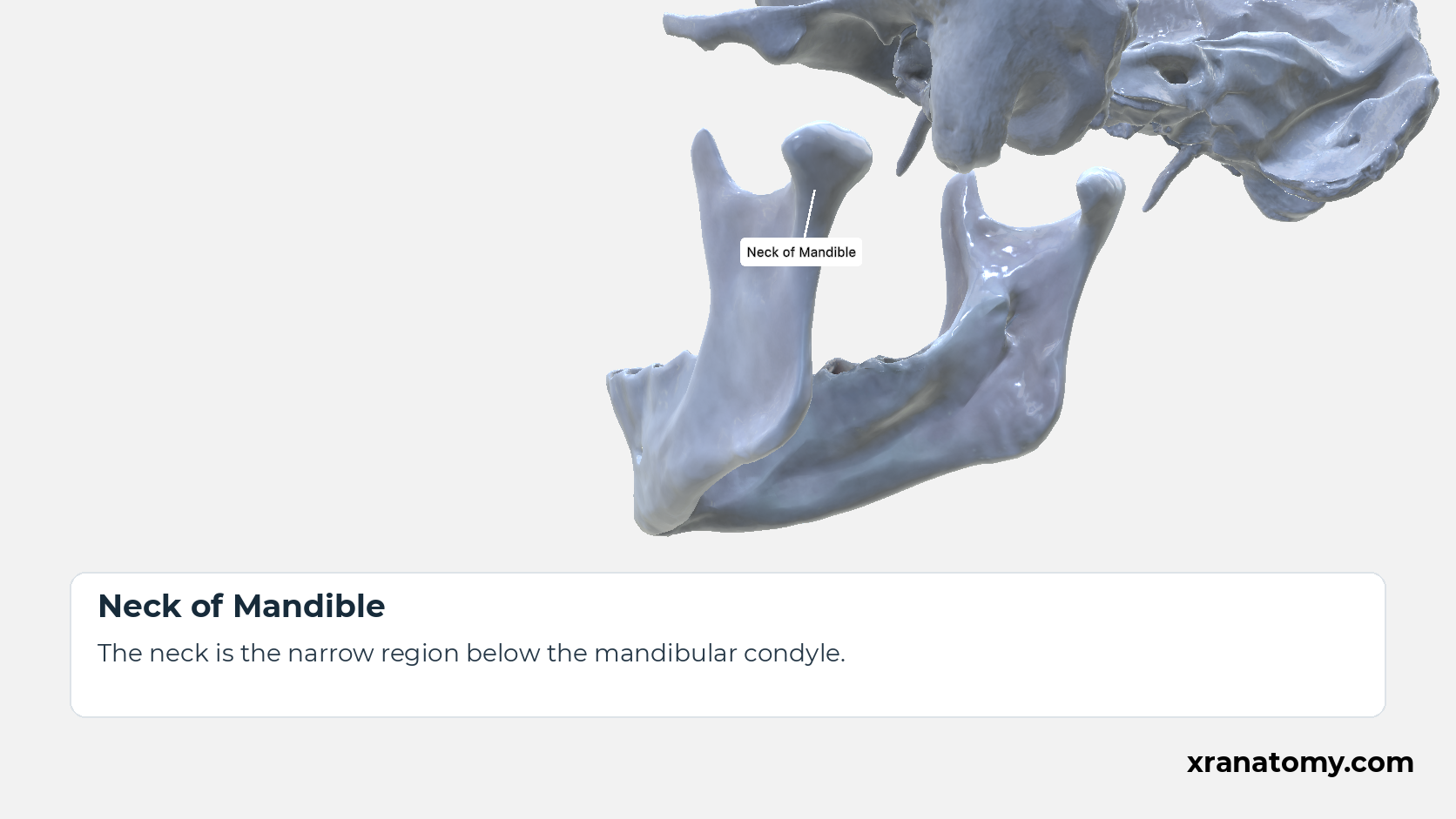
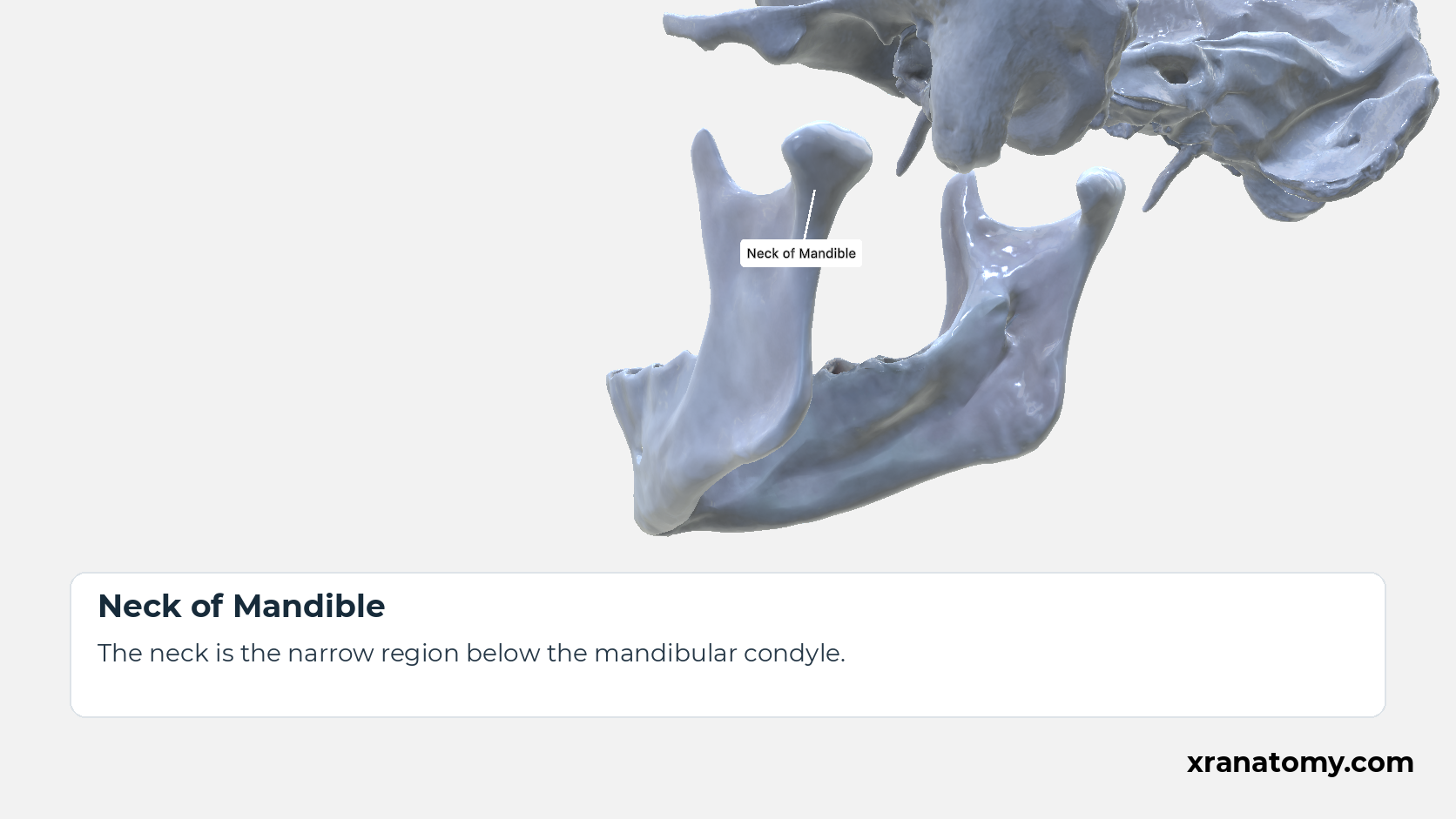
The neck is the narrow region below the mandibular condyle.
Illustration by XR Anatomy.
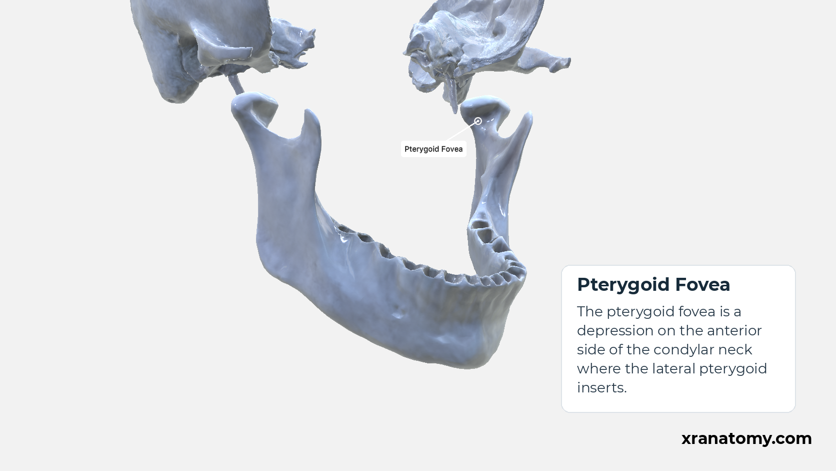
Pterygoid Fovea
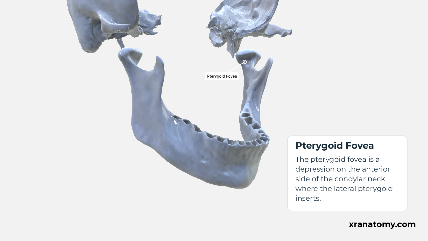
The pterygoid fovea is a depression on the anterior side of the condylar neck where the lateral pterygoid inserts.
Illustration by XR Anatomy.
Condylar Process
The condylar process is the posterior upper projection of the mandibular ramus. It consists of the mandibular condyle, which participates in the temporomandibular joint, and the narrower neck beneath it, whose anterior surface bears the pterygoid fovea.Show less
Illustration by XR Anatomy.
Mandibular Condyle
The mandibular condyle is the rounded, convex head of the condylar process. It forms the temporomandibular joint with the temporal bone, and its articular surface meets the inferior surface of the articular disc.
Illustration by XR Anatomy.
Neck of Mandible
The neck is the narrow region below the mandibular condyle.
Illustration by XR Anatomy.
Pterygoid Fovea
The pterygoid fovea is a depression on the anterior side of the condylar neck where the lateral pterygoid inserts.
Illustration by XR Anatomy.
Coronoid Process of Mandible
The coronoid process is the thin, triangular projection at the anterior upper end of the mandibular ramus. It provides the main insertion for the temporalis muscle, with some masseter attachment on its lateral surface; the mandibular notch separates it from the condylar process behind.Show less
Illustration by XR Anatomy.
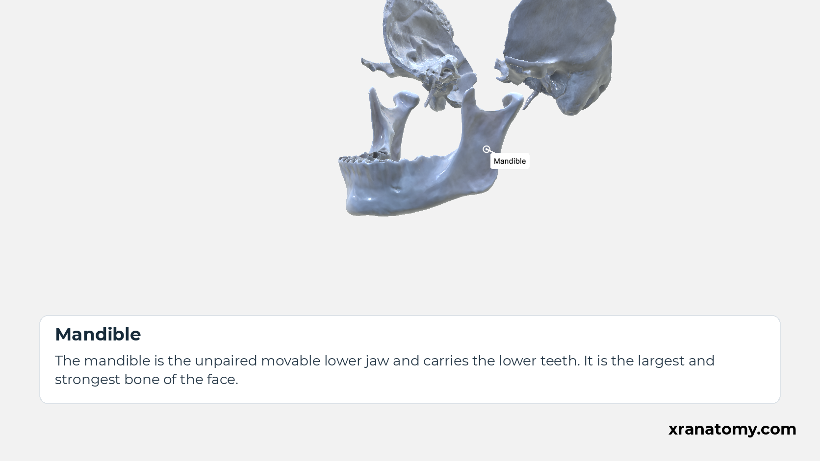
Mandible
The mandible is the unpaired movable lower jaw and carries the lower teeth. It is the largest and strongest bone of the face.
Illustration by XR Anatomy.
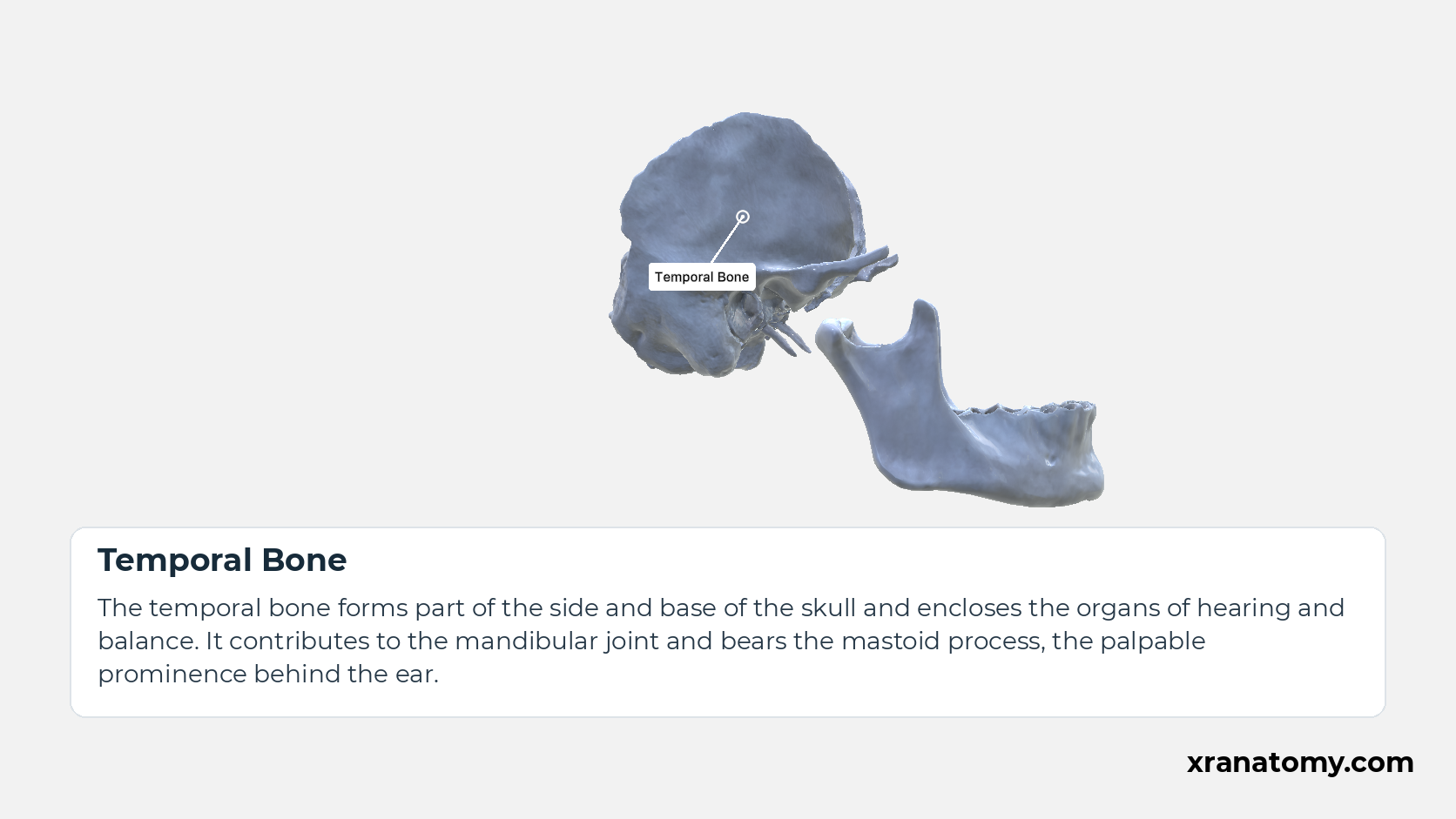
Temporal Bone
The temporal bone forms part of the side and base of the skull and encloses the organs of hearing and balance. It contributes to the mandibular joint and bears the mastoid process, the palpable prominence behind the ear.
Illustration by XR Anatomy.
 ↺ drag to rotate · scroll to zoom
Condylar Process of the Mandible
Condylar Process of the Mandible
Drag or use the viewer controls to inspect the condyle, neck, and pterygoid fovea from different angles.
Sourced from 3D Osteology.
Thicker than the coronoid process, the condylar process includes a mandibular condyle supported by a neck. The anterior surface of the neck contains the pterygoid fovea.
In depth · 3 sections · 1 min read3 more sections on this part3 sections open — reading In depth- Mandibular Condyle
- Neck of Mandible
- Pterygoid Fovea
Mandibular Condyle
The mandibular condyle is the rounded, convex head of the condylar process. It forms the temporomandibular joint with the temporal bone, and its articular surface meets the inferior surface of the articular disc.
Neck of Mandible
The neck is the narrow region below the mandibular condyle.
Pterygoid Fovea
The pterygoid fovea is a depression on the anterior side of the condylar neck where the lateral pterygoid inserts.
Practice · Active recall · 8 questions Recall: Ramus of Mandible
Why this helps
Testing yourself before revealing the answer (active recall) strengthens memory far more than re-reading — so each prompt stays hidden until you choose to reveal it.
Each ramus rises from which part of the mandibular body?
posterior part
Each ramus rises from the posterior part of the body at the angle. Its main features are the lateral surface, medial surface, inferior border, mandibular notch, coronoid process, and condylar process.
Review this sectionHow well did you know it? · press 1–4
In depth · 1 question Recall: Angle of Mandible
The mandibular angle is the lower back junction between the body and .
ramus
The angle is the lower back junction between the body and ramus. Its angle varies in adults.
Review this sectionHow well did you know it? · press 1–4
Nice work — revisit in a few days.
Lower oblique ridges on the lateral surface of the mandibular ramus mark attachment.
masseter
Most of the lateral surface of the ramus is flat and smooth; lower oblique ridges mark masseter attachment. These ridges form the masseteric tuberosity.
Review this sectionHow well did you know it? · press 1–4
The medial surface of the mandibular ramus includes the inferior alveolar foramen, lingula, canal, and groove.
mylohyoid
The medial surface has the inferior alveolar foramen, together with its lingula, canal, and mylohyoid groove. The lower part also bears the pterygoid tuberosity.
Review this sectionHow well did you know it? · press 1–4
Nice work — revisit in a few days.
In depth · 1 question Recall: Inferior Border
The lower border of each mandibular ramus continues into the mandibular .
body
The inferior border is thick, fairly straight, and continuous with the lower border of the mandibular body.
Review this sectionHow well did you know it? · press 1–4
Nice work — revisit in a few days.
In depth · 1 question Recall: Mandibular Notch
The mandibular notch lies between the coronoid and processes.
condylar
The mandibular notch is a deep curve between the coronoid and condylar processes. It carries the masseteric nerve and vessels.
Review this sectionHow well did you know it? · press 1–4
Nice work — revisit in a few days.
The coronoid process of the mandible is thin, triangular, and flattened .
side to side
The coronoid process is thin, triangular, and flattened side to side. Most of the temporalis inserts on it, and masseter also reaches its lateral surface. The medial side bears the temporal crest.
Review this sectionHow well did you know it? · press 1–4
The condylar process of the mandible includes a mandibular condyle supported by a .
neck
Thicker than the coronoid process, the condylar process includes a mandibular condyle supported by a neck. The anterior surface of the neck contains the pterygoid fovea.
Review this sectionHow well did you know it? · press 1–4
Nice work — revisit in a few days.
Related Structures
Mandible
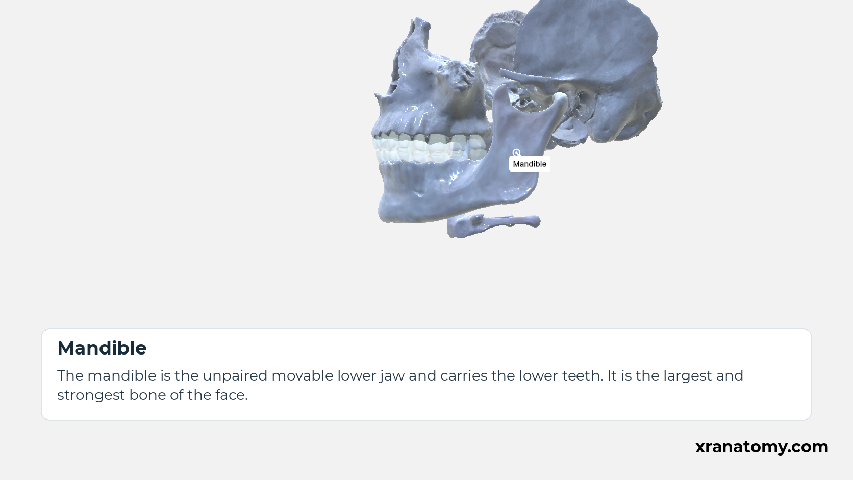
The mandible is the unpaired movable lower jaw and carries the lower teeth. It is the largest and strongest bone of the face.
Study Mandible and Related Bones in this fixed image. Use the thumbnails to move between the 2D views; each view highlights one structure. The image does not rotate.
Illustration by XR Anatomy.
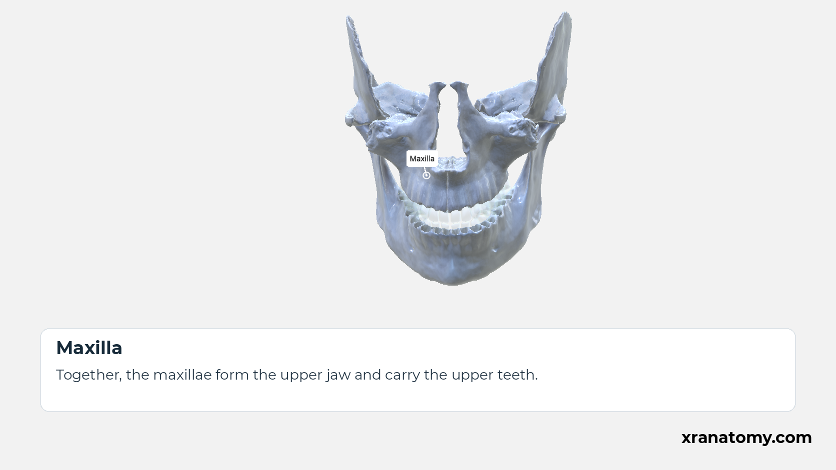
Maxilla
Together, the maxillae form the upper jaw and carry the upper teeth.
Illustration by XR Anatomy.
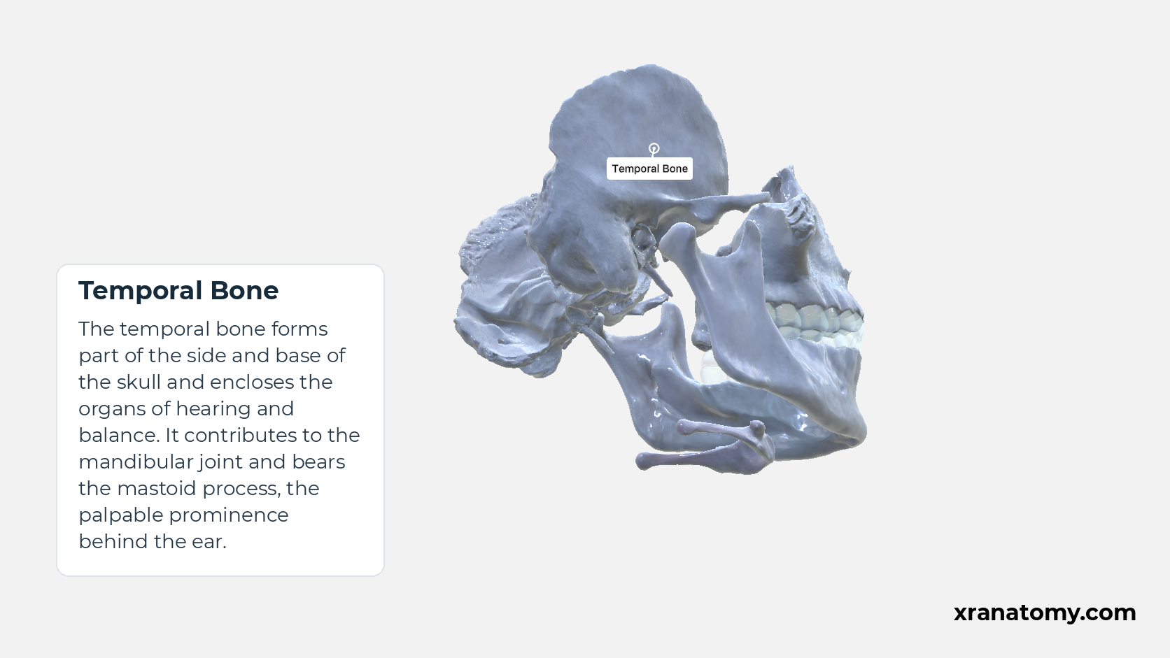
Temporal Bone
The temporal bone forms part of the side and base of the skull and encloses the organs of hearing and balance. It contributes to the mandibular joint and bears the mastoid process, the palpable prominence behind the ear.
Illustration by XR Anatomy.
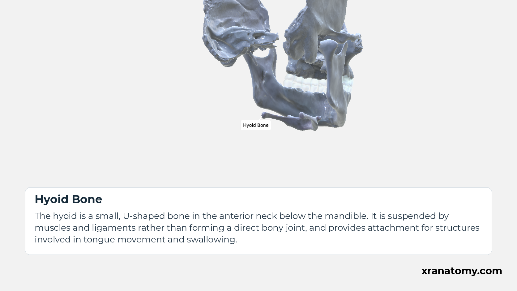
Hyoid Bone
The hyoid is a small, U-shaped bone in the anterior neck below the mandible. It is suspended by muscles and ligaments rather than forming a direct bony joint, and provides attachment for structures involved in tongue movement and swallowing.Show less
Illustration by XR Anatomy.
The maxilla faces the mandible across the dental arches. The temporal bone forms the temporomandibular joint with the mandible. Suprahyoid muscles link the mandible and hyoid bone without a direct bony joint.
Practice · Active recall · 1 question Recall: Related Structures
The maxilla faces the mandible across the dental .
arches
The maxilla faces the mandible across the dental arches. The temporal bone forms the temporomandibular joint with the mandible. Suprahyoid muscles link the mandible and hyoid bone without a direct bony joint.
Review this sectionHow well did you know it? · press 1–4
Nice work — revisit in a few days.
Bibliography
Every claim on this page is sourced against the standard anatomical references.
- FIPAT. Terminologia Anatomica, 2nd edition, Part 2: Bones. https://cdn.dal.ca/content/dam/dalhousie/pdf/library/FIPAT/TA2/FIPAT-TA2-Part-2.pdf
- Gray H. Anatomy of the Human Body. 21st ed. Philadelphia: Lea & Febiger; 1924.
- Drake RL, Vogl AW, Mitchell AWM. Gray's Anatomy for Students. Elsevier.
- Goyushov S, Tözüm MD, Tözüm TF. Assessment of morphological and anatomical characteristics of mental foramen using cone beam computed tomography. Surg Radiol Anat. 2018;40(10):1133-1139. doi:10.1007/s00276-018-2043-z. https://pubmed.ncbi.nlm.nih.gov/29802432/
- Santini A, Alayan I. A comparative anthropometric study of the position of the mental foramen in three populations. Br Dent J. 2012;212(4):E7. doi:10.1038/sj.bdj.2012.143. https://pubmed.ncbi.nlm.nih.gov/22349415/
- Cornelius CP, Ehrenfeld M. The Use of MMF Screws: Surgical Technique, Indications, Contraindications, and Common Problems in Review of the Literature. Craniomaxillofac Trauma Reconstr. 2010;3(2):55-80. https://pmc.ncbi.nlm.nih.gov/articles/PMC3052677/
- Shah SP, Mehta D. Mandibular Retromolar Foramen and Canal - A Systematic Review and Meta-Analysis. Ann Maxillofac Surg. 2020;10(2):444-449. doi:10.4103/ams.ams_19_20. https://pmc.ncbi.nlm.nih.gov/articles/PMC7944007/
- Breeland G, Aktar A, Patel BC. Anatomy, Head and Neck, Mandible. [Updated 2023 Apr 1]. In: StatPearls [Internet]. Treasure Island (FL): StatPearls Publishing; 2026 Jan-. https://www.ncbi.nlm.nih.gov/books/NBK532292/
