Anatomy
Right atrium
The right atrium, the heart's collecting chamber for the body, receives the venae cavae and the coronary sinus and opens into the right ventricle.
Blood returning from the body reaches the right atrium before moving into the right ventricle. This chamber also receives most of the blood returning from the heart’s own muscle. Its internal landmarks help show where these returning streams enter and how they relate to the opening into the ventricle.
Smooth and muscular atrial regions
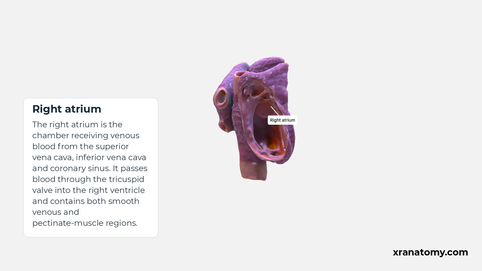
Right atrium
The right atrium is the chamber receiving venous blood from the superior vena cava, inferior vena cava and coronary sinus. It passes blood through the tricuspid valve into the right ventricle and contains both smooth venous and pectinate-muscle regions.Show less
Study Smooth and muscular atrial regions in this fixed image. Use the thumbnails to move between the 2D views; each view highlights one structure. The image does not rotate.
Illustration by XR Anatomy.
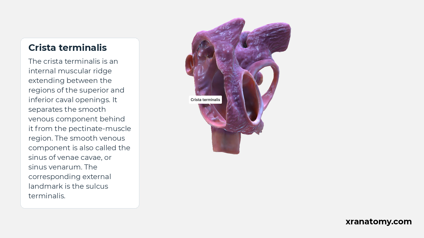
Crista terminalis
The crista terminalis is an internal muscular ridge extending between the regions of the superior and inferior caval openings. It separates the smooth venous component behind it from the pectinate-muscle region. The smooth venous component is also called the sinus of venae cavae, or sinus venarum. The corresponding external landmark is the sulcus terminalis.Show less
Illustration by XR Anatomy.
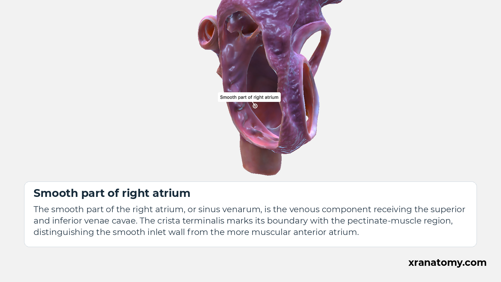
Smooth part of right atrium
The smooth part of the right atrium, or sinus venarum, is the venous component receiving the superior and inferior venae cavae. The crista terminalis marks its boundary with the pectinate-muscle region, distinguishing the smooth inlet wall from the more muscular anterior atrium.Show less
Illustration by XR Anatomy.
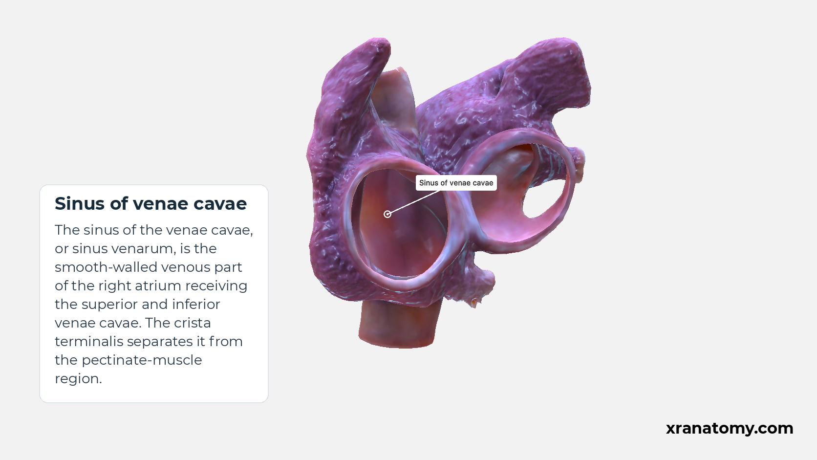
Sinus of venae cavae
The sinus of the venae cavae, or sinus venarum, is the smooth-walled venous part of the right atrium receiving the superior and inferior venae cavae. The crista terminalis separates it from the pectinate-muscle region.
Illustration by XR Anatomy.
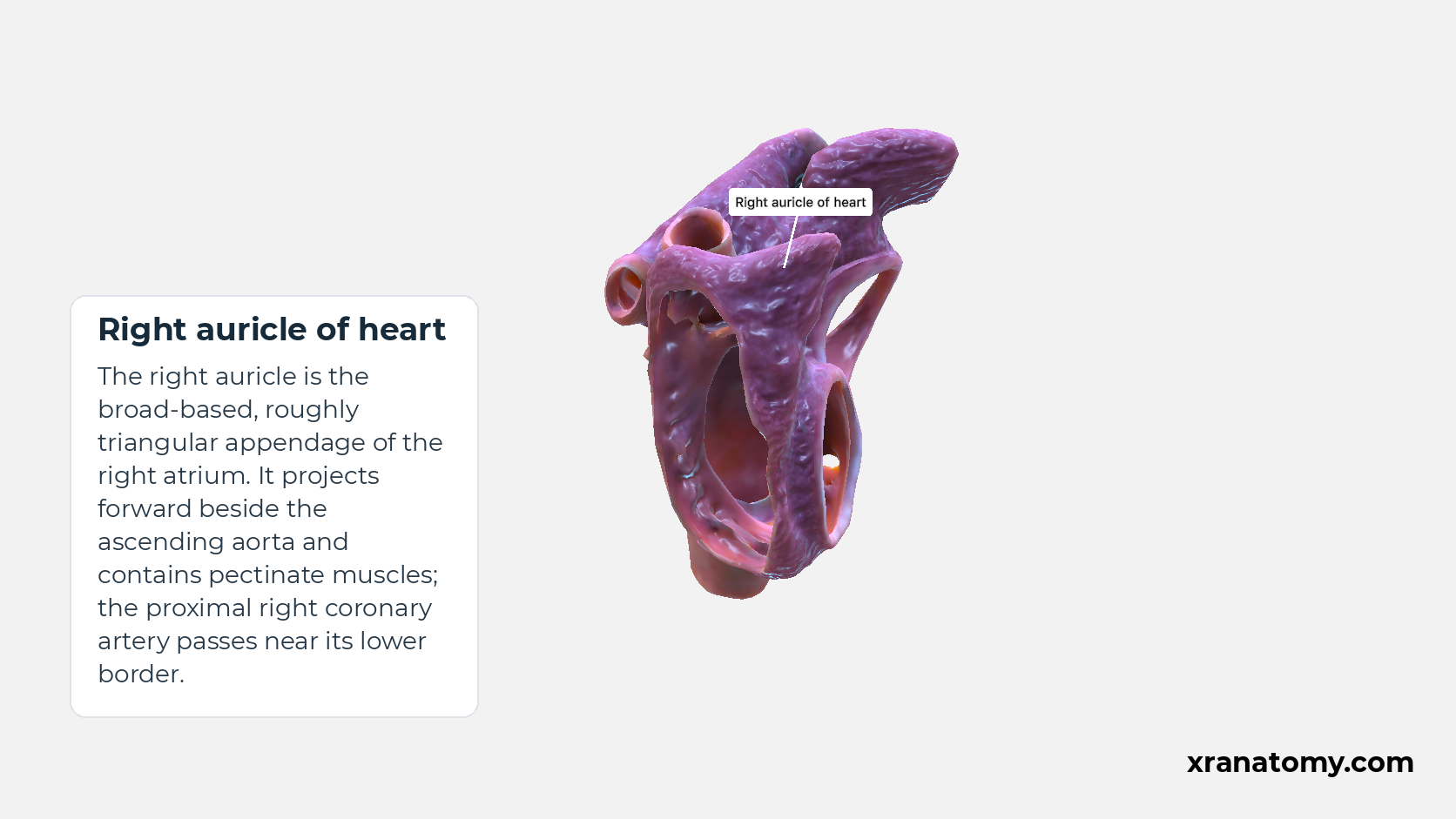
Right auricle of heart
The right auricle is the broad-based, roughly triangular appendage of the right atrium. It projects forward beside the ascending aorta and contains pectinate muscles; the proximal right coronary artery passes near its lower border.
Illustration by XR Anatomy.
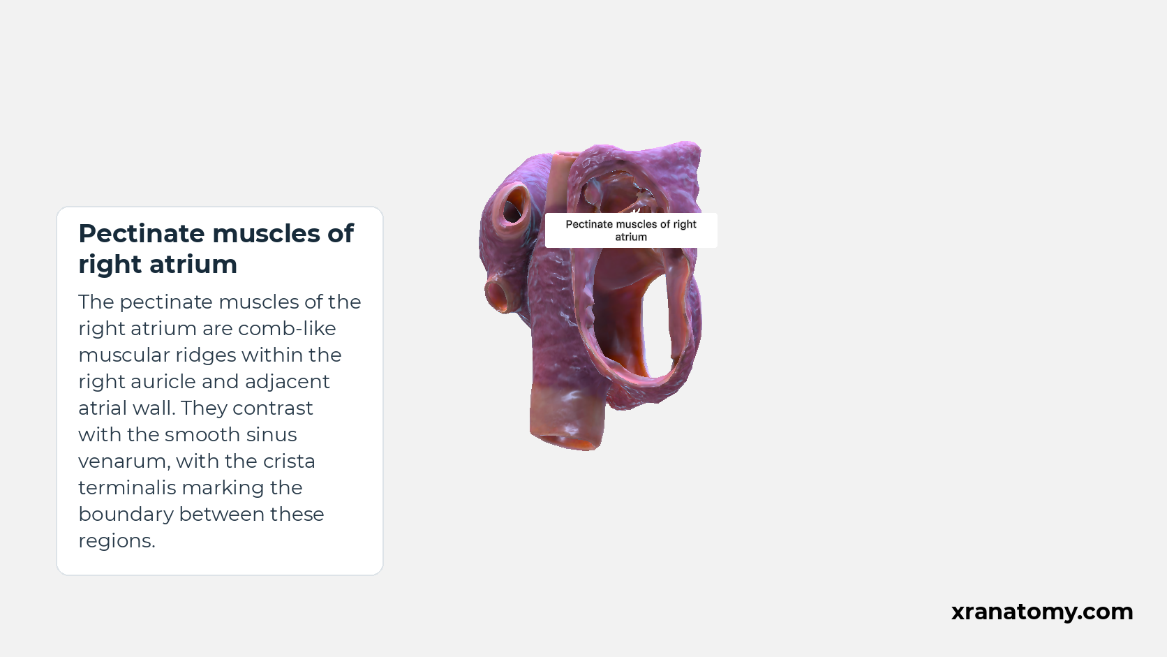
Pectinate muscles of right atrium
The pectinate muscles of the right atrium are comb-like muscular ridges within the right auricle and adjacent atrial wall. They contrast with the smooth sinus venarum, with the crista terminalis marking the boundary between these regions.
Illustration by XR Anatomy.
The chamber and its internal regions
The right atrium is a thin-walled chamber that opens into the right ventricle through the tricuspid opening. Its inner surface changes from one region to another. The smooth venous component receives the returning veins, while the muscular region and the pouch called the auricle have more prominent muscle ridges. Close to the atrioventricular opening, the vestibule forms the region leading towards the valve.
Crista terminalis and smooth venous component
A muscular ridge called the crista terminalis helps separate these regions. It runs between the areas of the superior and inferior caval openings. Behind the ridge lies the smooth venous component, also known as the sinus of venae cavae or sinus venarum. On the other side is the pectinate-muscle region. On the outside of the atrium, the corresponding landmark is a groove called the sulcus terminalis.
Right auricle and pectinate muscles
The right auricle, also called the right atrial appendage, projects from the chamber as a roughly triangular pouch. Inside it, pectinate muscles form raised ridges with a comb-like appearance. These ridges continue onto the adjacent atrial wall, helping distinguish this muscular region from the smooth venous component.
Where the returning veins enter
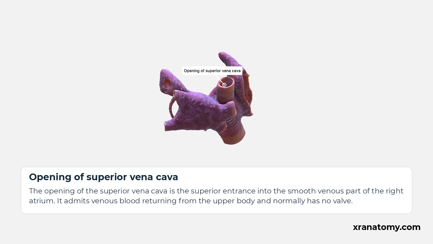
Opening of superior vena cava
The opening of the superior vena cava is the superior entrance into the smooth venous part of the right atrium. It admits venous blood returning from the upper body and normally has no valve.
Study Venous openings and their valves in this fixed image. Use the thumbnails to move between the 2D views; each view highlights one structure. The image does not rotate.
Illustration by XR Anatomy.
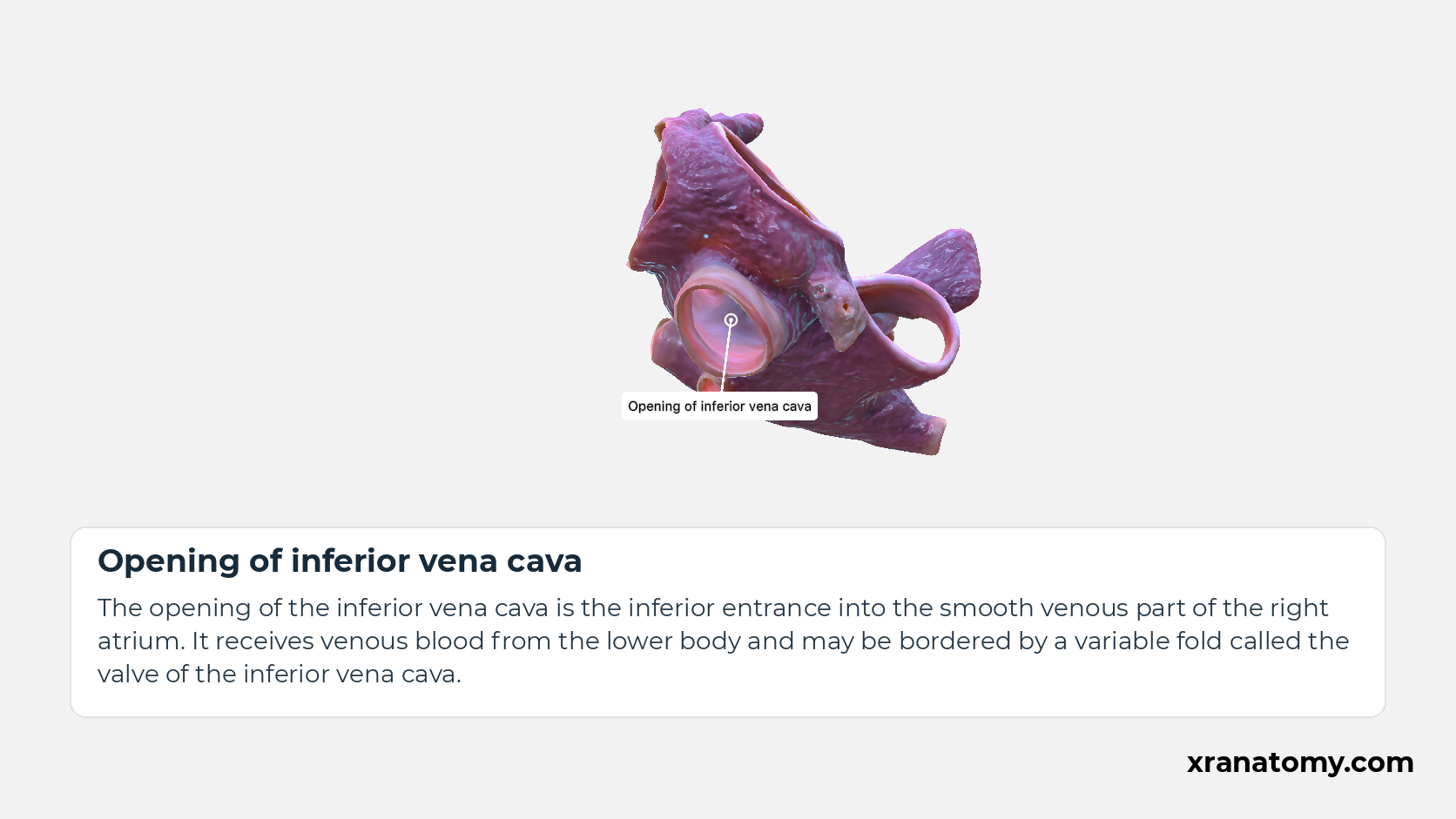
Opening of inferior vena cava
The opening of the inferior vena cava is the inferior entrance into the smooth venous part of the right atrium. It receives venous blood from the lower body and may be bordered by a variable fold called the valve of the inferior vena cava.
Illustration by XR Anatomy.
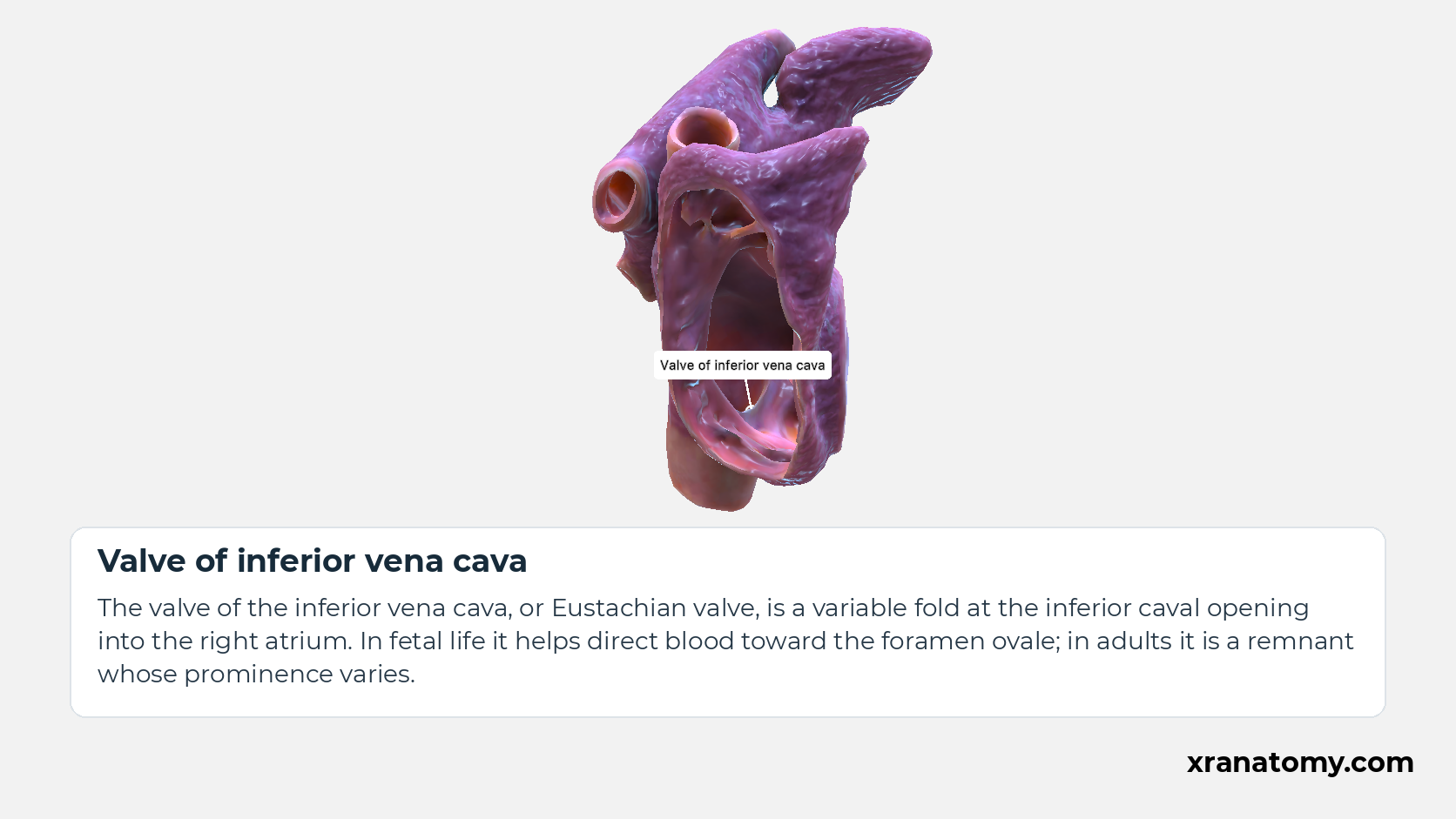
Valve of inferior vena cava
The valve of the inferior vena cava, or Eustachian valve, is a variable fold at the inferior caval opening into the right atrium. In fetal life it helps direct blood toward the foramen ovale; in adults it is a remnant whose prominence varies.Show less
Illustration by XR Anatomy.
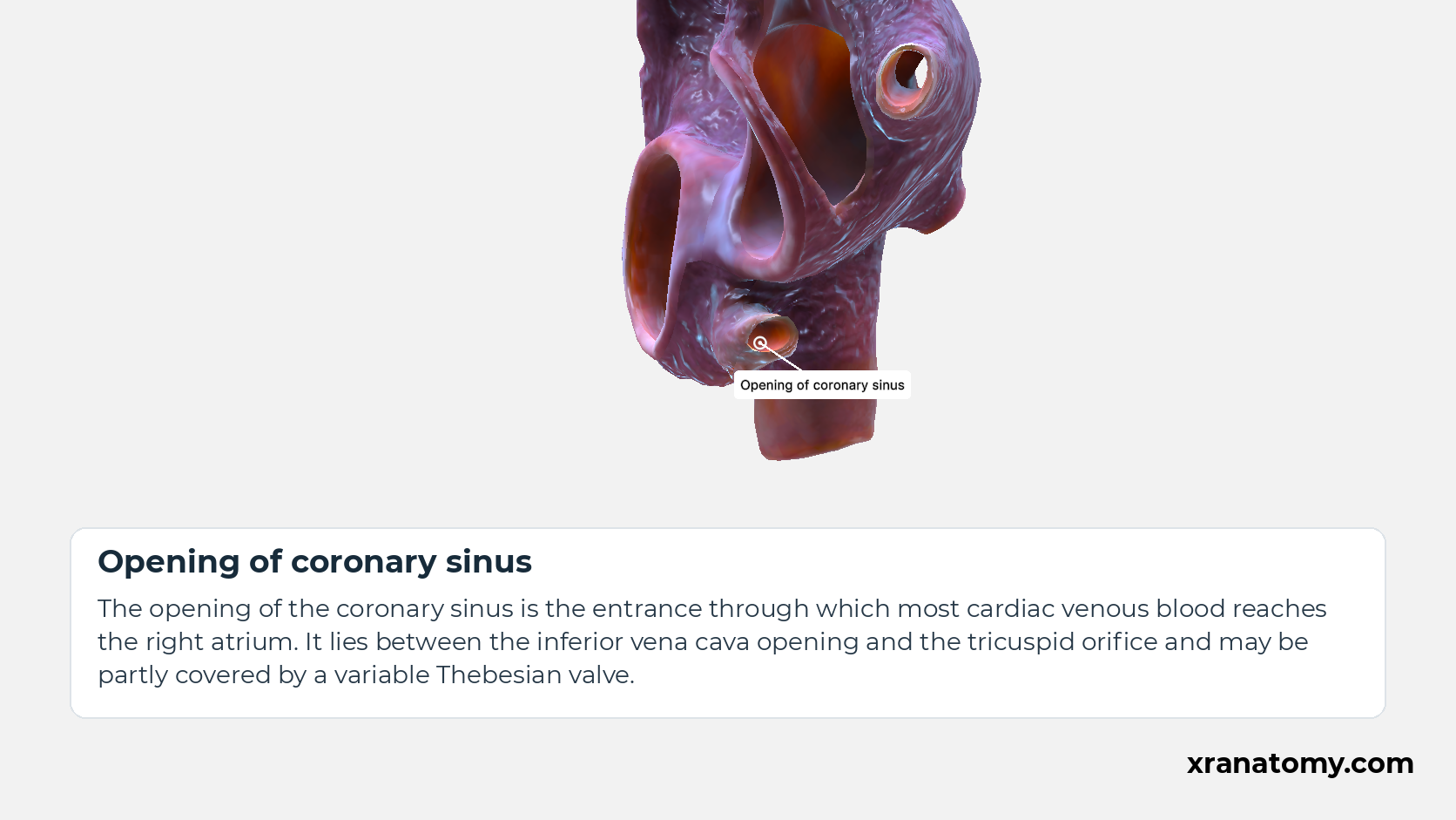
Opening of coronary sinus
The opening of the coronary sinus is the entrance through which most cardiac venous blood reaches the right atrium. It lies between the inferior vena cava opening and the tricuspid orifice and may be partly covered by a variable Thebesian valve.Show less
Illustration by XR Anatomy.
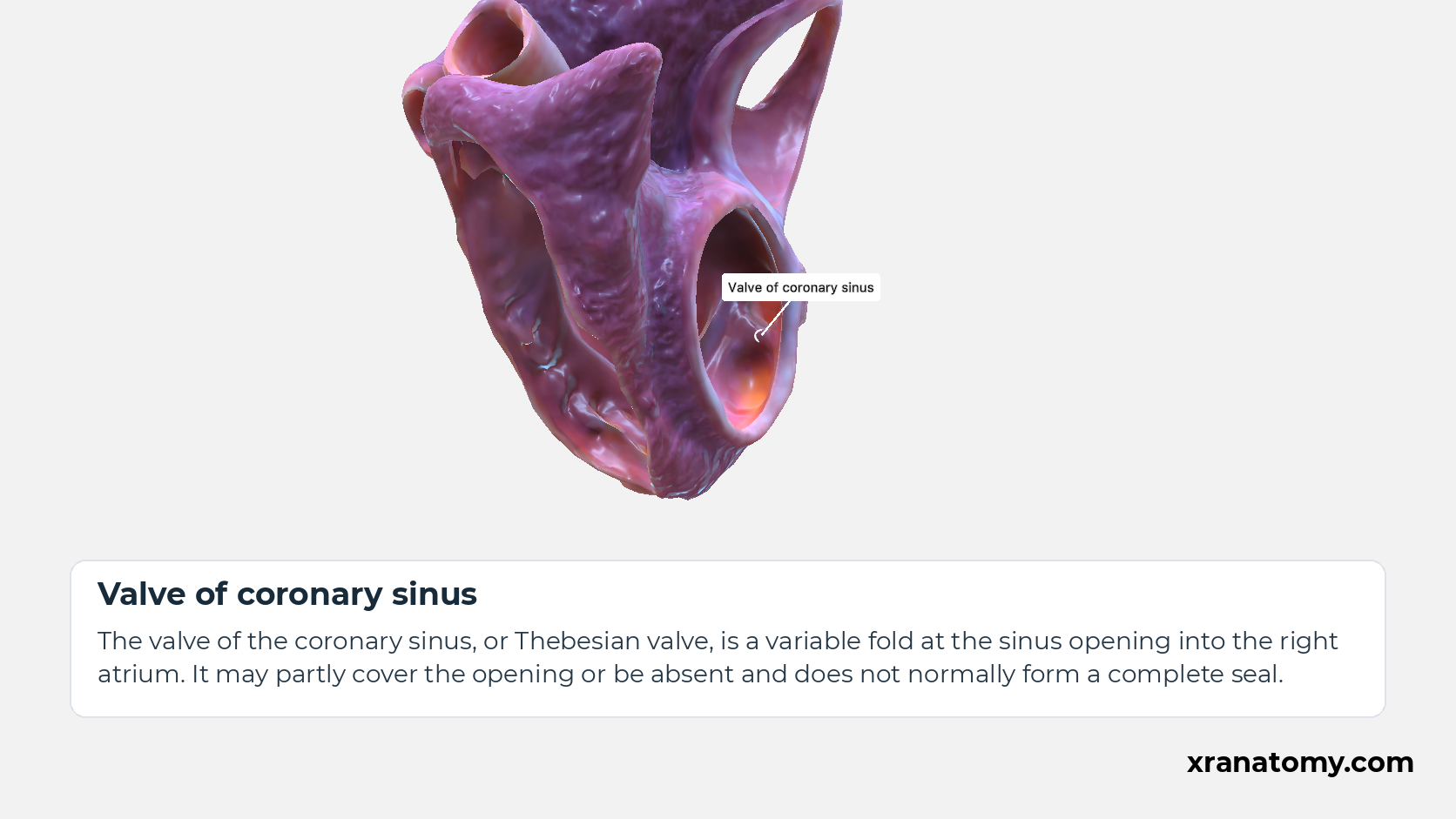
Valve of coronary sinus
The valve of the coronary sinus, or Thebesian valve, is a variable fold at the sinus opening into the right atrium. It may partly cover the opening or be absent and does not normally form a complete seal.
Illustration by XR Anatomy.
The returning veins open into the smooth venous region. The superior and inferior venae cavae bring blood back from the body, while the coronary sinus carries blood from much of the heart muscle. The coronary-sinus opening lies between the inferior caval opening and the right atrioventricular orifice, providing a useful landmark near the route into the ventricle.
In depth · 3 sections · 1 min read3 more sections on this part3 sections open — reading In depth- Caval openings
- Coronary-sinus opening
- Vestibule and septal landmarks
Caval openings
The superior and inferior venae cavae provide superior and inferior landmarks in the venous component of the right atrium. The valve of the inferior vena cava, also called the Eustachian valve, helps direct incoming blood towards the foramen ovale in fetal circulation. Its adult appearance is variable.
Coronary-sinus opening
At the opening of the coronary sinus into the right atrium, between the inferior caval opening and the right atrioventricular orifice, a fold may partly cover the orifice. This is the valve of the coronary sinus, or Thebesian valve. It varies in size and may be absent; it does not consistently seal the opening.
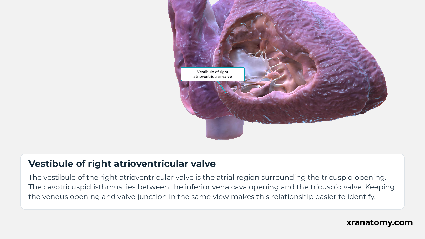
Vestibule of right atrioventricular valve
The vestibule of the right atrioventricular valve is the smooth right atrial region surrounding the tricuspid opening. It forms the transition from the receiving chamber to the valve leading into the right ventricle.
Study Vestibule and septal landmarks in this fixed image. Use the thumbnails to move between the 2D views; each view highlights one structure. The image does not rotate.
Illustration by XR Anatomy.
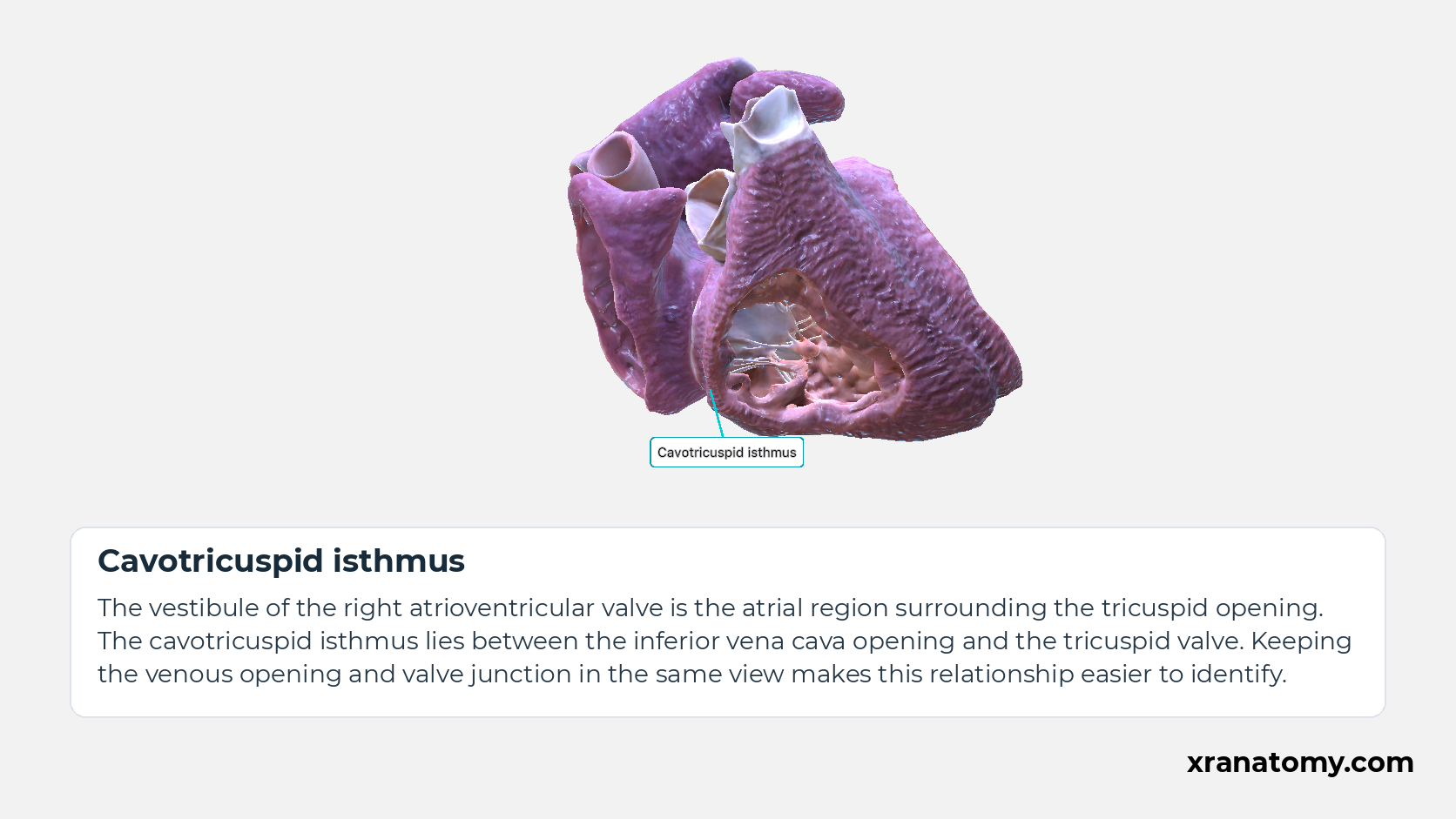
Cavotricuspid isthmus
The cavotricuspid isthmus is the strip of right atrial tissue between the inferior vena cava opening and the tricuspid valve. It forms part of the inferior atrial wall and is an important anatomical route for some atrial electrical circuits.Show less
Illustration by XR Anatomy.
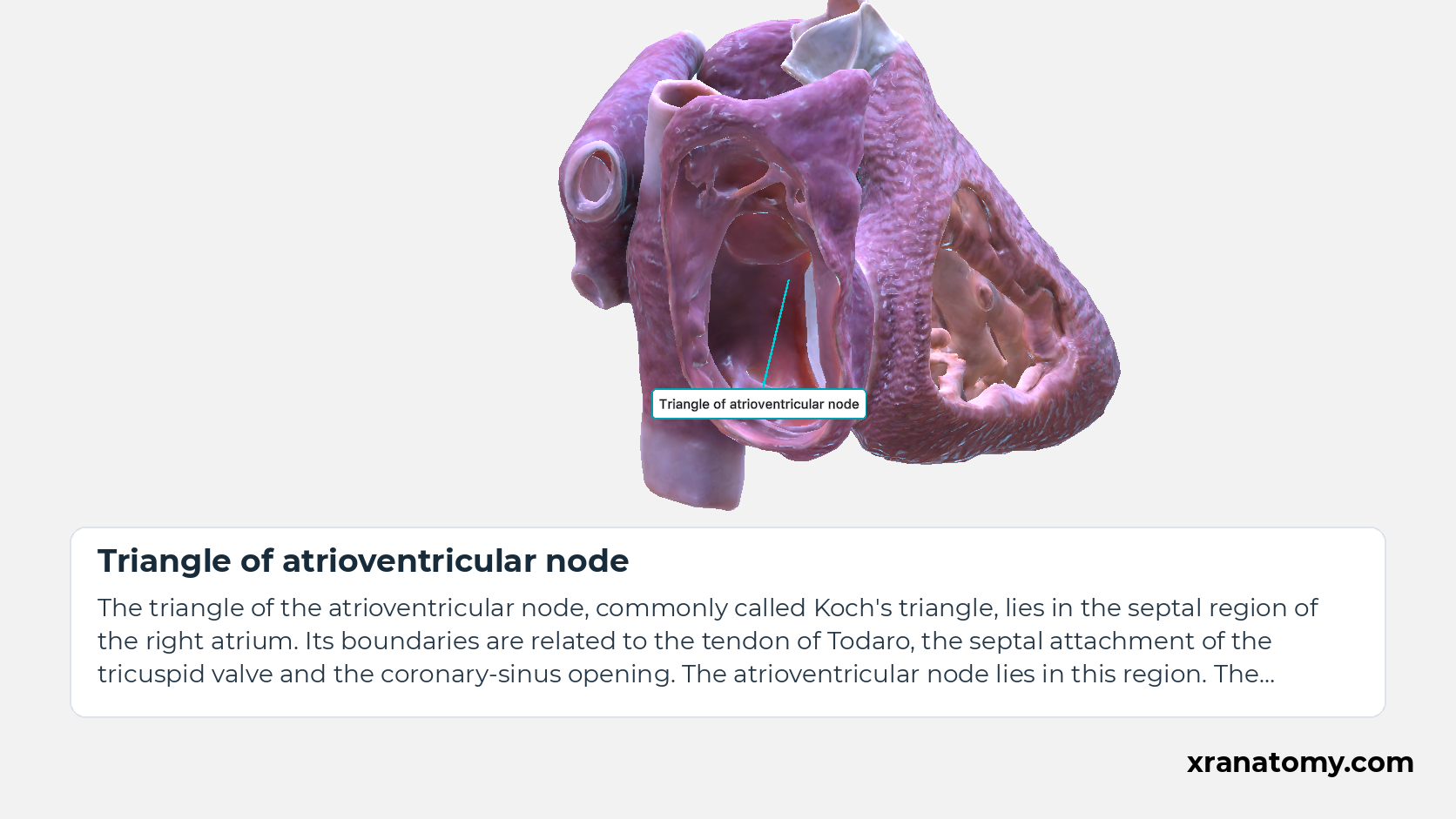
Triangle of atrioventricular node
The triangle of the atrioventricular node, or Koch's triangle, is a region on the septal side of the right atrium. It is bounded by the tendon of Todaro, the septal tricuspid attachment and the coronary sinus opening, with the atrioventricular node near its apex.Show less
Illustration by XR Anatomy.
In depth · continued · 1 more section- Vestibule and septal landmarks
Vestibule and septal landmarks
Vestibule and cavotricuspid isthmus
The vestibule of the right atrioventricular valve is the atrial region surrounding the tricuspid opening. The cavotricuspid isthmus lies between the inferior vena cava opening and the tricuspid valve. The relationship is clearest when the venous opening and the valve junction are seen together.
Triangle of the atrioventricular node
The triangle of the atrioventricular node, commonly called Koch's triangle, lies in the septal region of the right atrium. Its boundaries are related to the tendon of Todaro, the septal attachment of the tricuspid valve and the coronary-sinus opening. The atrioventricular node lies in this region. The nearby fossa ovalis is a landmark of the atrial septum.
